Decellularized cartilage material from pig costal cartilage as well as preparation method and application of decellularized cartilage material

A technology of decellularized cartilage and costal cartilage, applied in medical science, tissue regeneration, prostheses, etc., to achieve the effect of inhibiting polarization reaction, reducing residual DNA content, and reducing the risk of immune rejection

Inactive Publication Date: 2021-11-05
PLASTIC SURGERY HOSPITAL CHINESE ACAD OF MEDICAL SCI
View PDF5 Cites 2 Cited by
  • Summary
  • Abstract
  • Description
  • Claims
  • Application Information

AI Technical Summary

Problems solved by technology

Acellular materials have been widely used in clinical operations, such as joint and bronchial surgery, human-derived acellular dermal matrix materials, pig-derived acellular heart valves, etc., but no acellular cartilage material has been used to fill costal cartilage defects. Related research

Method used


Image

  • Decellularized cartilage material from pig costal cartilage as well as preparation method and application of decellularized cartilage material
  • Decellularized cartilage material from pig costal cartilage as well as preparation method and application of decellularized cartilage material
  • Decellularized cartilage material from pig costal cartilage as well as preparation method and application of decellularized cartilage material

Examples


Embodiment 1

[0024] Example 1: Preparation of decellularized cartilage material from porcine costal cartilage

[0025] Purchase 6 Bama miniature pigs, 5 months old, male, weighing 12-13kg. All animals were provided by the Animal Center of Plastic Surgery Hospital, received humane care in this experiment, and were approved by the Hospital Animal Ethics Committee.

[0026] The following preparation method was used to prepare the decellularized cartilage material from porcine costal cartilage:

[0027] (1) After the pig is anesthetized, the hair in the operation area is shaved, disinfected with tincture of iodine, deiodized with alcohol, and covered with sterile towels;

[0028] After local infiltration anesthesia, a 10cm incision was made on the right side of the lower end of the sternum with a circular knife. The tangent line was parallel to the costal arch, and the skin, subcutaneous and muscle layers were cut in layers to expose the 6th, 7th, and 8th costal cartilages, and the 3rd costal...

Embodiment 2

[0037] Example 2: Implantation of decellularized cartilage material from porcine costal cartilage in costal cartilage defect area

[0038] Purchase 18 healthy New Zealand white rabbits, male, 3 months old, weighing about 2kg. All animals were provided by the Animal Center of Plastic Surgery Hospital, received humane care in this experiment, and were approved by the Hospital Animal Ethics Committee.

[0039] (1) Grouping

[0040] In order to compare the difference between the decellularized cartilage material prepared by the present invention and the prior art, two sets of experimental examples were set up. Experimental example 1 is the decellularized cartilage material prepared according to the preparation method of Example 1, and experimental example 2 is the decellularized cartilage material prepared according to the prior art. In the decellularized cartilage material prepared by conventional methods, lye is used as the decellularized solution in the decellularized step, su...

Embodiment 3

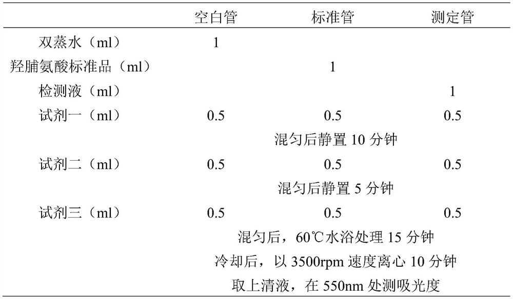
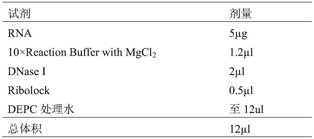
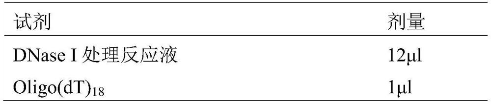
[0045] The following properties of the blank control group and the filling group of the two experimental examples were detected respectively: CT reconstruction of costal cartilage, determination of DNA content, determination of total collagen content, determination of glycosaminoglycan (GAG) content, determination of α-Gal content, degradation rate and porosity Rate determination, histological examination (including hematoxylin and eosin HE staining, Alcian Blue staining, Alizarin Red staining, Masson staining, Safranin O and fast green staining staining), scanning electron microscope measurement Cellular cartilage material pore size, culture and passage of rabbit costal chondrocytes, toxicity test of acellular cartilage material, Live / Dead cell activity test, macrophage polarization reaction (including reagent preparation, PMA-induced THP-1 cells to M0, THP -1 Induces polarization into M1 / M2, detects related proteins in cell supernatant by ELISA, detects cell surface markers b...


PUM

PropertyMeasurementUnit
thicknessaaaaaaaaaa
absorbanceaaaaaaaaaa

Abstract

The invention relates to a decellularized cartilage material from pig costal cartilage and a preparation method and application of the decellularized cartilage material. The preparation method comprises a decellularization step: specifically, a phosphate buffer solution containing trypsin and disodium ethylene diamine tetraacetate is adopted as a decellularized solution, a costal cartilage material is added into the decellularized solution and put into a shaking table, the volume ratio of the decellularized solution to the costal cartilage material is 20: 1, the rotating speed of the shaking table is set to be 150r / min, the action lasts for 72 hours, the temperature is 37 DEG C, and the decellularized solution is replaced every 24 hours. The prepared decellularized cartilage material basically retains the structure and function of an extracellular matrix, a stable environment is provided for cell proliferation and tissue regeneration, most collagenous fiber components in the extracellular matrix are retained, in the costal cartilage defect repairing process, cell migration differentiation and tissue regeneration can be induced, and mechanical support is provided, so that the effect of improving local thoracic depression and deformity is achieved.

Description

technical field [0001] The invention relates to a decellularized cartilage material from pig costal cartilage, a preparation method and an application thereof, and belongs to the technical field of biomaterials. Background technique [0002] With the advantages of no immune rejection, sufficient quantity, good plasticity, good histocompatibility and stable shape, autologous costal cartilage is widely used in the field of plastic surgery, such as auricle reconstruction surgery and costal cartilage rhinoplasty. In the reconstructive operation of auricle reconstruction, 2-3 autologous costal cartilages need to be sculpted and spliced ​​into the auricle shape bracket. Cartilage loss will be left after the autologous costal cartilage is taken, and the loss of multiple costal cartilages may lead to a more obvious thorax Morphological abnormalities, such as partial depression of the chest wall, asymmetric development of bilateral thorax, etc., affect the appearance. In order to re...

Claims


Application Information

Patent Timeline
Patent Type & Authority Applications(China)
IPC IPC(8): A61L27/36
CPCA61L27/3654A61L27/3687A61L27/3691A61L2430/06
Inventor 王长琛潘博蒋海越
Owner PLASTIC SURGERY HOSPITAL CHINESE ACAD OF MEDICAL SCI
Why Eureka
Try Eureka