Free cell marking and separating probe as well as related product and application thereof

A technology of free cells and cells, applied in the field of biomedicine, can solve problems such as less than 330-380

Pending Publication Date: 2022-01-04
沈剑萍
View PDF1 Cites 0 Cited by
  • Summary
  • Abstract
  • Description
  • Claims
  • Application Information

AI Technical Summary

Problems solved by technology

All the methods reported so far are basically: only 1-2 fetal trophoblast cells can be detected per 1ml of maternal blood, which is much lower than 330-380

Method used


Image

  • Free cell marking and separating probe as well as related product and application thereof
  • Free cell marking and separating probe as well as related product and application thereof
  • Free cell marking and separating probe as well as related product and application thereof

Examples


Embodiment 1

[0086] Example 1 Preparation of free cell capture probes with the function of displaying green fluorescent protein

[0087] 1. Add 3ml of fetal bovine serum (FBS) to 27ml of DMEM-10 medium (DMEM medium containing 10% fetal bovine serum); make it into DMEM-20 medium, add to 225cm 2 in a glass culture flask (flask).

[0088] 2. Loosen the cap of the flask that was added with the medium, and put it in a 37°C cell culture incubator to balance for 15 minutes; thaw the frozen 293FT cells (Shanghai Pituo Biotechnology Co., Ltd.) in a 37°C water bath.

[0089] 3. Add the thawed 293FT cells to the flask with balanced medium in step 2, and culture for 2-3 days until the cells are full.

[0090] 4. Wash the cells once with 1-2ml of trypsin, then add 1-2ml of trypsin and put it back in the cell culture incubator at 37°C for 7 minutes, and neutralize the trypsin with 9ml of DMEM-10.

[0091] 5.2-5×10 6 The cells were cryopreserved in separate tubes, passaged at a ratio of 1:15 of the ov...

Embodiment 2

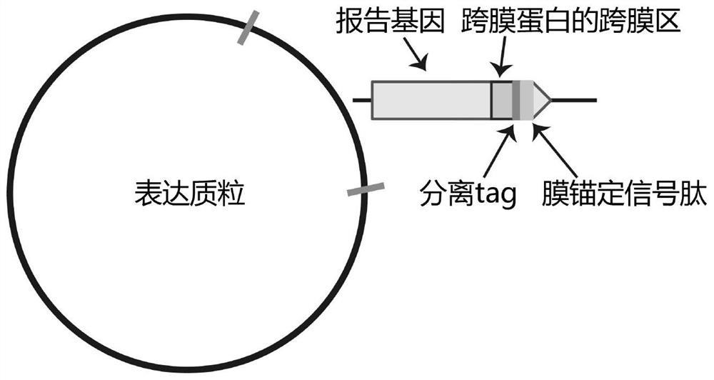
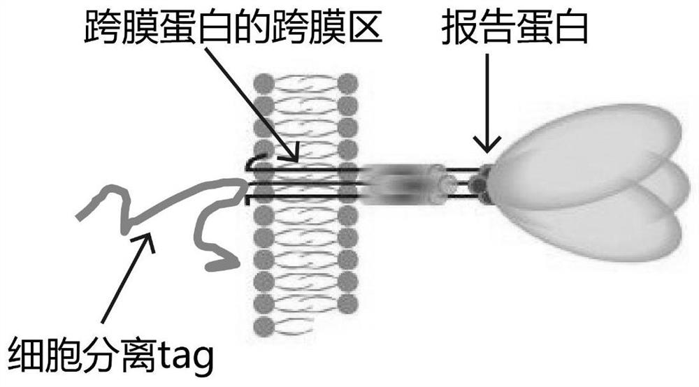
[0109] Example 2 Preparation of green fluorescent protein free cell capture probe with cell surface localization function 1. Use human hepatocyte cDNA as template with plasmid, primer 1 in Table 1 as primer, Table 2 and Table 3 as reaction system and The reaction procedure is to amplify the DNA sequence of the SMIM1 gene except the 1-40 amino acid residues in the intracellular part plus the N-terminal signal peptide by PCR reaction.

[0110] Table 1

[0111]

[0112] Table 2

[0113] mixed primer 1μl 2×SYBR mix 10μl human liver cDNA 1μl Sterilized water 8μl

[0114] table 3

[0115]

[0116] 2. Using the plasmid pClneoEGFP as a template, using primer 2 in Table 1 as a primer, and using Table 4 and Table 5 as a reaction system and a reaction program, amplify the DNA sequence of the EGFP gene without the stop codon by PCR reaction.

[0117] Table 4

[0118] mixed primer 1μl 2×SYBR mix 10μl PclneoEGFP plasmid ...

Embodiment 3

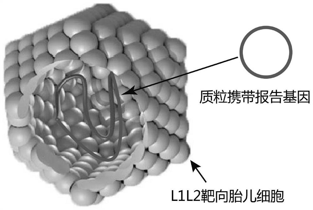
[0141] Example 3 Free cell capture probes label fetal trophoblast cells in the peripheral blood of pregnant women

[0142] 1. Use EDTA anticoagulant tubes to draw 10ml of blood from 5 pregnant women who are pregnant with male babies and 5 pregnant women who are pregnant with female babies at 12 weeks; then draw 10ml of blood from 5 non-pregnant women. Take 1×10 3 Cancer cells (female cervical cancer cell lines) derived from epithelial tissue were mixed into the blood of these 5 non-pregnant women; 10ml of blood from each of 5 men was taken and 1×10 3 Cancer cells derived from epithelial tissue (male prostate cancer cell line); after mixing red blood cell lysate with blood volume 1:1, stand at 4°C for 10 minutes (the operation is carried out in a sterile environment) to break the red blood; use 1ml Gently rinse 3 times with DPBS;

[0143] 2. After resuspending with 200 μl of DMEM medium containing 10% fetal bovine serum placed in a 37°C cell culture incubator for 30 minutes, ...


PUM


Abstract

The invention discloses a free cell marking and separating probe as well as a related product and application thereof, and particularly aims at fetal trophoblast cells or epithelial tissue-derived tumor cells. A free cell capture probe comprises human papilloma virus-like particles and reporter genes carried by the human papilloma virus-like particles and/or protein tag expression genes used for separating target cells. Whether a blood sample of a subject contains fetal trophoblast cells or not can be effectively detected by utilizing the characteristic that the human papilloma virus-like particles selectively infect the placental trophoblast cells, and the aim of separating the fetal trophoblast cells can be achieved after optimization. The probe can also be used for identifying or separating epithelial tissue-derived cells such as epithelial tissue-derived tumor cells in a sample by utilizing the epitheliophilic characteristic of the probe. The method is ingenious in conception, safe and reliable, and has very high sensitivity and specificity.

Description

technical field [0001] The invention relates to the field of biomedicine, in particular to a free cell labeling and separation probe and related products and applications. Background technique [0002] Prenatal diagnosis in early pregnancy is very important to detect genetic defects such as genetic or chromosomal abnormalities in the fetus. Currently, there are three widely used methods for prenatal screening: amniocentesis, chorionic villus sampling, and cell-free fetal DNA (cfDNA) testing. However, the current three technologies have certain limitations. Amniocentesis: professional technicians insert a needle into the amniotic sac of the fetus, take 20-30ml of amniotic fluid, and then conduct cytogenetic tests on the fetal cells in it, usually at 16 weeks of pregnancy; chorionic villus sampling: professional technicians obtain a small amount of placenta A biopsy sample is taken and analyzed for genetic testing, usually at 8-12 weeks. These two detection methods can prov...

Claims


Application Information

Patent Timeline
Patent Type & Authority Applications(China)
IPC IPC(8): C12N5/10C12N15/85C12N15/65C07K14/025C12Q1/02G01N21/64
CPCC12N5/0605C12N5/0693C12N15/85C12N15/65C07K14/005G01N21/6428G01N33/5005C12N2710/20023C12N2510/00C12N2509/00
Inventor 沈剑萍
Owner 沈剑萍
Why Eureka
Try Eureka