Ultrafine iron oxide nanoparticle-based magnetic resonance imaging T1 contrast agent

A technology of iron oxide nanometer and magnetic resonance imaging, which is applied in the direction of nanomagnetism, nanotechnology for materials and surface science, nanotechnology, etc., and can solve problems such as T2 weighted image signal interference

Pending Publication Date: 2022-01-11
セラベスト カンパニーリミテッド
View PDF1 Cites 0 Cited by
  • Summary
  • Abstract
  • Description
  • Claims
  • Application Information

AI Technical Summary

Problems solved by technology

Although iron oxide nanoparticles synthesized by pyrolysis method have been reported to have better MRI T2 contrast than those developed by co-precipitation method, T1-weighted images are preferred over T2-weighted images for clinical applications because T1-weighted images are more accurate, and T2-weighted images suffer from severe signal interference

Method used


Image

  • Ultrafine iron oxide nanoparticle-based magnetic resonance imaging T1 contrast agent
  • Ultrafine iron oxide nanoparticle-based magnetic resonance imaging T1 contrast agent
  • Ultrafine iron oxide nanoparticle-based magnetic resonance imaging T1 contrast agent

Examples


Embodiment 1-1

[0080] Example 1-1: Preparation of 3nm ultrafine iron oxide nanoparticles

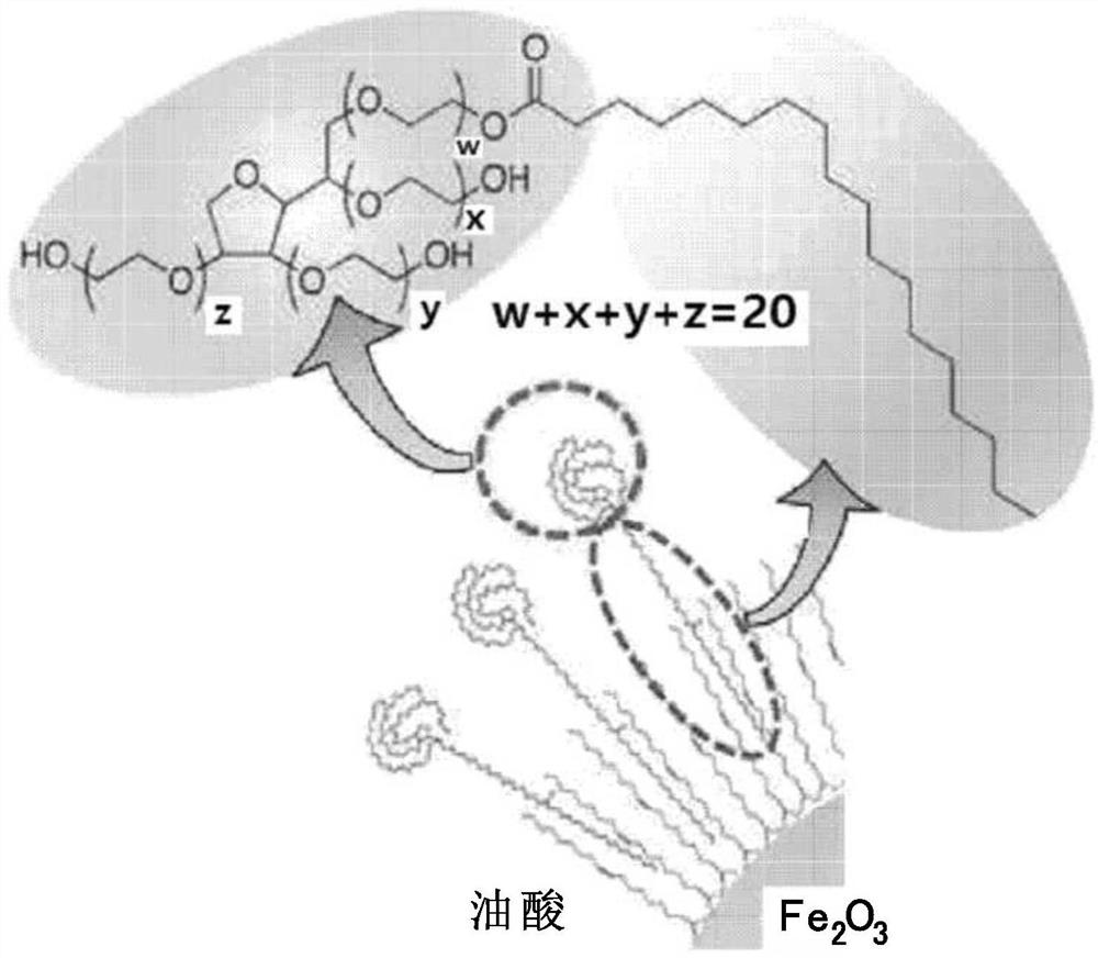
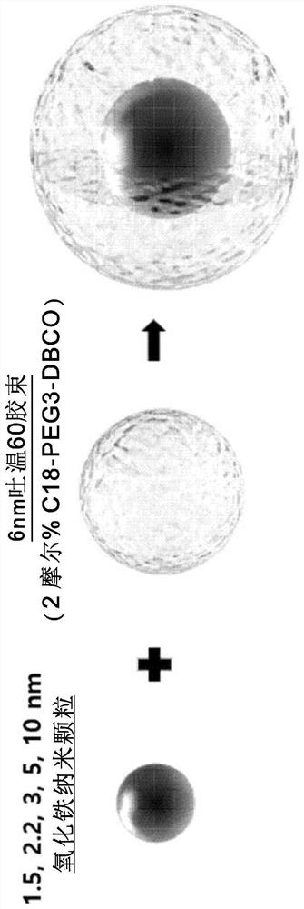
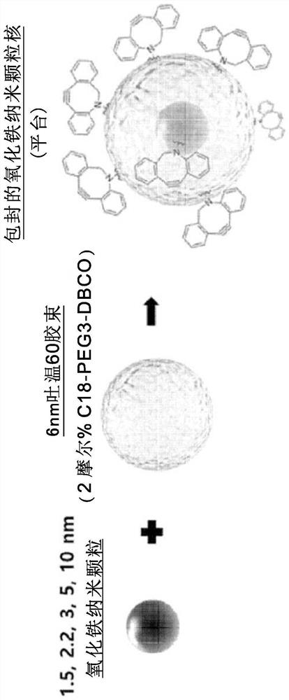
[0081]1.8 g (2 mmol) iron oleate, 0.57 g (2 mmol) oleic acid and 1.61 g (6 mmol) oleyl alcohol were mixed with 10 g diphenyl ether. The mixture was placed in a round bottom flask. The flask was evacuated at 80°C for about 1 hour to remove air. Thereafter, an inert environment was created under argon flow. The reaction was performed while increasing the temperature to 250°C at a rate of 10°C / min. The color of the reaction mixture turned black as the reaction proceeded. Reacting at 250°C for 30 minutes gave 3 nm nanoparticles, which were rapidly cooled and washed with excess acetone. The obtained precipitate was dispersed in chloroform or hexane as an organic solvent.

Embodiment 1-2

[0082] Example 1-2: Preparation of 5nm ultrafine iron oxide nanoparticles

[0083] 1.8 g (2 mmol) iron oleate and 0.28 g (1 mmol) oleic acid were mixed with 10 g octadecene. The mixture was placed in a round bottom flask. The flask was evacuated at 80°C for about 1 hour to remove air. Thereafter, an inert environment was created under argon flow. The reaction was carried out while raising the temperature to 317°C at a rate of 10°C / min. The color of the reaction mixture turned black as the reaction proceeded. The reaction was carried out at 317°C for 30 minutes to obtain about 5 nm nanoparticles, which were rapidly cooled and washed with excess acetone. The obtained precipitate was dispersed in chloroform or hexane as an organic solvent.

Embodiment 1-3

[0084] Example 1-3: Preparation of 10 nm ultrafine iron oxide nanoparticles

[0085] 1.8 g (2 mmol) iron oleate and 0.28 g (1 mmol) oleic acid were mixed with 10 g octadecene. The mixture was placed in a round bottom flask. The flask was evacuated at 80°C for about 1 hour to remove air. An inert environment was created under a stream of argon. The reaction was carried out while raising the temperature to 315°C at a rate of 10°C / min. The color of the reaction mixture turned black as the reaction proceeded. Reacting at 315°C for 30 minutes gave about 10 nm nanoparticles, which were rapidly cooled and washed with excess acetone. The obtained precipitate was dispersed in chloroform or hexane as an organic solvent.


PUM

PropertyMeasurementUnit
diameteraaaaaaaaaa
diameteraaaaaaaaaa
particle diameteraaaaaaaaaa

Abstract

Provided is a magnetic resonance imaging T1 contrast agent comprising: a fine iron oxide nanoparticle core; and a micelle for encapsulating the core particles, wherein the micelle comprises a non-ionic surfactant containing: a hydrophilic residue containing at least two chains; and a hydrophobic residue consisting of at least one C10-C30 hydrocarbon chain. The T1 contrast agent of the present invention is a novel fine iron oxide nanoparticle-based T1 contrast agent that can replace conventional gadolinium-based T1 contrast agents, is not harmful to the human body, and exhibits a uniform contrast effect due to rapid distribution in blood and the uniform size. In addition, the T1 contrast agent enables image observation for at least 1 hour, or up to a maximum of 2 hours and is excreted through the kidneys and liver, and thus can address the problems of conventional gadolinium-based contrast agents.

Description

technical field [0001] The invention relates to a magnetic resonance imaging (Magnetic Resonance Imaging, MRI) T1 contrast agent based on ultrafine iron oxide nanoparticles, more particularly, to a magnetic resonance imaging based on surface-hydrophilized iron oxide nanoparticles of uniform size Imaging T1 contrast agent. Background technique [0002] Magnetic Resonance Imaging (MRI) is a method of acquiring the anatomical information, physiological information and biochemical information of the body by using the phenomenon that the spin of hydrogen atoms relaxes in a magnetic field. MRI is one of the non-invasive diagnostic tools currently used for real-time imaging of body organs in living humans and animals. [0003] Due to its diverse and precise applications in biological sciences and medicine, MRI is performed by introducing foreign bodies into the body to increase image contrast. These materials are called contrast agents. Superparamagnetic and paramagnetic materia...

Claims


Application Information

Patent Timeline
Patent Type & Authority Applications(China)
IPC IPC(8): A61K49/18
CPCA61K49/1809A61K49/1839A61K49/186A61K49/1833A61K49/1812A61K2123/00A61K49/1887B82Y5/00B82Y25/00B82Y30/00B82Y40/00
Inventor 李润相玄泽焕郑载民李载成朴志龙金奎完申在烉高根培李活朴银我
Owner セラベスト カンパニーリミテッド
Why Eureka
Try Eureka