Manufacturing method of animal tiny tissue paraffin section
A paraffin section and production method technology, applied in the biological field, can solve the problems of lack of standardized and reliable technical guidance, difficulty in the production of microscopic tissue pathological sections, etc., and achieve the effects of preventing floating, not easy to lose, and high definition.
- Summary
- Abstract
- Description
- Claims
- Application Information
AI Technical Summary
Problems solved by technology
Method used
Image
Examples
Embodiment 1
[0086] Example 1 Making mouse ovary paraffin sections
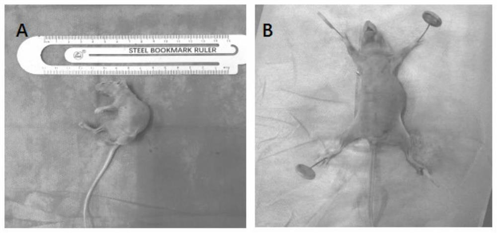
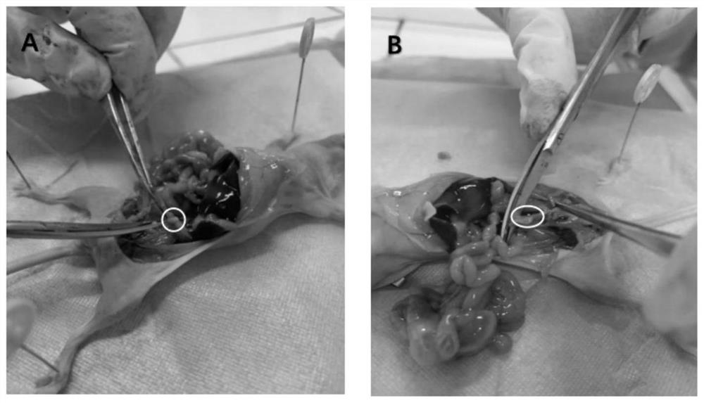
[0087] like Figure 1-2 , 5, 8-9, 13, 16-17, 20-21, kill the mice and dissect the mice, correctly identify the mouse ovarian tissue and quickly and completely collect the material, obtain 2 mouse ovarian tissues, and put them into 10 % neutral formaldehyde fixative for 48h. After the fixation is completed, remove the mouse ovarian tissue from the fixative and place it in a wide-mouthed bottle, tie the bottle mask with gauze and fasten it with a thread, and use a rubber tube to drain the tap water into the wide-mouthed bottle to allow the water to overflow from bottom to top. Achieve overflow flushing of tiny tissues.
[0088] After 2 hours of overflow rinsing, the mouse ovarian tissue was taken out, marked with eosin staining solution on the surface of the mouse ovary, and then wrapped with lens tissue to obtain tiny tissue wraps. The wrapping method is as follows: Wet the lens tissue with AF solution or water, and p...
Embodiment 2
[0099] Example 2 Preparation of mouse adrenal paraffin sections
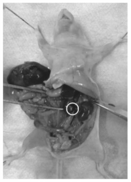
[0100] like image 3 , 6 , 8, 10, 14, 16, 18, 22-23, mouse adrenal paraffin sections were prepared according to the slicing method of the present invention.
[0101] S1. Sampling: Correctly identify the adrenal glands of mice and take samples quickly and completely. One mouse has two intact adrenal tissues.
[0102] S2. Fixation: Put the mouse ovarian tissue into 10% neutral formaldehyde fixative solution for 48 hours; after fixation, remove the mouse ovarian tissue and place it in a wide-mouth bottle, tie the bottle mask with gauze and fasten it with a thread, and use a rubber The tube drains tap water into the jar, allowing the water to overflow from bottom to top to flush the mouse ovarian tissue.
[0103] S3. Marking and wrapping: After rinsing, the surface of mouse ovarian tissue was marked with eosin staining solution; after marking, it was wrapped with lens tissue to obtain micro-tissue wrapping.
...
Embodiment 3
[0111] Example 3 Making mouse ovary paraffin sections
[0112] The mice were sacrificed and dissected. The mouse ovarian tissue was correctly identified and obtained quickly and completely. Two mouse ovarian tissues were obtained, which were placed in a 10% neutral formaldehyde fixative solution for 48 hours. After the fixation is completed, remove the mouse ovarian tissue from the fixative and place it in a wide-mouthed bottle, tie the bottle mask with gauze and fasten it with a thread, and use a rubber tube to drain the tap water into the wide-mouthed bottle to allow the water to overflow from bottom to top. Achieve overflow flushing effect on tiny tissues. After overflow washing for 10 minutes, the mouse ovarian tissue was taken out, and the surface of the mouse ovary was marked with eosin staining solution, and then wrapped with lens tissue to obtain tiny tissue wraps.
[0113] Put the micro-tissues into the embedding box, cover the cover of the embedding box, and carry...
PUM
| Property | Measurement | Unit |
|---|---|---|
| melting point | aaaaa | aaaaa |
| thickness | aaaaa | aaaaa |
Abstract
Description
Claims
Application Information
 - R&D
- Intellectual Property
- Life Sciences
- Materials
- Tech Scout
- Unparalleled Data Quality
- Higher Quality Content
- 60% Fewer Hallucinations
Browse by: Latest US Patents, China's latest patents, Technical Efficacy Thesaurus, Application Domain, Technology Topic, Popular Technical Reports.
© 2025 PatSnap. All rights reserved.Legal|Privacy policy|Modern Slavery Act Transparency Statement|Sitemap|About US| Contact US: help@patsnap.com


