High resolution Adama conversion micro-image analyzer

A technology of Hadamard transformation and microscopic image, applied in high sensitivity, high signal-to-noise ratio, can solve the problem of low resolution, achieve high resolution, avoid measurement error, and solve the effect of mechanical modulation accuracy

Inactive Publication Date: 2004-05-05
WUHAN UNIV
View PDF1 Cites 1 Cited by
  • Summary
  • Abstract
  • Description
  • Claims
  • Application Information

AI Technical Summary

Problems solved by technology

Patent ZL 94 1 07751.9 discloses a multifunctional Hadamard transform microscopic imager, which consists of a light source, a fluorescence microscope, image compression imaging, a Hadamard transform modulation template, a monochromator, a photomultiplier tube detector, and a computer. The patent uses a 15×17 two-dimensional template and photomultiplier tube for single-point detection, with low resolution, and each image consists of 255 pixels

Method used


Image

  • High resolution Adama conversion micro-image analyzer
  • High resolution Adama conversion micro-image analyzer
  • High resolution Adama conversion micro-image analyzer

Examples


Embodiment 1

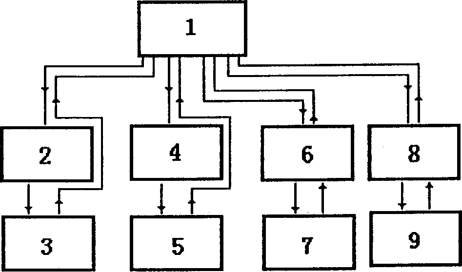
[0030] Embodiment 1, Hadamard transform microspectral image analyzer.

[0031] See figure 1 , figure 2 . The analysis sample is placed on the research-type epi-fluorescence microscope stage produced by Chongqing Optical Instrument Factory, and the image signal (transmitted light or fluorescence) of the analysis sample is collected by the microscope objective lens, and the Hadamard transform optical system on the fluorescence microscope The component total reflection mirror P is pushed to the optical path, and the image of the field of view is turned 90 degrees, and then enters the image composed of converging lens group L1, mirror M1, Hadamard transformation template I and diaphragm, collimating lens group L2, grating G, mirror M1, The cylindrical lens L3 passes through the slit S, namely the Hadamard transform microspectral image analyzer as shown in the figure. The lens group L1 compresses the image of the microscopic field of view, and the image is clearly imaged on the...

Embodiment 2

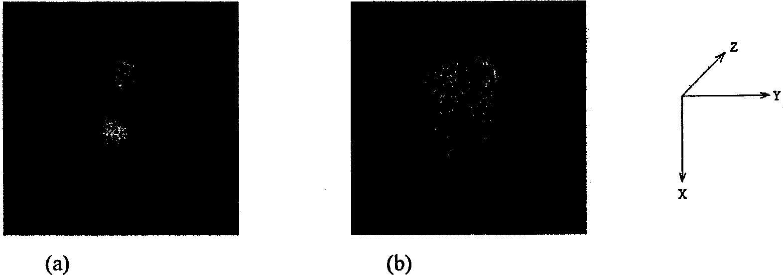
[0033] Example 2: Quantitative analysis of fluorescent images of human breast cancer cells, morphology of nuclei and DNA content.

[0034] Take direct smears of breast tumor puncture fluid, use normal human peripheral venous blood lymphocytes or normal epithelial cells in breast tumor specimens as standard diploid cells, and use acridine orange (AO, 50 μg / mL, containing 1% Triton X- 100) is a cellular DNA fluorescent probe, and the DNA content (ploidy) of human breast tumor cells is measured with a Hadamard transform microspectral image analyzer. At least 20 tumor site cells are analyzed for each patient, and two quantifications are measured for each cell. Data: Nuclei DNA content (in arbitrary units) and morphology (measured area in pixels). The measurement data shows that the Hadamard transform spectral image can correctly reflect the basic fluorescence distribution of cells, and obtain quantitative fluorescence intensity and distribution information. Compared with standard...

Embodiment 3

[0035] Example 3: Preliminary screening of anticancer drugs for cell cycle non-specific drugs.

[0036] Using human peripheral venous blood lymphocytes or mouse hepatocytes as materials, using fluorescent probe AO (acridine orange) to trace to determine whether the drug can enter the nucleus and its ability to bind to nuclear DNA, a method targeting nuclear DNA is proposed. A new method for preliminary screening of anticancer drugs based on cell cycle non-specific drugs. The specific method is to label human peripheral venous blood lymphocytes or mouse liver cells with fluorescent probe AO, and use Hadamard transform microscopic fluorescence image analyzer to detect single cells. The fluorescence intensity of the AO-DNA complex in the nucleus at emission peak 530nm. The fluorescence inhibition rate of the drug on the cellular DNA-AO complex ((F 0 -F) / F 0 ×100%), that is, the strength of the interaction between the drug and the cell DNA, is used as the basis for the prelimina...


PUM


Abstract

The present invention discloses a high-resolution high-sensitivity Adama transform microimage analysis instrument. Every image is formed by 260,000 pixels. Said analysis instrument contains 511 unit Adama template and high-sensitivity linear array CCD detector. It adopts the Adama transform space multichannel imaging technique and combines it with microscopic technology, can provide image of microbody and quantitative analysis result of image, and can be used for making microfluorescent spectrum image and microtransmission spectrum image analysis so as to create a quick, sensitive, accurate and applicable microscopic spectrum image quantitative analysis technique for cell molecular biology and other subject fields. Its measurement accuracy and its sensitivity are high, it is applicable to faint light signal detection, it can be used for measuring unicellular topography and cellular fluorescent intensily, and its analysis result reproducibility is good.

Description

technical field [0001] The invention relates to a high-resolution Hadamard transform microscopic image analyzer, which combines the 511 coding Hadamard transform multi-channel spatial imaging technology with the microscopic technology, uses the linear array CCD as the detector, and measures the tiny The new instrument for the spectral image of the object's microscopic fluorescence and microscopic transmitted light is a new type of measuring instrument with high sensitivity and high signal-to-noise ratio, especially suitable for the analysis of weak microscopic spectral images. It belongs to the technical field of medical testing instruments, the field of analytical testing technology, and the field of biochemical technology. It is the product of the combination of light modulation technology, instrument manufacturing, computer and modern instrument analysis. Background technique [0002] Hadamard Transform (Hadamard Transform) spectroscopy is a light modulation technique sim...

Claims


Application Information

Patent Timeline
IPC IPC(8): G01N21/17G01N21/64G01N33/483G01N33/52
Inventor 陈观铨唐宏武叶盛元潘伟正曾立波陈蓓
Owner WUHAN UNIV
Why Eureka
Try Eureka