Photo-acoustic functional brain imaging method and device

A brain function and imaging technology, which is applied in medical science, sensors, vaccination and ovulation diagnosis, etc., can solve the problems of inability to capture dynamic changes in brain function, high price, low time resolution, etc., and achieve fast and non-destructive brain function imaging with low cost , easy to apply and promote the effect

Inactive Publication Date: 2006-12-27
SOUTH CHINA NORMAL UNIVERSITY
View PDF0 Cites 40 Cited by
  • Summary
  • Abstract
  • Description
  • Claims
  • Application Information

AI Technical Summary

Problems solved by technology

Poor spatial resolution and temporal resolution are the fatal flaws of SPECT. Therefore, SPECT imaging is difficult to distinguish the complex neural structures in the brain, and it is also unable to capture the dynamic changes of brain function in a short period of time.
The spatial resolution of PET has improved compared to SPECT, but the temporal resolution is still low
However, fMRI reflects hemodynamic blood changes through the principle of blood oxygenation level dependent (BOLD), does not involve radioactive elements, and has better spatial and temporal resolution, but an important disadvantage of fMRI and PET is that it is expensive.

Method used


Image

  • Photo-acoustic functional brain imaging method and device
  • Photo-acoustic functional brain imaging method and device
  • Photo-acoustic functional brain imaging method and device

Examples


Embodiment 1

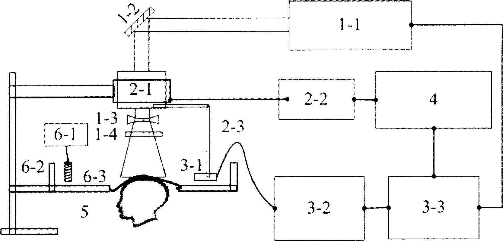
[0047] A five-week-old Kunming mouse weighing about 30 g was tested with the device of the present invention. Before the test, the hair on the head of the mice was depilated with a self-made depilatory solution (8% sodium sulfide glycerin paste), without damaging the epidermis and cranium of the brain. Anesthetize the mouse intravenously with 2% sodium pentobarbital at a dose of 30 mg / kg, then place the mouse on a three-dimensional lifting frame, fix the head with a fixer, and place it under the polyethylene film in the middle of the acoustic coupling pool , calibrate the scanning plane of the ultrasound probe so that it is on the same plane as the mouse brain.

[0048] In the experiment, a 532nm pulsed laser was selected, with a pulse width of 10ns, a repetition rate of 15Hz, and an output pulse energy density of 8mJ / cm 2 . The photoacoustic signal of the brain received by the ultrasonic detector is sent to the digital oscilloscope after being amplified. In the experiment,...

Embodiment 2

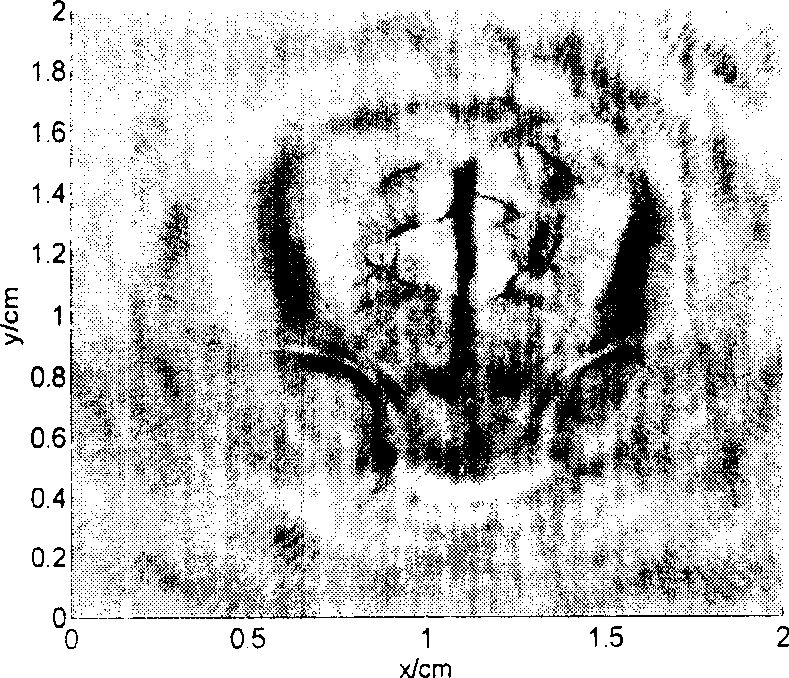
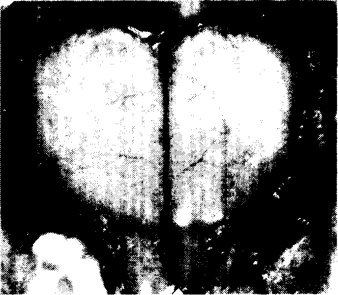
[0050] The device of the present invention is used to test a brain injury model mouse. Before the experiment, the mice were anesthetized by intravenous injection, the hair on the scalp was removed, and then a needle was artificially used to pierce the scalp and skull on the lower left side of the mouse's head, and inserted into the surface of the cerebral cortex to create a model of intracerebral injury and bleeding. After the mouse was fixed, according to the method of Example 1, a 1064nm pulsed laser was selected, with a pulse width of 10ns, a repetition rate of 15Hz, and an output pulse energy density of 8mJ / cm 2 , scan and collect the photoacoustic signal of the brain to obtain a photoacoustic tomographic image of the brain, as shown in FIG. 3( a ). After the experiment, the mice were sacrificed and dissected, and the anatomical diagram is shown in Figure 3(b). It can be seen from Fig. 3(a) that the area of ​​brain injury, the situation of blood vessel destruction, and th...

Embodiment 3

[0052] A drug-stimulated mouse was tested with the device of the present invention. The drug is acetazolamide (Acetazolamide), which is an isocyclic sulfonamide, is a carbonic anhydrase inhibitor, can make cerebral vasodilators, and is used for cerebral blood perfusion SPECT imaging to evaluate cerebral vascular reserve function. It is affirmed and called "ACZ Brain Stress Test", which can increase cerebral blood flow and dilate cerebral blood vessels. In the test, the same mouse was used to perform photoacoustic scanning according to the method of Example 1 before and after feeding the drug, and the resulting images were compared and compared. Feed it to mice with a dose of 25ul / g, and it will start to take effect after 30 minutes, and the drug effect will reach the peak after 2 hours. Thirty minutes after the experiment, the mice were awake and recovered. Figure 4(a) is the brain image before drug administration, and Figure 4(b) is the brain image after drug administration...


PUM

PropertyMeasurementUnit
Wavelengthaaaaaaaaaa

Abstract

The invention relates to an opto-acoustic brain function imaging method. The method realizes imaging of brain blood vessel structure distribution observation and function monitoring via the opto-acoustic tomographic image of the cortex blood vessels. Brain damage and cerebral hemorrhage variation degree can be monitored, and the parameters such as cerebral blood flow, brain oxygen consumption and brain oxygen saturation etc can be reflected via the blood vessel form distribution, blood diameter variation and opto-acoustic signal intensity variation of blood vessels. The laser generation component, acoustic signal acquisition component and computer among the devices realizing the mentioned method are electrically connected in turn. The rotary scanning mechanism is electrically connected with the computer. The sample fixation components are connected with the acoustic coupling components in turn. The invention is characterized by convenient operation and sensitive and quick performance, can make imaging analysis of blood vessels without damage, and then monitor brain function, which can thus provide imaging basis for brain function judgment.

Description

technical field [0001] The present invention relates to medicine and medical devices. In more detail, it is a method of photoacoustic brain functional imaging. [0002] The invention also relates to a device for carrying out the above-mentioned method. Background technique [0003] Brain functional imaging is a new medical imaging that uses brain local imaging technology to observe and analyze the high-level functional activities of the brain without any damage, and then evaluate the brain function. [0004] The first thing in brain science research is experimental research, especially the study of the dynamic process of the living brain under non-invasive conditions. Currently, methods commonly used in brain functional imaging include single photon emission computed tomography (SPECT: Single Photon Emission Computed Tomography), positron emission tomography (PET: Positron Emission Tomography) and nuclear magnetic resonance functional imaging (f...

Claims


Application Information

Patent Timeline
IPC IPC(8): A61B5/00A61B10/00
Inventor 邢达杨思华
Owner SOUTH CHINA NORMAL UNIVERSITY
Why Eureka
Try Eureka