Preparing method and use of chitosan-containing nano fibrous tissue recovery support

A nanofiber and tissue repair technology, which is applied in the fields of scaffolds, medical science, and prostheses, can solve the problems of PVA being unable to degrade and fail to meet the mechanical properties of tissue repair, and achieve good mechanical properties, high porosity, and the effect of promoting rehabilitation

Inactive Publication Date: 2007-02-28
DONGHUA UNIV
View PDF0 Cites 26 Cited by
  • Summary
  • Abstract
  • Description
  • Claims
  • Application Information

AI Technical Summary

Problems solved by technology

Chitosan is a deacetylation product of chitin, which has a structure similar to proteoglycan in natural extracellular matrix, and is a natural polymer biomaterial that is biorenewable, non-antigenic, biodegradable and has good biocompatibility , is widely used in wound dressings, drug release, and tissue engineering, but the preparation of tissue repair scaffolds by electrospun chitosan alone cannot meet the needs of mechanical properties in tissue repair
Recently, there have been attempts to use chitosan and polymer materials for electrospinning at home and abroad, such as m

Method used


Image

  • Preparing method and use of chitosan-containing nano fibrous tissue recovery support
  • Preparing method and use of chitosan-containing nano fibrous tissue recovery support
  • Preparing method and use of chitosan-containing nano fibrous tissue recovery support

Examples


Embodiment 1

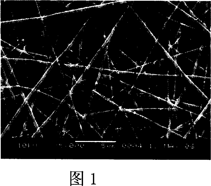
[0022]Dissolve 0.08 g of squid bone chitosan in 2 ml of HFIP / TFA (V / V, 9 / 1) mixed solvent, stir magnetically until completely dissolved, dissolve 0.08 g of P(LLA-CL) in 2 ml HFIP, stir magnetically until completely dissolved, After the two are completely dissolved, they are mixed at a volume ratio of 1:1, stirred evenly by magnetic force to obtain 4 ml of a mixed spinning solution with a total mass volume ratio of 4%. Inhale the mixed solution into the syringe, control the advance speed of the micro-injection pump to 0.6ml / h, select a No. 7 stainless steel needle and connect it to a 20KV high voltage, and use a grounded aluminum foil to receive the fiber filament at a distance of 13cm from the needle tip. After 6 hours, a disordered nanofiber film with an average diameter of about 205 nm was formed on the aluminum foil.

Embodiment 2

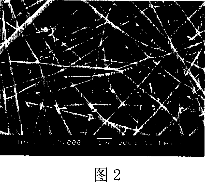
[0024] Dissolve 0.16 g of squid bone chitosan in 2 ml of HFIP / TFA (V / V, 9 / 1) mixed solvent, stir magnetically until completely dissolved; dissolve 0.08 g of P(LLA-CL) in 1 ml HFIP, stir magnetically until completely dissolved After the two are completely dissolved, mix by volume ratio 1: 2 (chitosan / P(LLA-CL)), magnetic stirring and mixing evenly obtain the total concentration of 8% mixed spinning solution 3ml, chitosan in its solute The mass ratio of / P(LLA-CL) was 1:2. Inhale the mixed solution into the syringe, control the advance speed of the micro-injection pump to 1ml / h, select a No. 9 stainless steel needle and connect it to a 20KV high voltage, and use a grounded aluminum foil to receive the fiber filament at a distance of 15cm from the needle tip. A film of disordered nanofibers was received on the aluminum foil after 3 hours.

Embodiment 3

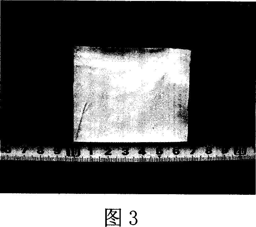
[0026] Dissolve 0.02 g of crab shell chitosan in 1 ml of HFIP / FA (V / V, 9 / 1) mixed solvent, stir magnetically until completely dissolved; dissolve 0.02 g of P(LLA-CL) in 1 ml HFIP, stir magnetically until completely dissolved Dissolving; after the two are completely dissolved, uniformly mix, magnetically stir and mix evenly to obtain chitosan / P(LLA-CL) with a volume ratio of 1:1 and a total concentration of 2% mixed spinning solution 2ml. Inhale the mixed solution into the syringe, control the advance speed of the micro-injection pump to 0.5ml / h, select a No. 9 stainless steel needle and connect it to a 20KV high voltage, and use a grounded aluminum foil to receive the fiber filament at a distance of 20cm from the needle tip. After 4 hrs a film made of disordered nanofibers was received on the aluminum foil. In this scheme, the chitosan solution obtained by HFIP / FA (V / V, 9 / 1) has a high viscosity, and there are a few droplets during the spinning process of the mixed solution. ...


PUM


Abstract

The invention discloses a preparing method and application of nanometer fiber tissue restoring rack with chitose, which comprises the following steps: dissolving chitose in the composite solution with hexafluoroisopropanol or trifluoroacetic acid and hexafluoroisopropanol or aminic acid, dissolving the mixture of polylactic acid-polycaprolactone in the hexafluoroisopropanol, stirring evenly to obtain the nanometer fiber spinning solution, adjusting pressure at 5-30KV and injection speed at 0.5-2 ml/h and receiving distance at 10-30cm.

Description

technical field [0001] The present invention relates to a preparation technology of a biomaterial scaffold for tissue repair, in particular to an electrospun chitosan-containing P(LLA-CL) (polylactic acid-polycaprolactone blend) nanofiber tissue repair Preparation method of scaffold. Background technique [0002] Defects and abnormal functions of tissues and organs are common diseases. The usual solution is organ transplantation, but there is a shortage of donors, which can easily lead to problems such as immune diseases. Emerging tissue engineering can use the principles of biology and engineering to prepare substitutes to replace damaged tissues and organs or restore their functions to achieve therapeutic purposes [Langer, Robert, vacanti et al, Tissue Engineering. Science 260.920-9261993.] . The three elements of tissue engineering are bioactive scaffolds, seed cells, and tissue construction techniques. Currently, the screening of suitable biodegradable bioactive scaff...

Claims


Application Information

Patent Timeline
IPC IPC(8): A61L27/26A61L27/58A61L27/54A61L27/56A61F2/82
Inventor 莫秀梅陈峰何创龙
Owner DONGHUA UNIV
Why Eureka
Try Eureka