Methods and compositions for achieving hemostasis and stable blood clot formation

a technology of hemostasis and blood clot formation, applied in the field of methods and compositions for achieving hemostasis and blood coagulation, can solve the problems of significant morbidity, current available approaches have limitations in effectiveness, safety, applicability and ease of use, and tissue sealants are not hemostats per se, etc., to achieve rapid action, promote coagulation, and produce hemostasis

Active Publication Date: 2019-12-31
ENDOMEDIX
View PDF116 Cites 8 Cited by
  • Summary
  • Abstract
  • Description
  • Claims
  • Application Information

AI Technical Summary

Benefits of technology

[0008]The invention provides a “tunable” biopolymer system that addresses heretofore unmet needs in the field of hemostasis. In this system, two processed natural polysaccharides in the form of separate solutions are simultaneously mixed and applied onto a bleeding site, where they form a hydrogel device in situ. The hydrogel can form and produces hemostasis within seconds after application. The system may also be designed or “tuned” to produce hydrogels and facilitate hemostasis more slowly depending on the specific method of use. Rapid hemostasis can also contribute to the reduction of major complications in some surgical procedures by facilitating a shorter procedure time. It is believed that the rapid hemostasis acts to quickly stop bleeding and the hydrogel comprises specific intrinsic properties resulting in the simultaneous capture, aggregation and concentration of platelets and red blood cells to initiate the cascade of coagulation, which is accomplished without the use of exogenously added clotting agents, drugs or other chemicals. The resulting hemostatic action assures that the device does not act simply as a tamponade or sealant that might allow the pooling of blood and formation of a hematoma under a sealant.
[0009]The hydrogel itself forms without the use of a third chemical or synthetic cross-linking agent and thus avoids the potential for irritation and cytotoxicity or the interference with predictable rapid biodegradation associated with cross-linking agents. Furthermore, due to the internal chemistry, the hydrogel itself gently and predictably contracts over time and does not swell, even when immersed in blood or body fluids. This feature permits the hemostats described herein to be used in brain, spinal or other surgical procedures in which swelling of a hemostatic agent can lead to mass effects and subsequent complications, which themselves require medical or surgical intervention.
[0010]Furthermore, the hydrogel provided herein is essentially transparent, so that a surgeon can visually verify its action and effect in real time. This feature allows surgeons to proceed with the surgical procedure and close the site while verifying hemostasis, in contrast to hemostats which are opaque.
[0011]Furthermore, the hydrogel provided herein is cohesively strong and thus can quickly establish its initial tamponade effect. Furthermore, the hemostat is also adherent and does not migrate away from the application site. The hydrogel forms in intimate continuous contact with the bleeding site so that all sources of bleeding are exposed and treated to the hemostatic action of the device.
[0077]The hemostatic composition, when applied to the location, can fill a cavity at the location without inducing compression of tissue surrounding the cavity when the hemostatic composition is exposed to physiological fluid or a body fluid. It is understood that hemostasis can be achieved from about 5 seconds to about 120 seconds, from about 10 seconds to about 120 seconds, from about 15 seconds to about 120 seconds, from about 5 seconds to about 60 seconds, from about 10 seconds to about 60 seconds, from about 15 seconds to about 60 seconds, from about 5 seconds to about 30 seconds, from about 10 seconds to about 30 seconds, from about 15 seconds to about 30 seconds, from about 5 seconds to about 15 seconds, or from about 10 seconds to about 15 seconds, after the hemostatic hydrogel composition is applied to the location.

Problems solved by technology

Significant blood loss can be fatal or cause significant morbidity, and surgeons and medical staff routinely compensate for blood loss with transfusions and the use of recirculating devices.
However no single approach is effective or suitable under all circumstances, and the currently available approaches have limitations in effectiveness, safety, applicability and ease of use.
However, tissue sealants are not hemostats per se and allow the pooling of blood underneath the seal in cases of moderate bleeding that may result in the formation of a hematoma.
Hemostatic products which require time-consuming application or which produce slow effective action can be costly both in terms of financial costs due to greater operating room time and in terms of post procedure patient morbidity.
Some techniques and products have limited effectiveness in certain applications, which may require a surgeon to reapply the product or switch to another product, which can be both time consuming and costly.
Under certain circumstances standard ligature techniques are inappropriate or not feasible.
However, the efficacy of these products is often variable and their use has been associated with adverse effects which can provide significant challenges during a surgical procedure.
Such challenges become especially pronounced in certain surgical procedures such as brain and spinal surgeries.
For example, certain sponges derived from porcine material can absorb up to 45-times their weight in blood and fluids resulting in significant swelling of the product that can lead to a patient experiencing adverse reactions such as pain, paresis or paralysis that can require revision surgery to address.
Gelatin-based devices typically swell during use and have been reported to cause mass effects leading to the development and progression of hemiparesis that required revision surgery to address the situation.
Most of these devices rely on absorption of blood and body fluids to produce hemostasis, which results in swelling and can lead to the reported mass effects.
The slow action of available absorbable hemostats, which are often used multiple times per case, can add to the length of surgical procedures, adding to expense and post-operative morbidity.

Method used


Image

  • Methods and compositions for achieving hemostasis and stable blood clot formation
  • Methods and compositions for achieving hemostasis and stable blood clot formation
  • Methods and compositions for achieving hemostasis and stable blood clot formation

Examples


example 1

Making an Exemplary Acrylated Chitosan Composition

[0339]This example describes a method for making an acrylated chitosan for use in creating a hemostatic composition.

[0340]An exemplary acrylated chitosan was prepared as follows:

Step 1—Preparation of a Chitosan Intermediate

[0341]4.0 g of raw chitosan (Mw=162 kDa, PDI=2.3, DDA=79%) starting material was dissolved in 200 g of 1% (v / v) acetic acid solution for 30 minutes at 25° C. to produce a mixture. The mixture was then heated to 100° C. and held at this temperature for 2 hours.

Step 2—Preparation of the Acrylated Chitosan Intermediate

[0342]The mixture comprising the chitosan intermediate was then cooled to 50° C. Thereafter 5.3 g of acrylic acid was added and the mixture was then heated to 100° C. and held at this temperature for 60 minutes. The reaction was then cooled to 28° C. and then quenched by the addition of NaOH until a final pH value of 12.4 was achieved.

Step 3—Preparation of the Acrylated Chitosan Composition

[0343]The mixt...

example 2

Making an Exemplary Oxidized Dextran Composition

[0345]This example describes a method for making an oxidized dextran for use in creating a hemostatic composition.

[0346]An exemplary oxidized dextran was prepared as follows:

Step 1—Preparation of the Oxidized Dextran Intermediate

[0347]A dextran solution was prepared by dissolving 5.0 g of raw dextran starting material (Mw=129 kDa, PDI=4.58) in 400 g of distilled water for 30 minutes at 25° C. Thereafter a sodium periodate solution was prepared by dissolving 3.28 g of sodium periodate in 50 g of distilled water. The sodium periodate solution was then added to the dextran solution and the resulting mixture was stirred for 24 hours.

Step 2—Preparation of the Oxidized Dextran Composition

[0348]The mixture comprising the oxidized dextran intermediate was then purified by dialysis for 8 hours in order to remove salts and low molecular weight components from the mixture. The purified oxidized dextran was recovered by precipitation from ethanol ...

example 3

Making an Exemplary Hemostatic Composition

[0352]This example describes a method for making a hemostatic composition for use in reducing / stopping bleeding at a tissue lesion.

[0353]An exemplary hemostatic composition was prepared as follows:

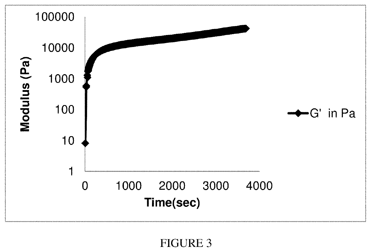
[0354]The acrylated chitosan of Example 1 and oxidized dextran of Example 2 were each solubilized into a buffered saline solution to give a final concentration of 7.5% (w / w) and 7.5% (w / w), respectively. Equal amounts of the aqueous solution of the oxidized dextran provided in Example 2 (concentration: 7.5% w / w) and the aqueous solution of acrylated chitosan provided in Example 1 (concentration: 7.5% w / w) were loaded in a dual barrel syringe. A mixer was attached to the top of the dual barrel syringe. The two solutions were then passed through the mixer simultaneously. The hydrogel was observed to form instantaneously at the tip of the mixer. The combined solution was further aerosolized under pressure.

[0355]The burst strength of the acrylated chit...


PUM

PropertyMeasurementUnit
PDIaaaaaaaaaa
PDIaaaaaaaaaa
PDIaaaaaaaaaa

Abstract

Provided is tunable biopolymer hydrogel produced from two processed natural polysaccharides for use as a hemostat. If desired, the hydrogel formation can be tuned so that the hydrogel forms within seconds when applied to a tissue lesion. The resulting hydrogel can adhere to tissue and, without swelling, produce hemostasis within seconds after application to tissue of interest. The hydrogel also captures, aggregates and concentrates platelets and red blood cells at the site of the tissue lesion thereby initiating a clotting cascade at the site of the lesion. The hemostat can be used to prevent blood loss during surgical procedures, for example, during brain, spine or other surgical procedures where hemostasis is desirable, and is particularly useful during surgical procedures where swelling of the hemostat (e.g., in the brain or spine) would be detrimental to the subject.

Description

FIELD[0001]The invention relates generally to methods and compositions for achieving hemostasis and blood coagulation, and more specifically relates to methods and compositions for achieving hemostasis and blood coagulation during surgical procedures.BACKGROUND[0002]Bleeding and blood loss are problems that must be addressed during and at the conclusion of surgery and other medical procedures. Significant blood loss can be fatal or cause significant morbidity, and surgeons and medical staff routinely compensate for blood loss with transfusions and the use of recirculating devices. A number of different techniques and products are currently used by surgeons to stop bleeding and effect hemostasis in surgery and medical procedures. However no single approach is effective or suitable under all circumstances, and the currently available approaches have limitations in effectiveness, safety, applicability and ease of use.[0003]The most commonly used approaches involve closing a tissue or b...

Claims


Application Information

Patent Timeline
Patent Type & Authority Patents(United States)
IPC IPC(8): A61L24/08C08B37/02A61L24/04A61L24/00C08B37/08
CPCC08B37/003C08B37/0021A61L24/043A61L24/0036A61L24/0015A61L24/08A61L24/0031A61L24/0005A61L2400/04A61L26/008A61L26/0085A61L26/0066A61L26/009A61L2400/06A61L2300/64A61L2300/418C08L5/08C08L5/02
Inventor MODAK, PIYUSHNADIG, MALAVIKARUSSO, RICHARD
Owner ENDOMEDIX
Why Eureka
Try Eureka