Endothelial cell growth factor, methods of isolation and expression

a growth factor and endothelial cell technology, applied in the field of growth factor, can solve the problems of loss of biological activity, no highly suitable mitogenic factor exists for this type of application, and no mitogenic activity has been purified or characterized

Inactive Publication Date: 2002-09-26
FERRARA NAPOLEONE +2
View PDF8 Cites 10 Cited by
  • Summary
  • Abstract
  • Description
  • Claims
  • Application Information

AI Technical Summary

Problems solved by technology

At present, no highly suitable mitogenic factor exists for this type of application.
To date, however, this mitogenic activity has not been purified or characterized.
Although losses in biological activity were encountered, presumably because of the acid conditions and solvent used, these were not serious enough to prevent the detection of bioactive fractions.
These results suggest that the factor is a glycoprotein with strong affinity for concanavalin A. However, the poor biological recovery of the factor from that type of affinity chromatography resin makes concanavalin A unsuitable as a step of purification.
However, the novel growth factor did not induce any appreciable mitogenic effect on corneal endothelial cells, lens epithelial cells, BHK-21 fibroblasts, adrenal cortex cells, or keratynocytes.
First, FGF lacks the hydrophobic signal sequences that govern secretion.sup.
Although the present study clearly established that this novel growth facto is mitogenic for capillary endothelial cells, it is not yet known whether it can stimulate other events linked to angiogenesis.
A disadvantage observed during the culturing of the bovine cells is that these cells produce a large amount of different proteins, which are secreted from the cell.
A disadvantage of culturing the murine cells AtT-20 is that the structural integrity of the cell culture layer is not high.

Method used


Image

  • Endothelial cell growth factor, methods of isolation and expression
  • Endothelial cell growth factor, methods of isolation and expression
  • Endothelial cell growth factor, methods of isolation and expression

Examples


example 1

Culture of Follicular Cells and Media Collection

[0238] Primary cultures of bovine pituitary FC were established as previously described (20, 41). In one embodiment in the culturing the 20% fetal bovine serum in reference 20 was reduced to 10%. Concentrations of 5 to 20% should be effective. Also no DNAase is used. All other components are the same. At confluency, cells were passaged into large scale tissue culture plates in the presence of low glucose Dulbecco's modified Eagle's medium (DMEM) supplemented with 10% fetal bovine serum, 2 mM glutamine and antibiotics. Shortly after reaching confluency the cultures were extensively washed with PBS in order to remove serum components. The cells were then incubated in a serum-free medium consisting of DMEM plus transferrin (10 .mu.l / ml), insulin (5 .mu.g / ml), selenium (10.sup.-8 M), 2 mM glutamine and antibiotics. After three or four days, the medium was collected and replaced with fresh serum free medium. The collected medium was centrif...

example 2

Concentration of Conditioned Medium

[0239] Four to six liter batches of conditioned medium (CM) were subjected to ammonium sulfate precipitation. Ammonium sulfate (500 g / L) was added under constant stirring, until the salt was completely in solution. After 8-12 hours in the cold room, the material was centrifuged (20,000 .times.g, 45 min at 4.degree. C.). The supernatant was discarded and the pellet was resuspended with 10 mM Tris / Cl, pH 7.2, 50 mM NaCl and dialyzed at 4.degree. C. against the same buffer for 8-12 h. The final volume was 50-60 fold less than the original.

[0240] Alternatively, the CM is concentrated using ultrafiltration using an Amecon stir cell (2.5 liter unit) using a membrane having a molecular weight cut off of 10,000 daltons with similar results.

example 3

Heparin-Sepharose Affinity Chromatography

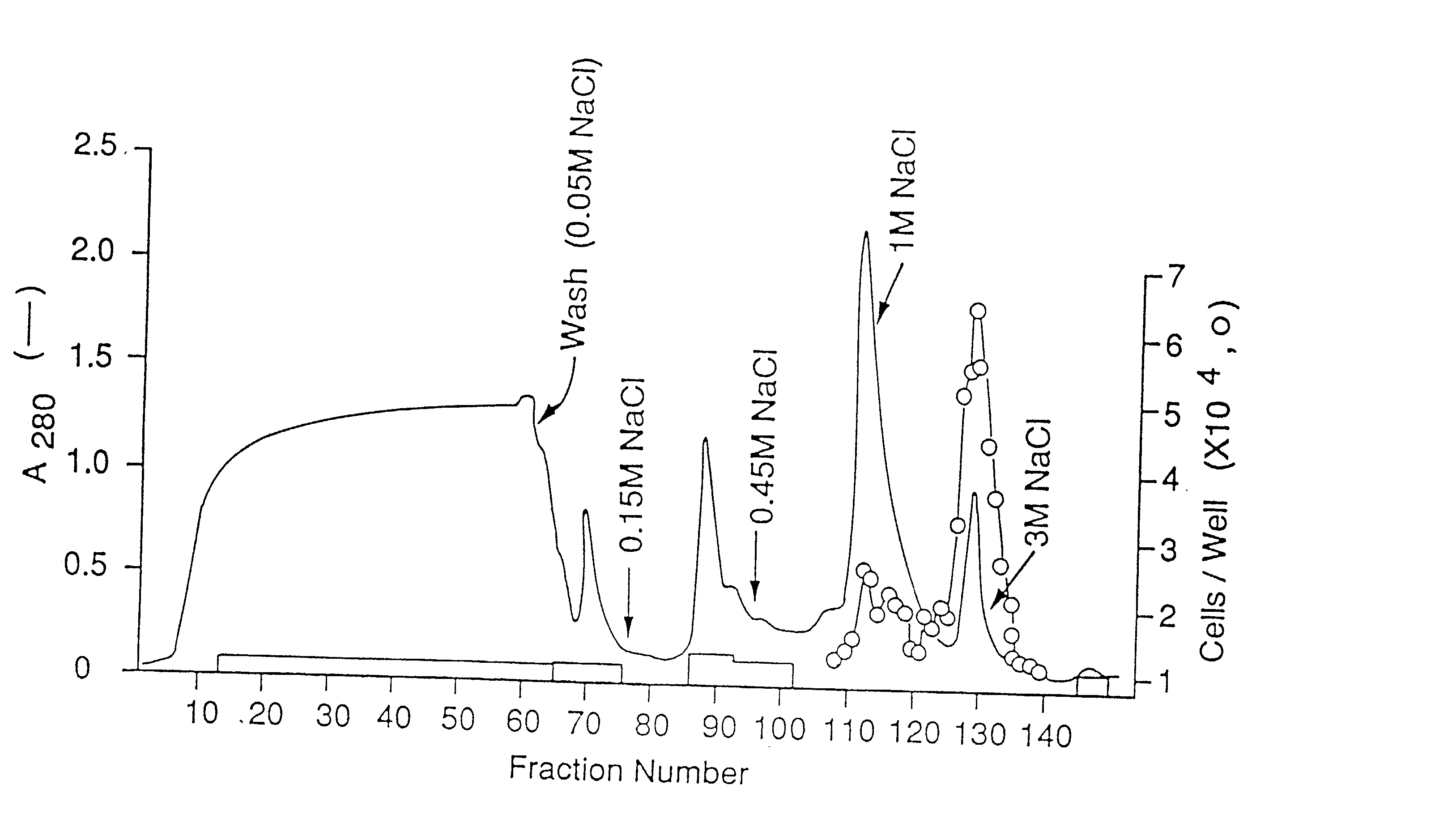
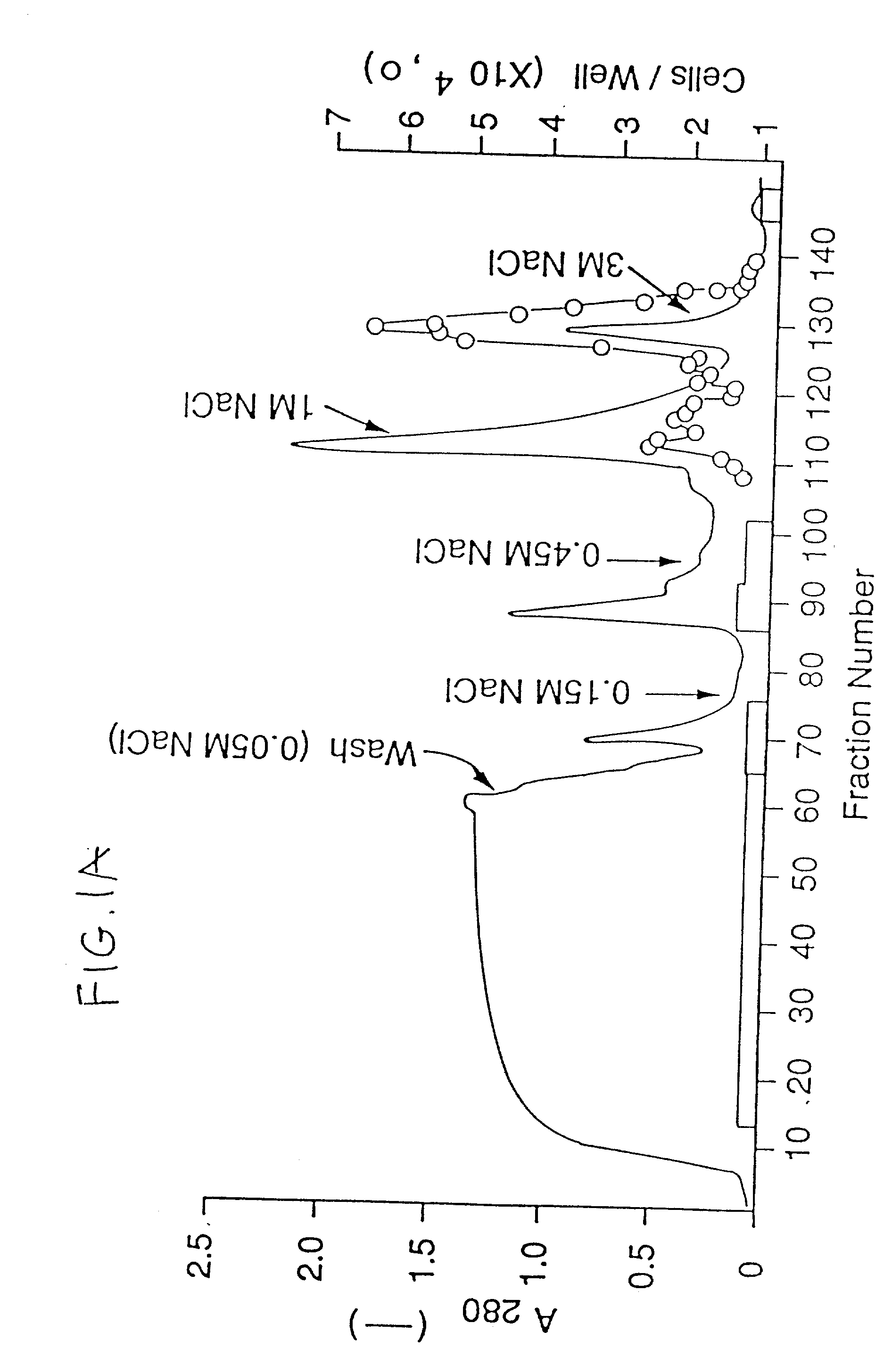
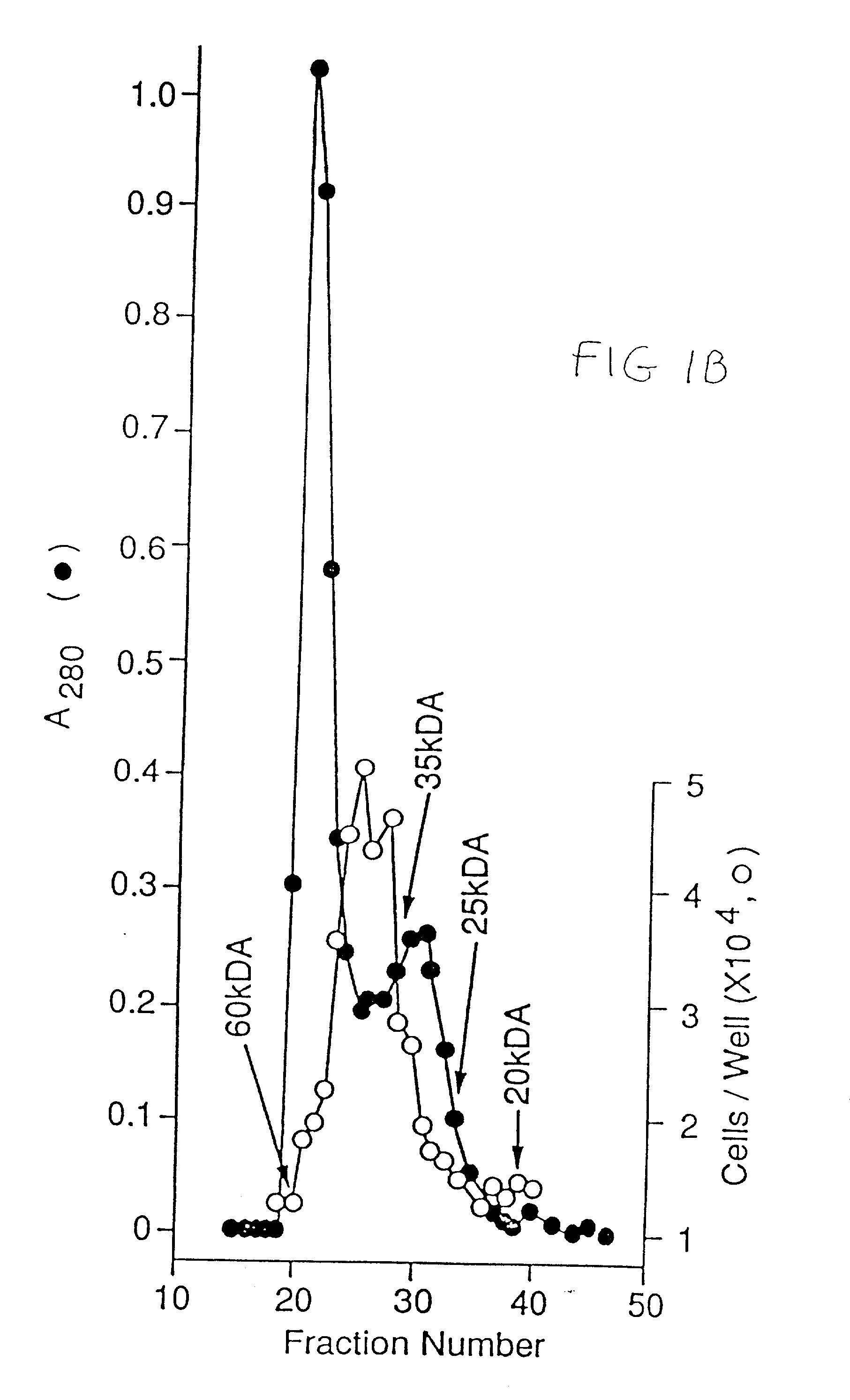
[0241] The concentrated CM was applied to a H-S column (14) (10 ml) preequilibrated with 10 mM Tris / Cl, pH 7.2, 50 mM NaCl. The column was then washed with the same buffer until the absorbance at 280 nm was negligible and then eluted stepwise with 10 mM Tris / Cl pH 7.2 containing 0.15, 0.9 nd 3 M NaCl. The flow rate was 1.5 ml / min. Fractions of 1.5 ml were collected and aliquots, diluted with 0.2% gelatin in PBS, were tested for mitogenic activity on endothelial cells.


PUM

PropertyMeasurementUnit
molecular weightaaaaaaaaaa
concentrationaaaaaaaaaa

Abstract

A novel growth factor specific for vascular endothelial cells has been identified in conditioned medium of bovine pituitary derived folliculo stellate cells. This factor, named folliculo stellate derived growth facto (FSdGF) or vascular endothelial growth factor (VEGF), was purified to homogeneity by a combination of heparin sepharose affinity chromatography, Bio Gel P-60 exclusion chromatography, Mono S ion exchange chromatography and hydrophobic chromatography on a C4 reverse phase HPLC column. The factor is also found in the murine AtT-20 cell line. Alternatively, the growth factor is purified by a first reverse phase HPLC using acetonitrile gradient followed by a second reverse phase HPLC using an isopropanol gradient. FSdGF, having a molecular weight of about 43,000 da, was characterized as a glycoprotein composed of two homologous sub units with MW of about 23 kDa. FSdGF was a potent mitogen for vascular endothelial cells with activity detectable at 10 pg / ml and saturation at 500 pg / ml. It did not stimulate the proliferation of other cell types such as bovine corneal endothelial cells, adrenal cortex cells, granulosa cells, BALB / MK cells or BHK-21 cells. Microsequencing revealed an amino terminal sequence containing no significant homology to any known protein. The release of FSdGF by pituitary cells and its unique target cell specificity indicate that FSdGF is useful in angiogenesis.

Description

[0001] The present application is a continuation-in-part of pending U.S. patent application Ser. No. 346,165, filed May 2, 1989, which is a continuation-in-part of pending U.S. patent application Ser. No. 328,181, filed Mar. 24, 1989, which are both incorporated by reference in its entirety.BACKGROUND OF INVENTION[0003] 1. Field of the Invention[0004] The present invention relates to a novel growth factor for vascular endothelial cells identified in media condition of cultured bovine pituitary follicular cells and of murine tumor cells. The invention also relates to isolation and purification of the growth factor.[0005] 2. Description of the Problem and Related Art[0006] Numerical references in parenthesis in the text refer to the publications listed below in the Reference Section.[0007] Angiogenesis is a multi-step phenomenon which involves capillary endothelial cell proliferation, migration and tissue infiltration (1). It plays a central role in a variety of physiological and path...

Claims


Application Information

Patent Timeline
Patent Type & Authority Applications(United States)
IPC IPC(8): A61K38/00C07K14/52
CPCC07K14/52A61K38/1866
Inventor FERRARA, NAPOLEONEGOSPODAROWICZ, DENISPLOUET, JEAN
Owner FERRARA NAPOLEONE
Why Eureka
Try Eureka