Tissue structure identification in advance of instrument

a tissue structure and tissue technology, applied in the field of tissue structure identification in advance of a tissue penetrating medical instrument, can solve the problems of severing injury, affecting the healing effect of patients, and important tissue damage inadvertently

a tissue structure and tissue technology, applied in the field of tissue structure identification in advance of a tissue penetrating medical instrument, can solve the problems of severing injury, affecting the healing effect of patients, and important tissue damage inadvertently

US20050027199A1Inactive Publication Date: 2005-02-03CLARKE DANA S

Image

  • Tissue structure identification in advance of instrument
  • Tissue structure identification in advance of instrument
  • Tissue structure identification in advance of instrument

Examples


Embodiment Construction

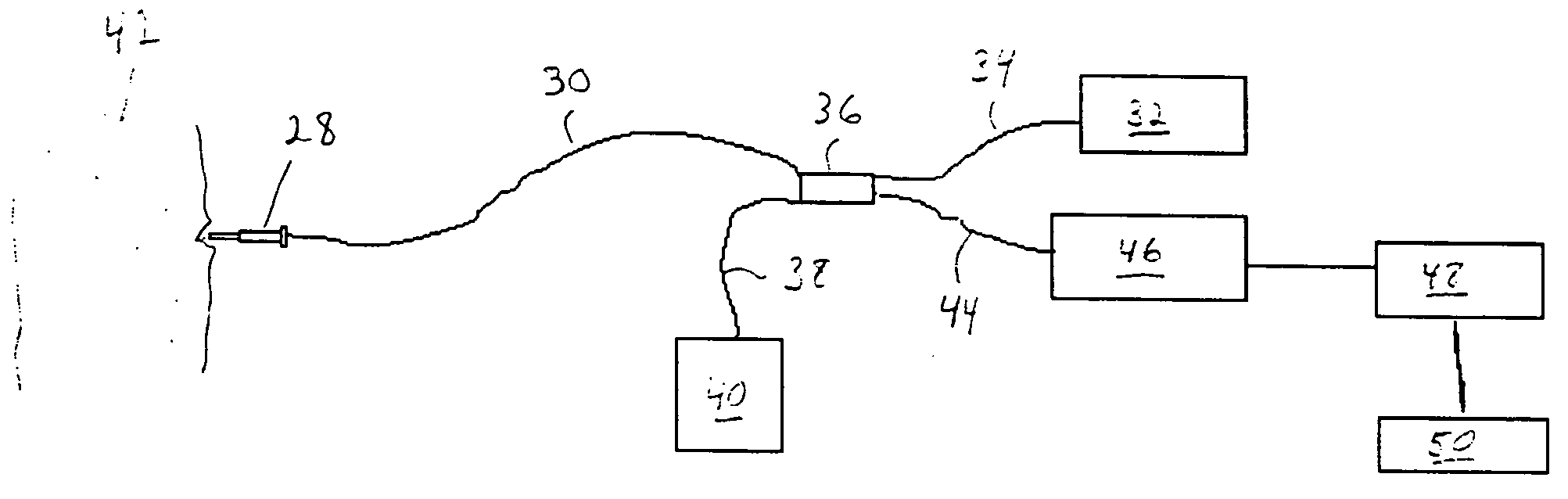
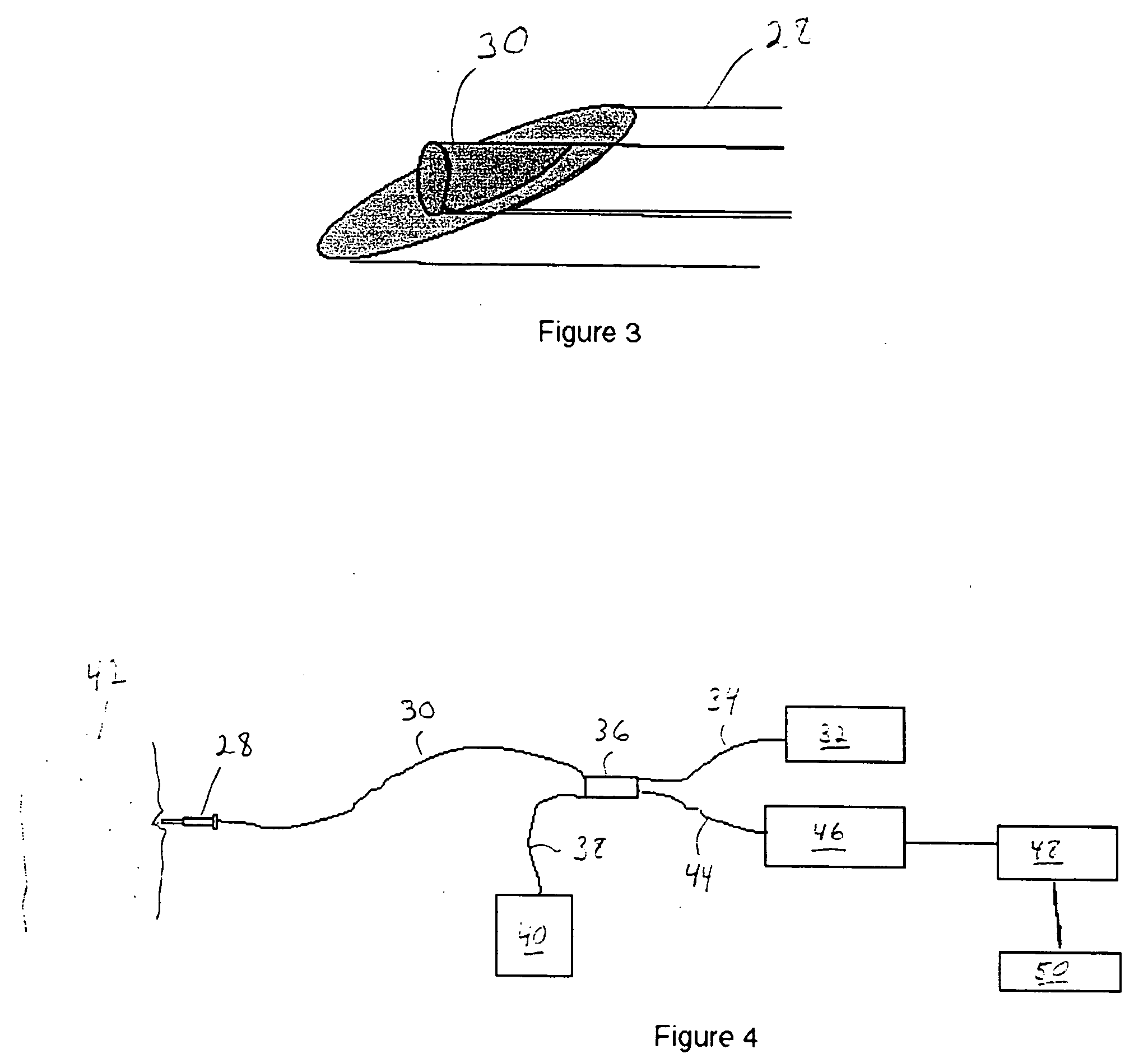
The method and apparatus of the present invention uses optical fibers aligned along a needle, scalpel or other mechanical cutting tool to both transmit and receive optical information directly in front of the cutting surface. OCDR is used to determine the depth of boundaries immediately in advance of a cutting surface such as a needle tip or scalpel blade. This information can be used to alert either a medical professional or a surgical robot to the presence of an upcoming boundary and possibly identify the tissue.

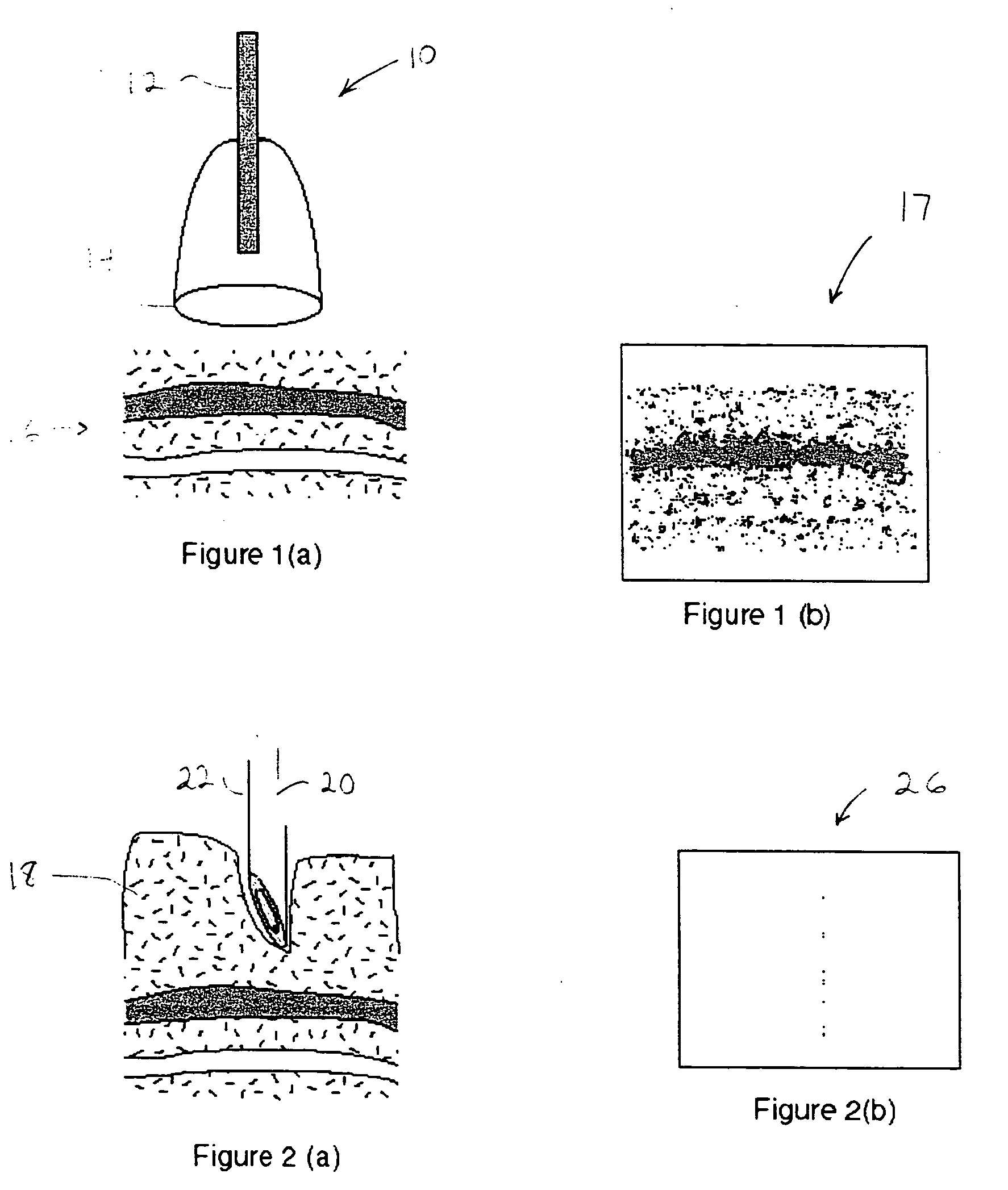
The basic concept of prior art OCT 10 is illustrated by FIGS. 1(a) and 1(b), which shows a typical test fiber 12 and associated distal optic 14 in relation to a tissue sample 16 to be scanned. This prior art system, which does not mechanically penetrate the tissue, can produce a two-dimensional tomographic image 17 of relatively limited depth of, for example, 1-2 millimeters.

In contrast, the apparatus for identifying tissue structures ahead of a mechanical tissue pene...


PUM


Abstract

A method and apparatus for identifying tissue structures in advance of a mechanical medical instrument during a medical procedure. A mechanical tissue penetrating medical instrument (22) has a distal end for penetrating tissue in a penetrating direction. An optical wavefront analysis system (32-50) provides light to illuminate tissue ahead of the medical instrument and receives light returned by tissue ahead of the medical instrument. An optical fiber (30) is coupled at a proximal end to the wavefront analysis system and attached at a distal end to the medical instrument proximate the distal end of the medical instrument. The distal end of the fiber has an illumination pattern directed substantially in the penetrating direction for illuminating the tissue ahead of the medical instrument and receiving light returned therefrom. The wavefront analysis system provides information about the distance from the distal end of the medical instrument to tissue features ahead of the medical instrument.

Description

FIELD OF THE INVENTION This invention relates to medical procedures and apparatuses for performing medical procedures and, in particular, to methods and apparatuses for identifying tissues structures in advance of a tissue penetrating medical instrument. BACKGROUND OF THE INVENTION During invasive medical procedures employing a tissue penetrating medical instrument, such as a needle or a scalpel, there is a need to identify tissue structures in advance of the medical instrument so as to control the depth of penetration of the tip or cutting edge of the medical instrument. This may be done to ensure that the penetration is to the right depth, but usually more importantly, this is done to ensure that the medical instrument does not penetrate too deeply and thereby damage tissue structure unnecessarily. During surgery important tissue is often damaged inadvertently. For example, biopsy patients often report a loss of sensation or motion control due to nerve damage sustained during s...

Claims


Application Information

Patent Timeline
03 Feb 2005
Publication
US20050027199A1
IPC
A61B5/00; A61B17/00; A61B17/32; A61B17/34; A61B19/00; G01N21/85
CPC
A61B5/0066; A61B5/0084; A61B5/6848; A61B5/6849; A61B17/3211; A61B17/34; G01N21/8507; A61B2017/00061
Inventors
CLARKE, DANA S.