
Chondrocyte therapeutic delivery system

a chondrocyte and delivery system technology, applied in the field of chondrocyte therapeutic delivery system, can solve the problem of no teaching in the art that chondrocytes can be used to express therapeutic effects, and achieve the effect of convenient optimization

Active Publication Date: 2005-03-10
DEPUY SYNTHES PROD INC
View PDF68 Cites 63 Cited by
  • Summary
  • Abstract
  • Description
  • Claims
  • Application Information

AI Technical Summary

Benefits of technology

[0006] Chondrocytes offer several unique advantages as vehicles for expressing therapeutic agents over other cell types. For example, chondrocytes do not require vascular support, therefore can readily be used in environments that have a reduced, or non-existent vascularization system. Furthermore, chondrocytes are able to survive in harsh in vivo environments, including ischemic tissue, as well as low pH and low oxygen surroundings. In addition, there is a reduced likelihood of malignancy due to the anti-angiogenicity properties of normal chondrocytes. Chondrocytes also possess an immune privileged property which reduces immune rejection of co-implanted allogenic or xenogenic tissue. Furthermore, chondrocytes are more easily scalable compared to other normal untransformed cell strains.
[0017] The biocompatible substrate that is implanted can be a gel matrix substrate, such as an alginate, polysaccharide, and agarose. This genetically altered chondrocyte-gel matrix substrate can be injected to a target region. Alternatively, the genetically altered chondrocyte-gel matrix substrate can be solidified prior to implantation to a target region. When the chondrocyte-gel matrix substrate can be solidified prior to implantation, the dimensions of the implanted gel matrix substrate determine the concentration of chondrocytes within the gel matrix substrate that are available to express the therapeutic agent. The concentration of chondrocytes in the gel matrix substrate is in the range of about 10,000 to hundreds of millions of cells per ml of gel matrix having a volume in the range of about 0.1 to 10 ml. Preferably, the concentration of the chondrocytes is about 100,000 to about one million cells per ml of substrate. Preferably, the volume of the gel matrix substrate is less than one milliliter. It is understood that the dose necessary to treat a particular pathology could be easily optimized by either changing the cellular concentration for a given allowable volume, or by changing the amount of chondrocyte-gel implanted. The injury to be ameliorated can be a wound, a bone defect, a cartilage defect, a skin wound, and a torn ligament. The disorder that can be ameliorated can be a blood disorder, an autoimmune disorder, a hormonal disorder, an anti-inflammatory disorder, a fertility disorder, and an neurodegenerative disorder.

Problems solved by technology

Although chondrocytes have been cultured and placed into substrates such as hydrogels, for treatment, or repair of cartilage or bone defects, there is no teaching in the art that chondrocytes can be used to express therapeutic agents for the treatment of pathologies or injuries other than cartilage tissue.

Method used


Image

  • Chondrocyte therapeutic delivery system
  • Chondrocyte therapeutic delivery system
  • Chondrocyte therapeutic delivery system

Examples


example 1

In vitro Isolation and Culturing of Chondrocyte Cells

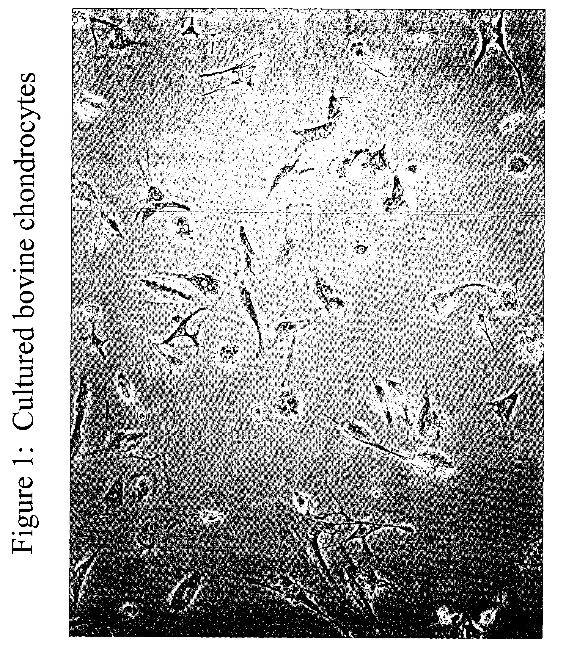
[0119] This example describes one of many possible methods of isolating and culturing chondrocyte cells. Articular cartilage was aseptically obtained from calf knee joints. Cartilage was shaved from the end of the bones using a scalpel. The tissue was finely minced in saline solution and submitted to collagenase and trypsin digestion for several hours at 37° C. until the tissue was completely digested and single cells were generated. The chondrocyte cells isolated from the cartilage tissue were then washed from enzyme and seeded onto tissue culture plastic dishes at a concentration of about 10,000 cells / cm2. The chondrocyte cells where expanded in 10% FBS containing DMEM culture medium Following expansion, the chondrocytes can be released from the vessel using a trypsin / EDTA treatment, centrifuged and concentrated or diluted to a desired concentration to achieve for example a seeding density of approximately 5,000 cells to about ...

example 2

In vitro Transfection of Chondrocyte Cells and Expression of a Marker Protein

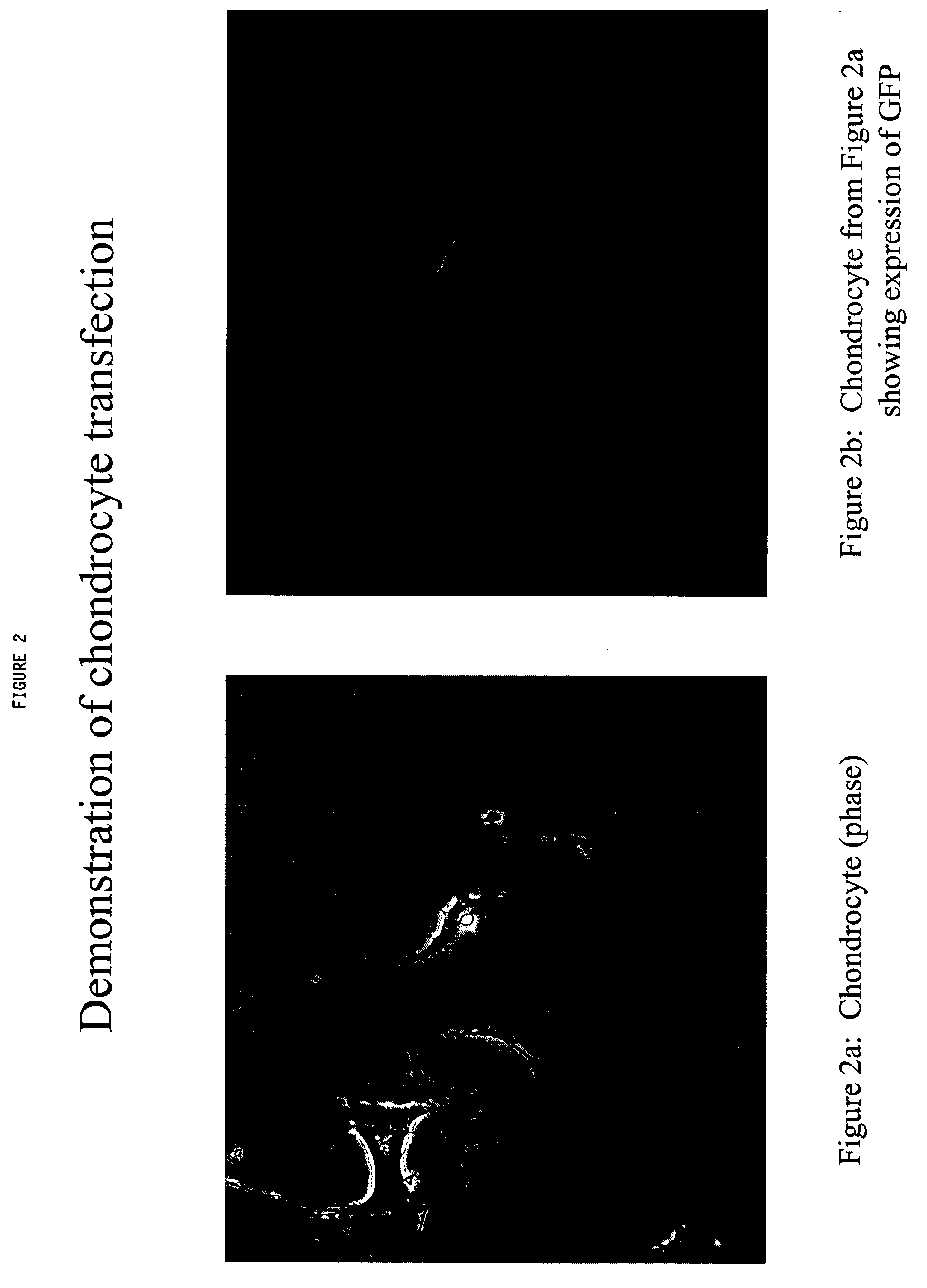
[0120] This example described the techniques used to transfect chondrocytes with a marker protein, Green fluorescent protein (GFP). A DNA vector carrying a reporter gene is used to establish proof of concept for the utility of normal articular chondrocytes as a drug delivery system for therapeutic application in ectopic sites. As a model, a DNA vector (pTracer-SV40) carrying a gene for green fluorescent protein was used as a surrogate to model a protein not normally present in chondrocytes, and of therapeutic value. If chondrocytes can be transfected by the model vector and expressed the foreign protein, it would demonstrated the ability of using this system for therapeutic protein. In the following example, a lipid formulation from Stratagene (GeneJammer) was used to introduced the foreign DNA into chondrocytes. Any other reagent or physical means of introducing DNA into cell would serve the same purpose....

example 3

In vitro Transfection of Human and Bovine Chondrocyte Cells and Expression of EPO Protein

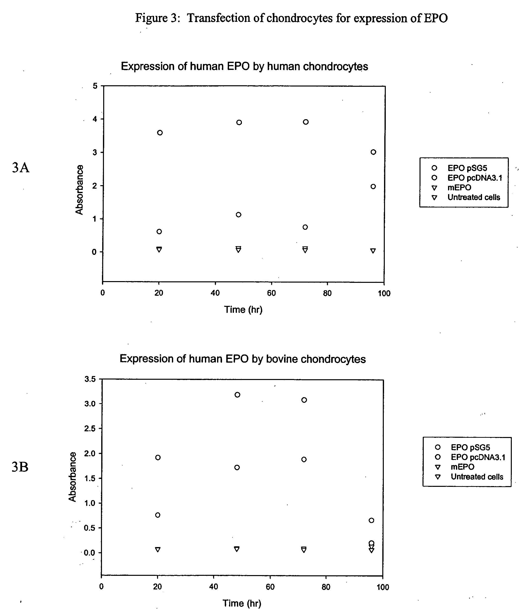
[0124] This example describes the techniques used to transfect chondrocytes with a therapeutic agent, human erythropoietin (EPO).

[0125] An EPO vector was generated which comprised the EPO gene (Genbank Accession No. 182198) inserted into the commercially available pSG5 backbone (Stratagene Inc.). The pSG5 plasmid contains ampicillin and kanamycin as a selection marker for bacteria. This construct also contains F1 filamentous phage origin and SV40 promoter for better expression. The 584 base pair EPO fragment was cloned into the NCO / Bam H1site site of pSG5. The size of this vector was about 4.6 kb.

[0126] A second EPO vector was generated which comprised the EPO gene inserted into the commercially available pcDNA3.1 backbone (Invitrogen Life Technologies). This particular construct has the antibiotic Neomycin as a selection marker for mammalian cells.

[0127] Each of the EPO vectors was transfec...


PUM

PropertyMeasurementUnit
volumeaaaaaaaaaa
volumeaaaaaaaaaa
volumeaaaaaaaaaa

Abstract

Methods and compositions for producing therapeutic agents using chondrocytes are provided. The genetically engineered chondrocytes can be used to express the therapeutic agent in a subject, including in an environment typically associated with chondrocytes and in an environment not typically associated with chondrocytes.

Description

FIELD OF THE INVENTION [0001] This invention relates to the treatment or amelioration of disorders or diseases using chondrocytes to deliver therapeutic agents. BACKGROUND OF THE INVENTION [0002] Chondrocytes are typically involved in cartilage repair. Cartilage is a structural support tissue that is found in the body in three main varieties. Hyaline, or articular cartilage, helps dissipate loads in joints. In articular cartilage, chondrocytes are encapsulated in a woven, mesh-like matrix of type II collagen and proteoglycans. Elastic cartilage provides flexible support to external structures, and is composed of chondrocytes embedded in a matrix of collagen and elastic fibers. Fibrocartilage aids in transferring loads between tendons and bone. It consists of an outer layer of collagen and fibroblasts that support and inner layer of chondrocytes that make type II collagen fibers. [0003] To date chondrocytes have been used to correct or repair cartilaginous defects. For example, by pl...

Claims


Application Information

Patent Timeline
Patent Type & Authority Applications(United States)
IPC IPC(8): A61K35/12A61P3/00A61K48/00A61P5/00A61P7/00A61P9/00A61P15/08A61P17/00A61P19/10A61P25/00A61P35/00A61P37/00C12N5/077
CPCA61K35/12C12N5/0655C12N2533/76C12N2533/74C12N2510/00A61P1/00A61P1/16A61P1/18A61P11/00A61P13/12A61P15/00A61P15/08A61P17/00A61P17/02A61P19/00A61P19/08A61P19/10A61P21/00A61P25/00A61P25/16A61P25/28A61P27/02A61P29/00A61P3/00A61P3/10A61P35/00A61P37/00A61P37/06A61P43/00A61P5/00A61P5/48A61P7/00A61P9/00
Inventor BINETTE, FRANCOISSTORY, BROOKS J.
Owner DEPUY SYNTHES PROD INC
Learn More