Protein fragment complementation assays in whole animals applications to drug efficacy, ADME, cancer biology, immunology, infectious disease and gene therapy

a technology of protein fragments and assays, applied in the field of protein fragment complementation assays in whole animal applications to drug efficacy, adme, cancer biology, immunology, infectious diseases and gene therapy, can solve the problems of inability to distinguish induced versus constitutive interactions, few convenient methods for studying proteins, and inability to detect high-throughput cdna libraries

Inactive Publication Date: 2005-04-14
ODYSSEY THERA INC
View PDF6 Cites 9 Cited by
  • Summary
  • Abstract
  • Description
  • Claims
  • Application Information

AI Technical Summary

Benefits of technology

[0016] The present invention seeks to provide the above-mentioned needs for which the prior art is silent. The present invention provides a general strategy for detecting protein interactions with other biopolymers including other proteins, nucleic acids, carbohydrates or for screening small molecule libraries for compounds of potential therapeutic value. In a preferred embodiment, the instant invention seeks to provide an oligomerization-assisted complementation of fragments of monomeric enzymes that require no other proteins for the detection of their activity. In one such embodiment, a protein-fragment complementation assay (PCA) based on reconstitution of dihydrofolate reductase activity by complementation of defined fragments of the enzyme in E. coli is hereby provided. This assay requires no additional endogenous factors for detecting specific protein-protein interactions (i.e. leucine zipper interactions) and can be conveniently extended to screening cDNA, nucleic acid, small molecule or protein design libraries for molecular interactions. In addition, the assay can also be adapted for detection of protein interactions in any cellular context or compartment and be used to distinguish between induced versus constitutive protein interactions in both prokaryotic and eukaryotic systems.
[0017] One particular strategy for designing a protein complementation assay (PCA) is based on using the following characteristics: 1) A protein or enzyme that is relatively small and monomeric, 2) for which there is a large literature of structural and functional information, 3) for which simple assays exist for the reconstitution of the protein or activity of the enzyme, both in vivo and in vitro, and 4) for which overexpression in eukaryotic and prokaryotic cells has been demonstrated. If these criteria are met, the structure of the enzyme is used to decide the best position in the polypeptide chain to split the gene in two, based on the following criteria: 1) The fragments should result in subdomains of continuous polypeptide; that is, the resulting fragments will not disrupt the subdomain structure of the protein, 2) the catalytic and cofactor binding sites should all be contained in one fragment, and 3) resulting new N- and C-termini should be on the same face of the protein to avoid the need for long peptide linkers and allow for studies of orientation-dependence of protein binding.
[0097] The present invention is pionneering as it is the first protein complementation assay displaying such a level of simplicity and versatility. The exemplified embodiments are protein-fragment complementation assays (PCA) based on mDHFR, where a leucine zipper directs the reconstitution of DHFR activity. Activity was detected by an E. coli survival assay which is both practical and inexpensive. This system illustrates the use of mDHFR fragment complementation in the detection of leucine zipper dimerization and could be applied to the detection of unknown, specific protein-molecular interactions in vivo.
[0111] PCA can be used to eliminate the need for traditional ADME approaches, by using whole-body imaging to detect the effect of the pharmaceutical agent on the target within living tissues. For example, fluorescence PCA has been used to detect activation of the erythropoietin receptor in living cells in response to erythropoietin or a peptide mimetic, EMP1 (Remy and Michnick). Cells that stably express the EPO receptor / PCA fragment constructs can be introduced into animals as described under methods. Suitable PCA reporters include but are not limited to luciferase, GFP, and beta-lactamase. The small molecule EPO mimetics would then be administered to the mice. If the agent causes activation of the receptor, the reporter will reassemble from its complimentary fragments, leading to a detectable signal. Fluorescence or luminescence can then detected by whole body imaging. The signal will persist as long as the pharmaceutical agent is active. If the mimetic is rapidly degraded or excreted, the signal will diminish. This is a “live” approach to ADME that does not require serial sampling of body fluids, and eliminates the need for analysis of the compound itself within the body fluids thereby saving time and money. With in vivo PCA, ADME only requires serial imaging of the animal over a period of time to establish the time course of activity of the agent. Similar approaches can be applied to other biopharmaceuticals such as cytokines, hormones, and mimetics of these agents. In addition, in vivo PCA can be applied to studies of efficacy and pharmacokinetics of receptor antagonists e.g. Herceptin, and like compounds. Finally these approaches can be applied to any agent that blocks a signaling pathway or a biomolecular event within cells, by simply tagging the appropriate genes with the reporter fragments, creating cell lines, and establishing the cells in animals. Moreover, in vivo PCA can be applied by creating transgenic mice that express the fusion constructs in every cell line, thereby enabling studies of the distribution of the pharmaceutical agent within tissues.

Problems solved by technology

Despite the importance of understanding protein assembly in biological processes, there are few convenient methods for studying protein-protein interactions in vivo4,5.
Limitations of this technique include the fact that the interaction must occur in a specific context (the nucleus of S. cerevisiae), and generally cannot be used to distinguish induced versus constitutive interactions.
Unfortunately, this strategy requires additional cellular factors (in this case ubiquitinases) and the detection method does not lend itself to high-throughput screening of cDNA libraries.
Secondly, this application describes a general strategy to develop molecular interaction assays from a large number of enzyme or protein detectors, all de novo designed assays, whereas the β-gal assay is not novel, nor are any general strategies or advancements over previosly well documented applications given.
Screening techniques for protein interactions, such as the yeast “two-hybrid” system, have transformed molecular biology, but can only be used to study specific types of constitutively interacting proteins or interactions of proteins with other molecules, in narrowly defined cellular and compartmental contexts and require a complex cellular machinery (transcription) to work.

Method used


Image

  • Protein fragment complementation assays in whole animals applications to drug efficacy, ADME, cancer biology, immunology, infectious disease and gene therapy
  • Protein fragment complementation assays in whole animals applications to drug efficacy, ADME, cancer biology, immunology, infectious disease and gene therapy
  • Protein fragment complementation assays in whole animals applications to drug efficacy, ADME, cancer biology, immunology, infectious disease and gene therapy

Examples


example 1

[0140] Experimental Protocol

[0141] DNA Constructs.

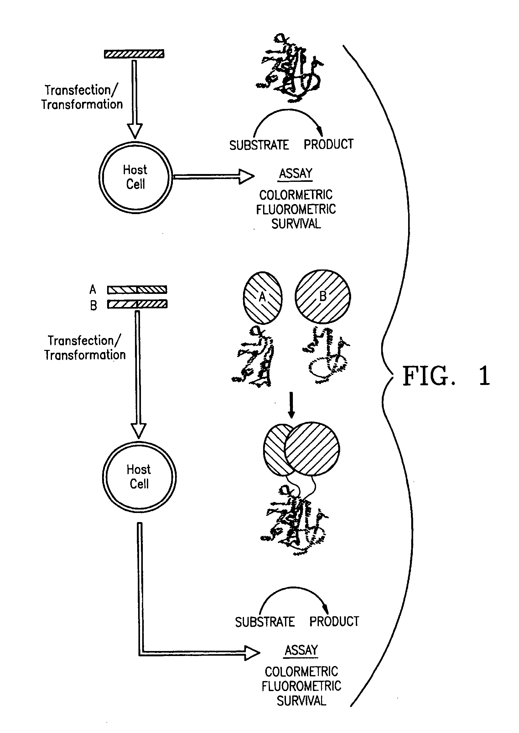
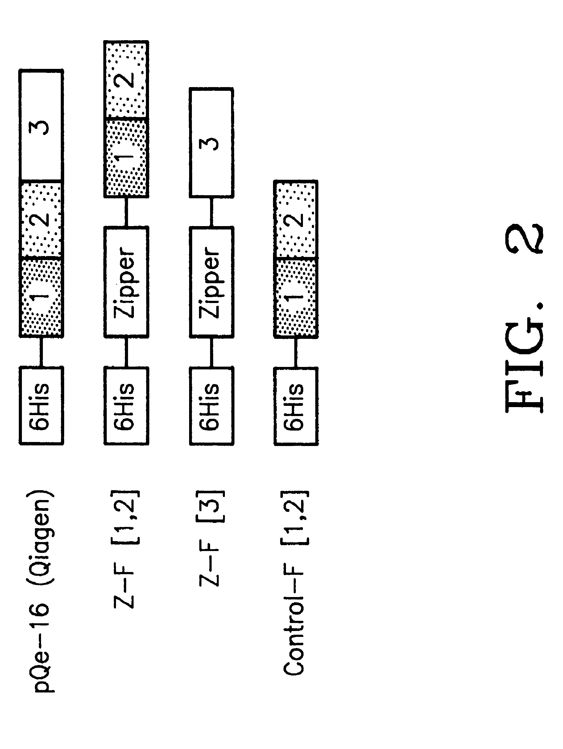
[0142] Mutagenic and sequencing oligonucleotides were purchased from Gibco BRL. Restriction endonucleases and DNA modifying enzymes were from Pharmacia and New England Biolabs. The mDHFR fragments carrying their own iN-frame stop codon were subcloned into pQE-32 (Qiagen), downstream from and iN-frame with the hexahistidine peptide and a GCN4 leucine zipper (FIG. 1; FIG. 2). All final constructs were based on the Qiagen pQE series of vectors, which contain an inducible promoter-operator element (tac), a consensus ribosomal binding site, initiator codon and nucleotides coding for a hexahistidine peptide. Full-length mDHFR is expressed from pQE-16 (Qiagen).

[0143] Expression Vector Harboring the GCN4 Leucine Zipper

[0144] Residues 235 to 281 of the GCN4 leucine zipper (a SalI / BamHI 254 bp fragment) were obtained from a yeast expression plasmid pRS3169. The recessed terminus at the BamHI site was filled-in with Klenow polymerase and th...

example 2

[0188] Applications of the PCA Strategy to Detect Novel Gene Products in Biochemical Pathways and to Map Such Pathways

[0189] Among the greatest advantage of PCA over other molecular interaction screening methods is that they are designed to be performed both in vivo and in any type of cell. This feature is crucial if the goal of applying a technique is to identify novel interactions from libraries and simultaneously be able to determine if the interactions observed are biologically relevant. The detailed example given below, and other examples at the end of this section illustrate how it is that validation of interactions with PCA is possible. In essence, this is achieved as follows. In biochemical pathways, such as hormone receptor-mediated signaling, a cascade of enzyme-mediated chemical reactions are triggered by some molecular event, such as by hormone binding to its membrane surface receptor. Enzyme interactions with protein substrates and protein-protein or protein-nucleic ac...

example 3

[0228] Other Examples of Protein Fragment Complementation Assays

[0229] Other examples of assays are herein examplified. The reason to produce these assays is to provide alternative PCA strategies that would be appropriate for specific protein association problems such as studying equilibrium or kinetic aspects of assembly. Also, it is possible that in certain contexts (for example, specific cell types) or for certain applications, a specific PCA will not work but an alternative one will. Further below are brief descriptions of each other PCAs embodiments.

[0230] 1) Glutathione-S-Transferase (GST) GST from the flat worm Schistosoma japonicum is a small (28 kD), monomeric, soluble protein that can be expressed in both prokaryotic and eukaryotic cells. A high resolution crystal structure has been solved and serves as a starting point for design of a PCA. A simple and inexpensive colorimetric assay for GST activity has been developed consisting of the reductive conjugation of reduced g...


PUM

PropertyMeasurementUnit
ODaaaaaaaaaa
pHaaaaaaaaaa
pHaaaaaaaaaa

Abstract

The instant invention describes a method for detecting protein-protein interactions in living organisms and / or cells, said method comprising: (a) synthesizing probe protein fragments from a protein which enables fluorescent or luminescent detection by dissecting the gene coding for the fluorescent or luminescent protein into a least two fragments; (2) constructing fusion proteins consisting of the probe protein fragments linked to protein domains that are to be tested for interactions; (3) coexpressing the fusion proteins; and (d) detecting reconstitution of the fluorescence or luminescence signal.

Description

[0001] This application is a continuation-in-part of pending U.S. application Ser. No. 10 / 353,090 filed Jan. 29, 2003; which application is a continuation of pending U.S. application Ser. No. 10 / 154,758 filed May 24, 2002; which is a continuation of U.S. Ser. No. 09 / 499,464 filed Feb. 7, 2000; and now U.S. Pat. No. 6,428,951; which is a continuation of U.S. Ser. No. 09 / 017,412 filed Feb. 2, 1998; and now U.S. Pat. No. 6,270,964. The entire contents of all those patents and applications are incorporated by reference herein.FIELD OF THE INVENTION [0002] The present invention relates to the determination of the function of novel gene products. The invention further relates to Protein fragment Complementation Assays (PCA). PCAs allow for the detection of a wide variety of types of protein-protein, protein-RNA, protein-DNA, Protein-carbohydrate or protein-small organic molecule interactions in different cellular contexts appropriate to the study of such interactions. BACKGROUND OF THE IN...

Claims


Application Information

Patent Timeline
Patent Type & Authority Applications(United States)
IPC IPC(8): C07K14/435C12N15/10G01N33/542G01N33/573G01N33/58G01N33/68
CPCA01K67/0271G01N33/6845A01K2267/0393C07K14/43595C07K2319/00C12N15/1055C12Q1/025G01N33/5008G01N33/5088G01N33/542G01N33/573G01N33/58G01N33/581G01N33/68G01N33/6803A01K2267/0331A61P43/00Y02A90/10
Inventor MICHNICK, STEPHEN WILLIAM WATSONMACDONALD, MARNIEWESTWICK, JOHN K.
Owner ODYSSEY THERA INC
Why Eureka
Try Eureka