Combined PET/MRI scanner

a dual-mode, combined technology, applied in tomography, instruments, applications, etc., can solve the problems of affecting the accuracy of pet imaging, so as to reduce the random coincidence and detector cross-talk, improve timing resolution, and reduce power consumption

Active Publication Date: 2005-05-26
BROOKHAVEN SCI ASSOCS
View PDF8 Cites 119 Cited by
  • Summary
  • Abstract
  • Description
  • Claims
  • Application Information

AI Technical Summary

Benefits of technology

[0031] As a result of the present invention, a positron emission tomograph (PET) is combined with a magnetic resonance imaging (MRI) device to allow both types of data to be collected simultaneously, and wherein the PET data is not detrimentally affected by the magnetic fields produced by the MRI scanner. Also, a method and apparatus for acquiring positron emission tomography (PET) images of the chemical and metabolic activity of a conscious animal and / or moving human subject is provided. The invention further provides a method and apparatus for performing PET studies on animals used for medical research and on non-compliant human subjects, without administering anesthesia and allows an animal and human subject freedom of movement to perform tasks during a PET scan. The invention also provides a method and apparatus that utilizes low-power consumption front-end electronics for generation and serial encoding of pulse signals from annihilation information for use with a conscious animal PET scanner. Another aspect of the present invention involves a compact, low power consumption Application Specific Integrated Circuit that includes the front-end electronics for data encoding to be used with a miniaturized wearable PET scanner. Moreover, the invention provides a method and apparatus that utilize improved timing resolution and selective shielding techniques to effectively reduce random coincidences and detector cross-talk.

Problems solved by technology

The radioisotope is unstable because it is positively charged and has too many protons.
The detector area commonly limits the spatial resolution obtainable in the reconstructed tomographic image.
PET imaging of animals, however, poses some problems.
Unfortunately, anesthesia profoundly disturbs the neurological state of the animal, complicating the interpretation of PET results.
The present state of the art PET scanners and methods of using PET have not addressed the problem of how to perform positron emission tomography on a conscious and awake animal or how to perform positron emission tomography on an awake animal while the animal performs some task.
In addition, human patients whose health management may benefit from the information provided by a PET scan are not always compliant with the requirement for maintaining a fixed position.
Non-compliance may also arise in patients that are unable to understand the necessity of remaining in a fixed position, such as in children and / or the mentally disabled.
The present state of the art PET scanners and methods of using PET have also not addressed the problem of how to perform positron emission tomography in human patients that are unable to maintain a fixed position, i.e., in a moving subject.
Moreover, although PET provides advanced functional information with a very high sensitivity, a major problem in PET imaging is the lack of anatomical information.
Even dedicated animal PET scanners with a spatial resolution of 1 mm in the reconstructed image do not provide sufficient morphological structure, especially in applications with novel, very specific tracers or cell trafficking studies.
However, when combining different imaging modalities such as PET and MRI for the purpose of simultaneous imaging there are many issues that arise.
For example, one major challenge is to develop PET detectors which can be used in a high magnetic field environment, to avoid susceptibility artifacts in the MR data due to the presence of the PET system and to eliminate electromagnetic interference effects between the PET and MR systems which could cause artifacts in either modality.
Technical difficulties include avoiding the use of conducting or ferromagnetic materials in the PET detector front end, maintaining the homogeneity of the main magnetic field and minimizing electromagnetic interference (EMI) between PET and MR signals.
In addition there are a number of practical issues.
Unfortunately, photomultiplier tubes (PMTs) and their associated electronics used for scintillation light detection of conventional PET detectors do not work in such high magnetic fields.
However, the long optical fibers transmit only a fraction (typically 20% or less) of the light produced in the scintillating crystals, which greatly reduces the energy resolution of the device.
This results in large background levels and severely limits the type of physiological data that can be extracted.
Here too, however, some of the front-end electronics of the proposed device are located outside of the magnetic field requiring electrical connection via relatively long coaxial cables which results in an increase in signal noise and distortion.
Thus, the challenge remains in providing a compact device that can send the signals generated by the detectors and their associated electronics to the data acquisition equipment with minimal noise and distortion.

Method used


Image

  • Combined PET/MRI scanner
  • Combined PET/MRI scanner
  • Combined PET/MRI scanner

Examples


Embodiment Construction

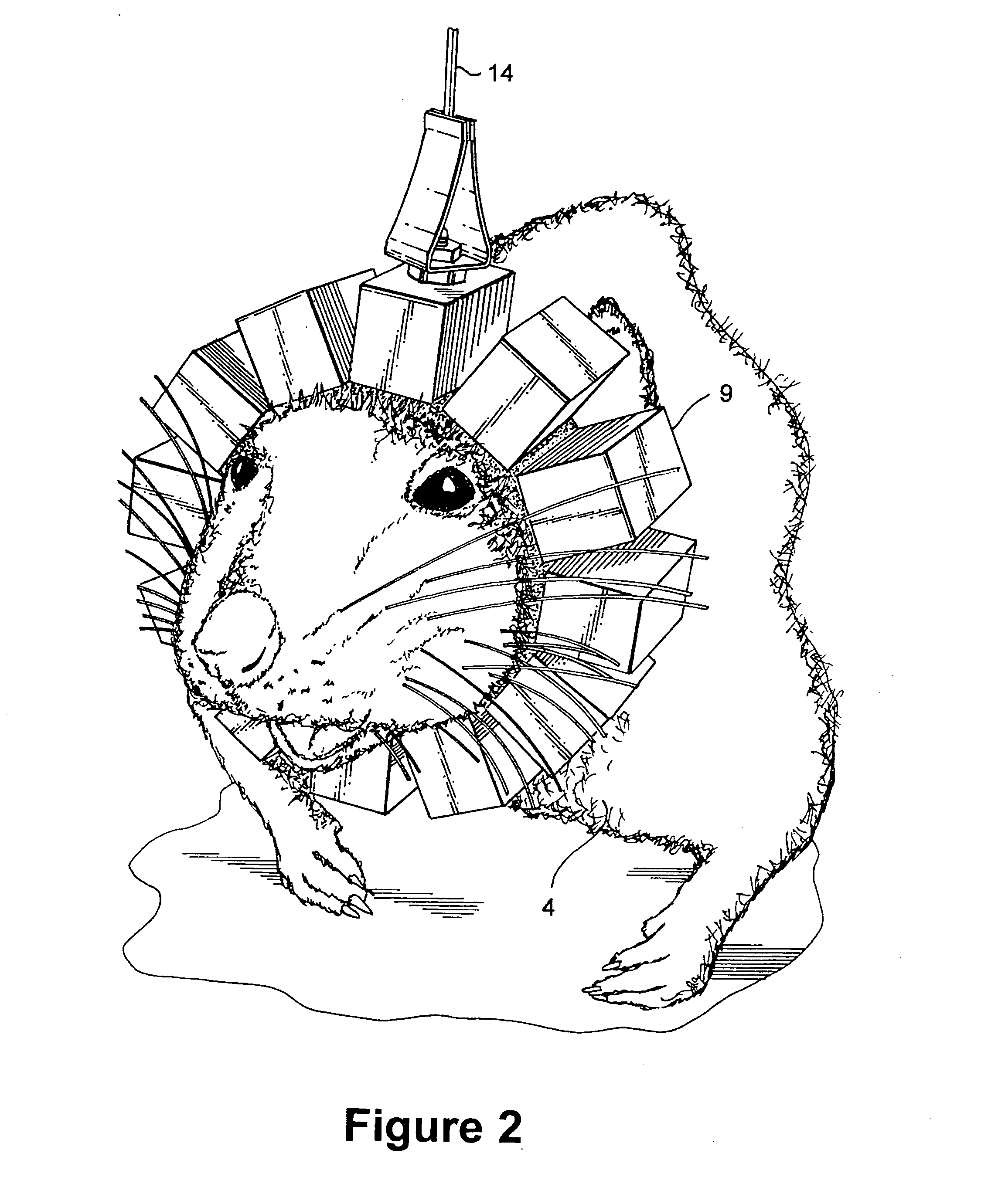
[0065] A compact conscious animal positron emission tomography scanner of the present invention addresses the need for imaging of metabolic or chemical activity of conscious animals, especially rats. Rats are commonly used as animal models in medical research because it is possible to genetically alter rats to isolate a specific trait, and to follow the trait through generations. The knowledge gained in these animal models can then be applied, for example, to human disease, cancer, drug research, and drug addiction.

[0066] An additional embodiment of the compact positron emission tomography scanner of the present invention includes the adaptation of the apparatus and methods of the present invention to the imaging of non-compliant moving human subjects.

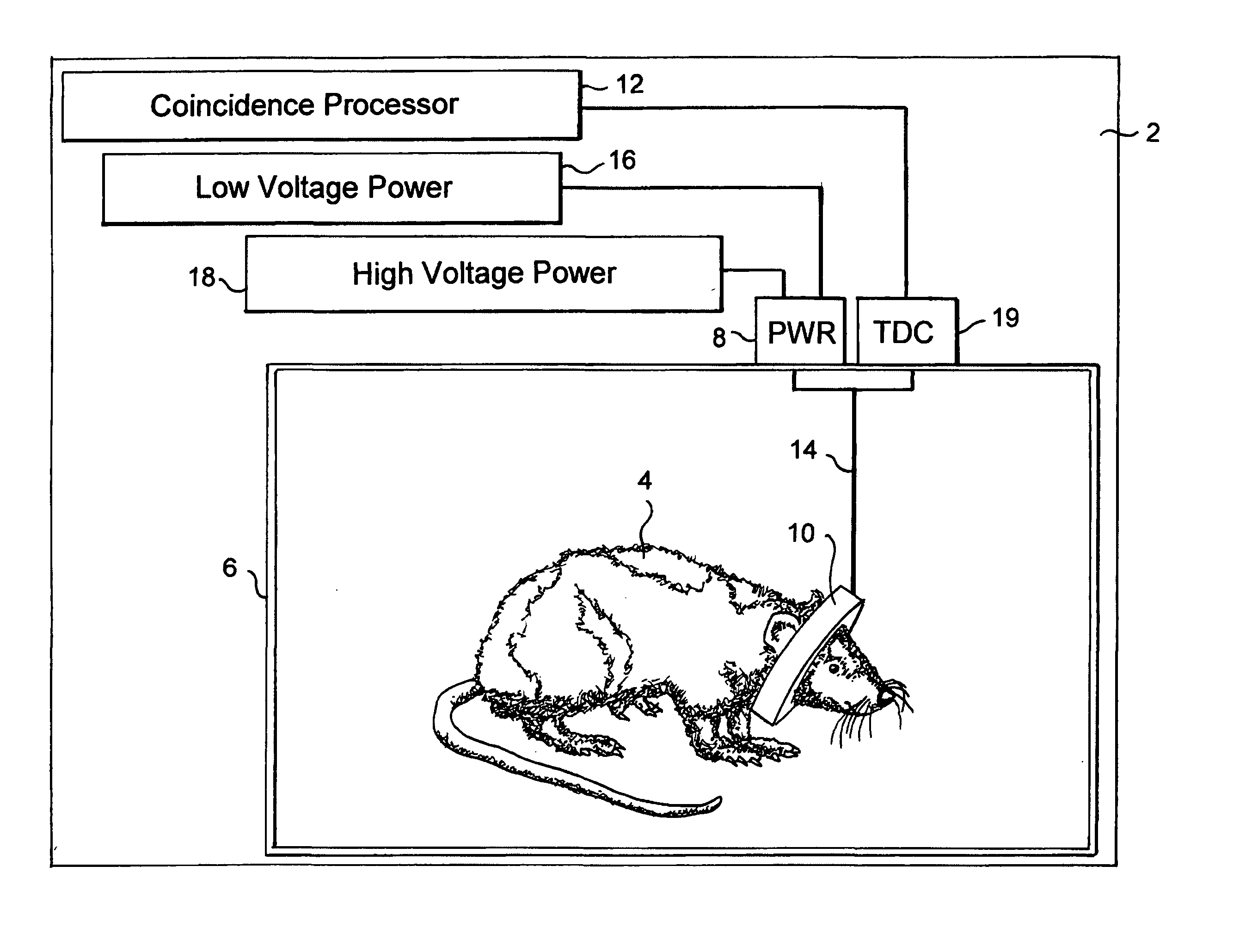
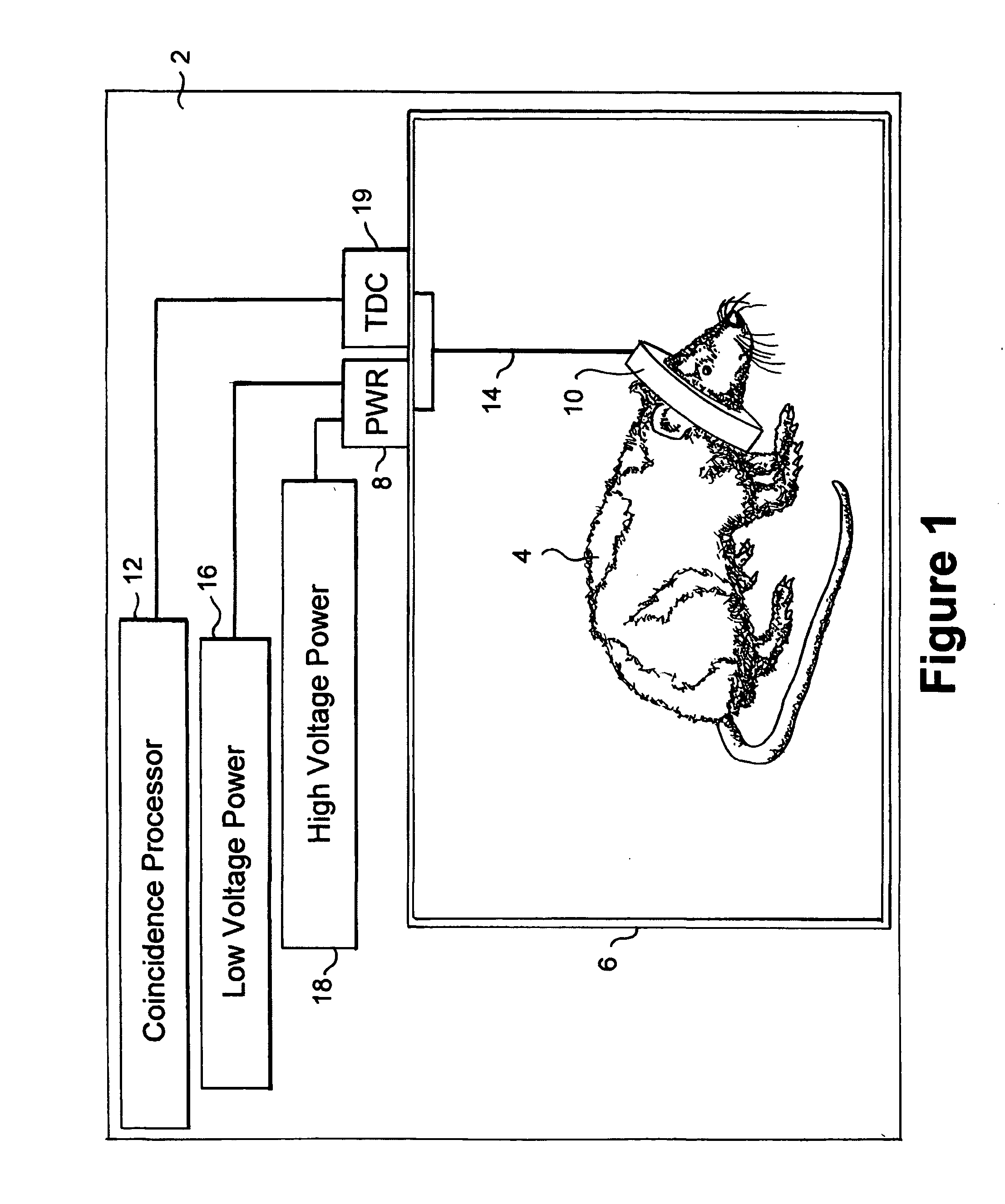
[0067] The method and apparatus formed in accordance with the present invention provides positron emission tomography (PET) images of chemical and metabolic activity within a conscious animal. FIG. 1 shows a block diagram of a prefer...


PUM


Abstract

A combined PET/MRI scanner generally includes a magnet for producing a magnetic field suitable for magnetic resonance imaging, a radiofrequency (RF) coil disposed within the magnetic field produced by the magnet and a ring tomograph disposed within the magnetic field produced by the magnet. The ring tomograph includes a scintillator layer for outputting at least one photon in response to an annihilation event, a detection array coupled to the scintillator layer for detecting the at least one photon outputted by the scintillator layer and for outputting a detection signal in response to the detected photon and a front-end electronic array coupled to the detection array for receiving the detection signal, wherein the front-end array has a preamplifier and a shaper network for conditioning the detection signal.

Description

CROSS-REFERENCE TO RELATED APPLICATIONS [0001] This application is a continuation-in-part of U.S. application Ser. No. 10 / 687,797 filed Oct. 16, 2003.[0002] This invention was made with support from the U.S. Government under Contract No. DE-AC02-98CH10886 awarded by the Department of Energy. The U.S. Government has certain rights in the invention.FIELD OF THE INVENTION [0003] The present invention relates to positron emission tomography (PET) scanners, and more particularly, to low-power, low-noise front-end electronics in compact conscious animal positron emission tomographs for use in imaging portions, such as the brain, of conscious animals. The present invention further relates to magnetic resonance imaging (MRI) scanners, and more particularly, to a combined dual modality PET / MRI scanner. BACKGROUND OF THE INVENTION [0004] Positron emission tomography (PET) is a non-invasive imaging technique that uses radioactive isotopes to map chemical or metabolic activity in living organis...

Claims


Application Information

Patent Timeline
Patent Type & Authority Applications(United States)
IPC IPC(8): A61B5/05G01T1/20G01T1/29
CPCA61B6/037A61B6/4417G01T1/2985G01R33/481A61B6/508A61B5/0035A61B5/0042A61B5/055A61B2503/40
Inventor SCHLYER, DAVID J.WOODY, CRAIG L.ROONEY, WILLIAM D.VASKA, PAULSTOLL, SEANPRATTE, JEAN-FRANCOISO'CONNOR, PAUL
Owner BROOKHAVEN SCI ASSOCS
Why Eureka
Try Eureka