
Method of, and computer software for, classification of cells into subpopulations

a cell and sub-population technology, applied in the field of cell classification, can solve the problems of obscuring or preventing observation, time-consuming analysis of individual cell responses, and insufficient measurement of cell cycle progression in assays, and achieve the effect of accurate classification of further sets of cells

Inactive Publication Date: 2005-09-22
GE HEALTHCARE LTD
View PDF9 Cites 63 Cited by
  • Summary
  • Abstract
  • Description
  • Claims
  • Application Information

AI Technical Summary

Benefits of technology

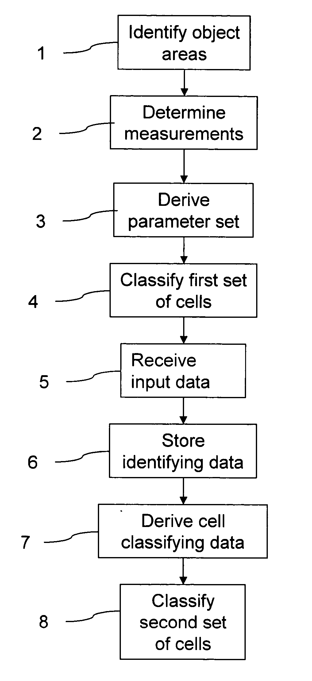
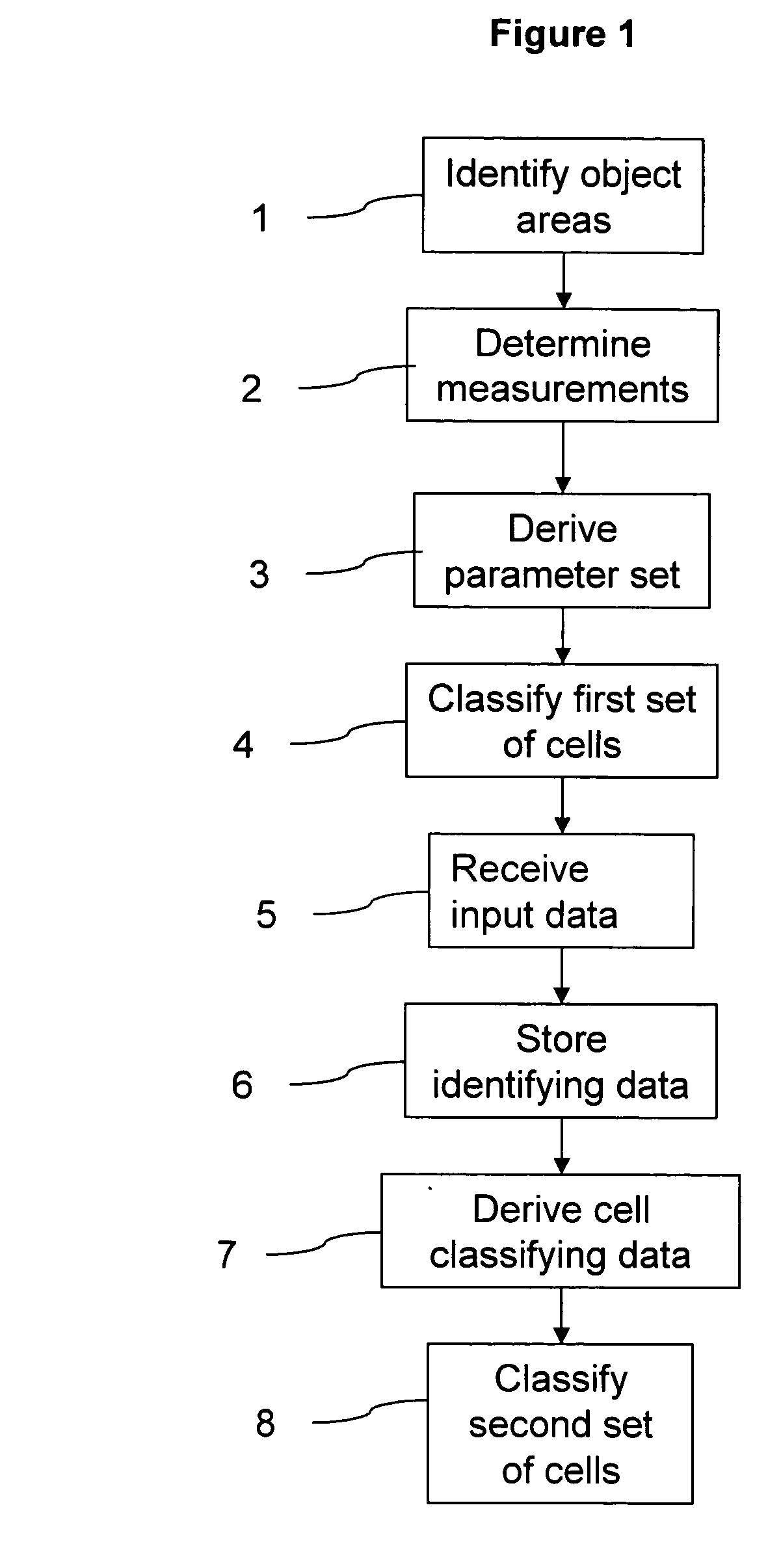
[0032] In an embodiment, the measurement(s) may be calculated automatically using a set of image analysis routines. The measurements for each object area may then be stored in memory in association with identification data, to build up a database of classifying data, which can later be applied with minimal user intervention to further sets of cells. High-throughput automated cell classification can thereby be achieved.
[0037] A method according to the present invention may take any measurement of the second set of cells from the image data without user intervention and as such will not require an operator to input any threshold or specify any measurement value relating to the second set of cells. The cell classifying data derived from the identifying data and the parameter set will therefore be derived from objective and accurate measurement data, facilitating accurate classification of further sets of cells.

Problems solved by technology

This variability may obscure or prevent the observation of biological activity important to compound screening.
However, analysis of the responses of individual cells will be time-consuming in the case of populations of large cell count.
Unlike measuring thymidine or BrdU incorporation, these assays do not provide any inherent measure of cell cycle progression, and give only a measure of cell mass ie. increase in cell number, relative to another population.
However, as for the colourimetric assays discussed above, these do not directly report cell cycle parameters and have not been widely adopted.
All of the above methods provide data on the overall proliferation within a cell population under examination, but do not identify the status of individual cells.
However, analysis by propidium iodide staining and flow cytometry is necessarily destructive and hence requires multiple samples to study cell cycle progression, which can become rate limiting where many hundreds of samples are to be analysed.
In addition, flow cytometry does not yield fine resolution of cell cycle position in G2 / M as the DNA content is the same in all cells.
The technique involves simply counting cells having a signal above a given threshold, and is unsuited for the detection of cell cycle phases other than mitosis.
However, although these specialised approaches provide valuable data on the mechanisms and components involved, they are not generic methods for monitoring the cell cycle.

Method used


Image

  • Method of, and computer software for, classification of cells into subpopulations
  • Method of, and computer software for, classification of cells into subpopulations
  • Method of, and computer software for, classification of cells into subpopulations

Examples


examples of

PRODUCTION OF STABLE CELL LINES

Example 1-Preparation of DNA Construct

[0171] i) The N-terminal third of the cyclin B1 mRNA (amino acids 1-171), encoding the cyclin B1 destruction box and the NES was amplified with HindIII and BamHI ends using standard PCR techniques and the following primers:

(SEQ ID NO: 1)5′- GGGAAGCTTAGGATGGCGCTCCGAGTCACCAGGAAC -3′(SEQ ID NO: 2)5′- GCCGGATCCCACATATTCACTACAAAGGTT -3′.

[0172] ii) The gene for wtGFP was amplified with primers designed to introduce restriction sites that would facilitate construction of fusion proteins. The PCR product was cloned into pTARGET (Promega) according to manufacturer's instructions and mutations (F64L / S175G / E222G) were introduced using the QuikChange site-directed mutagenesis kit (Stratagene). Constructs were verified by automated DNA sequencing. DNA encoding the mutant GFP was then cloned downstream of the cyclin B1 N-terminal region using BamHI and SaII restriction sites.

[0173] iii) The cell cycle dependent region of the...

example 2 -

Example 2-Effect of Cell Cycle Blocking Agents on GFP Fluorescence From Cell Cycle Phase Marker Using Transiently Transfected Cells

[0175] U2OS cells (ATCC HTB-96) were cultured in wells of a 96 well microtitre plate. Cells were transfected with a cell cycle reporter construct prepared according to Example 1, comprising a cyclin B1 promoter operably linked to sequences encoding the cyclin B1 D-box, the cyclin B1 CRS, and GFP in a pCORON4004 vector (Amersham Biosciences) using Fugene 6 (Roche) as the transfection agent.

[0176] Following 24 hours of culture, cells were exposed to the specific cell cycle blockers mimosine (blocks at G1 / S phase boundary) or demecolcine (blocks in M phase). Control cells were exposed to culture media alone.

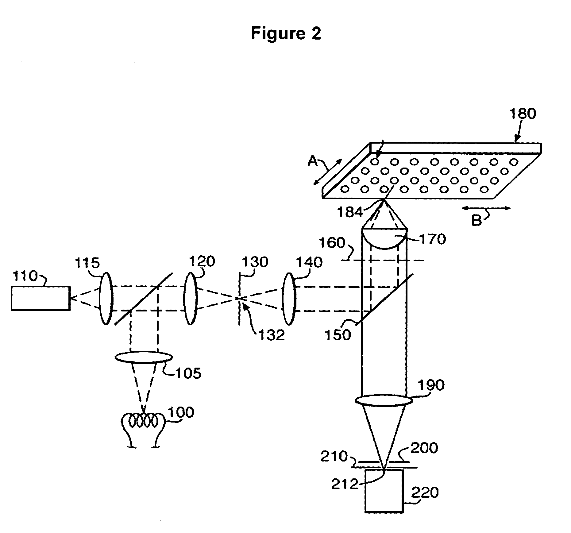
[0177] Cells were incubated for a further 24 hours and then analysed for nuclear GFP expression using a confocal scanning imager with automated image analysis (IN Cell Analysis System, Amersham Biosciences).

[0178] Cells exposed to demecolcine showed i...

example 3 -

Example 3-Microinjection of the Construct

[0179] HeLa cells were micro-injected with the construct prepared according to Example 1 and examined by time lapse microscopy. Differential interference contrast (DIC) images were made along with the corresponding fluorescence images. A cell in metaphase showed bright fluorescence in the nucleus. The same cell was imaged similarly at later times in anaphase and late anaphase. The DIC images showed the division of the cell into two daughter cells, the corresponding fluorescence images showed the loss of fluorescence accompanying destruction of the fluorescent construct as the cell cycle progresses.


PUM

PropertyMeasurementUnit
Sizeaaaaaaaaaa
Areaaaaaaaaaaa
Phaseaaaaaaaaaa

Abstract

A method of classifying cells into subpopulations using cell classifying data is described. The method comprises receiving and analyzing image data to identify object areas in the image data to determine, for at least one selected first cell, one or more measurements. A first parameter set is derived from the measurements for the first cell, the first parameter set comprising at least one of said one or more measurements. The first set of cells are classified into subpopulations, and identified to produce first identifying data. Cell classifying data for use in classifying a second set of cells into subpopulations is derived from the first parameter set and the first identifying data. A second set of cells is classified into subpopulations on the basis of one or more measurements taken for cells in the second set of cells, by use of the cell classifying data. The parameter sets of cells may be represented as vectors in an n-dimensional space.

Description

FIELD OF THE INVENTION [0001] The invention relates to methods of cell classification. Cells are imaged and classified into subpopulations. The invention further relates to apparatus and computer software adapted to carry out such a method. BACKGROUND OF THE INVENTION [0002] There is currently a need in drug discovery and development and in general biological research for methods and apparatus for accurately performing cell-based assays. Cell-based assays are advantageously employed for assessing the biological activity of chemical compounds. [0003] In addition, there is a need to quickly and inexpensively screen large numbers of chemical compounds. This need has arisen in the pharmaceutical industry where it is common to test chemical compounds for activity against a variety of biochemical targets, for example, receptors, enzymes and nucleic acids. These chemical compounds are collected in large libraries, sometimes exceeding one million distinct compounds. The use of the term chem...

Claims


Application Information

Patent Timeline
IPC IPC(8): G01N15/14G01N21/64G06K9/00
CPCG01N15/1475G01N21/6428G01N21/6452G06K9/00127G01N2015/1477G01N2015/1493G01N21/6458G06V20/69G01N15/1433
Inventor ARINI, NICKZALTSMAN, ALLAGOODYER, IANALEXANDROV, YURIYCYBUCH, JUREKSOLTYS, BOHDANDAGENAIS, LOUISROQUEMORE, LIZMURPHY, SAM
Owner GE HEALTHCARE LTD
Learn More