Agents for use in magnetic resonance and optical imaging

a technology of magnetic resonance and optical imaging, applied in the field of imaging agents, can solve the problems of poor resolution, limited nanoparticle application, and limited application of luminescent nanoparticles in vivo

Inactive Publication Date: 2005-10-06
RGT UNIV OF CALIFORNIA
View PDF10 Cites 56 Cited by
  • Summary
  • Abstract
  • Description
  • Claims
  • Application Information

AI Technical Summary

Benefits of technology

[0017] The advantages of these nanoparticles are several fold. As optical imaging agents, their non-bleaching luminescence is well described and renders them extremely useful for imaging applications requiring repeated interrogation of regions of interest. Attachment to specific molecules of interest can allow targeting of the particles to specific cell or tissue types, or to allow binding to other molecules. With the magnetic component, these particles can be used to obtain in vivo images using MRI, and then tissue can be collected for histological investigation using optical imaging. This method would be invaluable, for example, to allow a clinician to be certain that biopsied tissue came from the area of interest identified in an MRI image.

Problems solved by technology

The application of luminescent nanoparticles in vivo is limited, however, due to the shallow depth of penetration of light through tissues.
Clinical imaging methodologies such as magnetic resonance imaging (MRI) offer unlimited depth of interrogation, but poorer resolution than optical methods.
However, these nanoparticles are limited by the low level of incorporation of the magnetic material, and a possible decrease in the photoluminescent efficiency of the nanoparticle.

Method used


Image

  • Agents for use in magnetic resonance and optical imaging
  • Agents for use in magnetic resonance and optical imaging
  • Agents for use in magnetic resonance and optical imaging

Examples


example 1

Cd1-xMnxSe

[0058] Cd1-xMnxSe was synthesized from the organometallic complex Mn2(μ-SeMe)2—(CO)8 via a high-temperature pyrolysis process (Mikulec et al., 2000). This method was followed and Mn-doped CdSe nanoparticles were synthesized. The compound Mn2(μ-SeMe)2—(CO)8 was synthesized, which was confirmed by some of the characterization results such as infrared spectrum and chemical analysis. FIG. 6 shows the IR spectra of both the compound Mn2(μ-SeMe)2—(CO)8 and starting material of Mn(CO)5Br.

[0059] Cd1-xMnxSe nanoparticles were then prepared in a mixture of tri-n-octylphosphine oxide (TOPO) and hexadecylamine (HDA) by use of the synthesized compound of Mn2(μ-SeMe)2—(CO)8 as Manganese source. FIG. 7 shows the results from TEM measurement. As shown in FIG. 7A, the synthesized Cd1-xMnxSe nanoparticles were nearly monodispersed and uniform with a particle size 7.7 nm. FIG. 7B shows Manganese was contained in the particles. FIG. 7C shows a schematic of the surface covered with an organi...

example 2

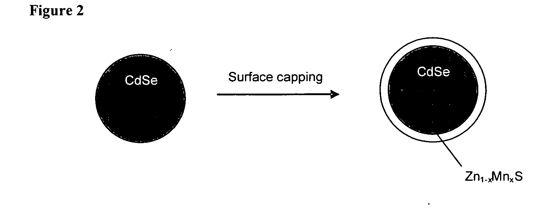
Doping Manganese in the Quantum Shell

[0061] The above-mentioned method is an effective one for incorporating Manganese into CdSe nanoparticles. However, limitations exist in the low level of Mn-doping and the possibility of decreasing the photoluminescent efficiency. To overcome these problems, a new material containing a CdSe core and a manganese-doped ZnS (or CdS) shell was developed. Compared with Cd1-xMnxSe particles, the new material has the following advantages: (1) Manganese could be doped with much higher concentration due to the comparable parameters of crystal lattice between Manganese and ZnS (or CdS), (2) there is improvement of the photoluminescent efficiency due to the ZnS (or CdS) shell, and (3) Manganese does not enter the crystal lattice of CdSe, accordingly, the doping will not affect the perfection of CdSe crystal lattice. FIG. 15A shows a comparison of the emission profiles between the dye of rhodamine 6 G and CdSe / ZnS nanoparticles. FIG. 15B depicts the XRD pat...

example 3

Preparation of Zn1-xMnxS / SiO2 Using Microemulsion Method

[0068] Strategy for the synthesis In typical synthesis, Zn1-xMnxS nanoparticles form in a reverse microemulsion phase composed of nonionic surfactant olyoxyethylene nonaphenyl ether (Igepal CO-520). The synthesis involves the following procedures: a dilute (NH4)2S solution is added to one microemulsion solution while a dilute Zn(NO3)2 and Mn(NO3)2 solution is added to the other. After equilibration, the two aliquot microemulsions are mixed and allowed to stand overnight. The silica coating is generated by the addition of ammonium hydroxide and tetraethoxysilane to the Zn1-xMnxS-contained microemulsion system. After 18 hours, the resulting Zn1-xMnxS / SiO2 nanoparticles are isolated by adding acetone to the microemulsion system followed by centrifugation and washing with cyclohexane and methanol for few times. Zn1-xMnxS / SiO2 nanoparticles of core-shell structure are thus obtained. To synthesize a nanocomposite of Zn1-xMnxS homoge...


PUM


Abstract

Semiconductor nanoparticles are doped with paramagnetic ions to serve as dual-mode optical and magnetic resonance imaging (MRI) contrast agents. These nanoparticles can be constructed in smaller diameters than typical MRI agents. The dual-modality nature allows the particles to be used for in vivo imaging by MRI, and then followed by histology with optical imaging techniques.

Description

CROSS-REFERENCE TO RELATED APPLICATIONS [0001] This application claims priority to U.S. provisional application No. 60 / 559,374 filed on Apr. 1, 2004 which is hereby incorporated by reference in its entirety.FIELD OF THE INVENTION [0002] The present invention relates generally to agents for use in imaging of biological samples. In particular, it relates to imaging agents with properties which allow their use in magnetic resonance imaging (MRI) as well as optical imaging techniques, both in vivo and in vitro. BACKGROUND OF THE INVENTION [0003] Luminescent nanoparticles have the potential to overcome problems encountered by organic small molecules in certain fluorescent tagging applications by combining the advantages of high photobleaching threshold, good chemical stability, and readily tunable spectral properties. [0004] Nanoparticles are small particles with a typical size of a few nanometers (Wolfgang et al., 2003). Due to their discrete energy level similar to that of atoms, nanop...

Claims


Application Information

Patent Timeline
Patent Type & Authority Applications(United States)
IPC IPC(8): A61K49/00A61K49/18H01L21/00
CPCA61K49/0002A61K49/0017A61K49/0065A61K49/0067A61K49/1878B82Y5/00
Inventor KAUZLARICH, SUSANLOUIE, ANGELIQUE
Owner RGT UNIV OF CALIFORNIA
Why Eureka
Try Eureka