Foam matrix embolization device

a foam matrix and embolization device technology, applied in the field of medical devices, can solve problems such as incomplete embolization of aneurysms, and achieve the effect of reducing coil stretching and improving sponge mechanical integrity

Inactive Publication Date: 2006-03-16
CORDIS CORP
View PDF31 Cites 66 Cited by
  • Summary
  • Abstract
  • Description
  • Claims
  • Application Information

AI Technical Summary

Benefits of technology

[0054] In accordance with yet another aspect of the present invention, there is provided an embolization device having an embolization coil and foam sleeve disposed about the coil periphery, which is radiopaque. The embolization device may be made visible under fluoroscopy by using an embolization coil made from a radiopaque material such as a platinum or tungsten alloy. Alternatively, the foam sleeve may be made radiopaque by incorporating a radiopaque filler material such tantalum or tungsten powder or a radiopaque iodine commonly used in contrast media such as RENOGRAFFIN. This radiopaque material aids in the

Problems solved by technology

These balloons do not generally conform to the aneurysm space, thereby leaving unoccupied space leading to an incomplete aneurysm embolization.
Often times

Method used


Image

  • Foam matrix embolization device
  • Foam matrix embolization device
  • Foam matrix embolization device

Examples


example 1

Polymer Solution Preparation

[0092] A 1% w / w solution of epsilon-caprolactone / glycolide copolymer in 1,4-dioxane was prepared as follows. 1.5 grams of 36 / 64 poly(epsilon-caprolactone-glycolic acid) copolymer, obtained from American Polymer Incorporated (American Polymer Inc., Birmingham, Ala.), were added to 148.8 grams of 1,4-dioxane (Fisher Scientific, Raritan, N.J.) in a 250 milliliter Erlenmeyer screwed cap flask. The mixture was stirred for 4 hours in 60° C. water bath set on a temperature controlled heating plate. The polymer solution was filtered through an extra coarse thimble filter to remove any undissolved solids.

example 2

Making 36 / 64 PCLUPGA Foamed Coils Using Freeze-Dry Method

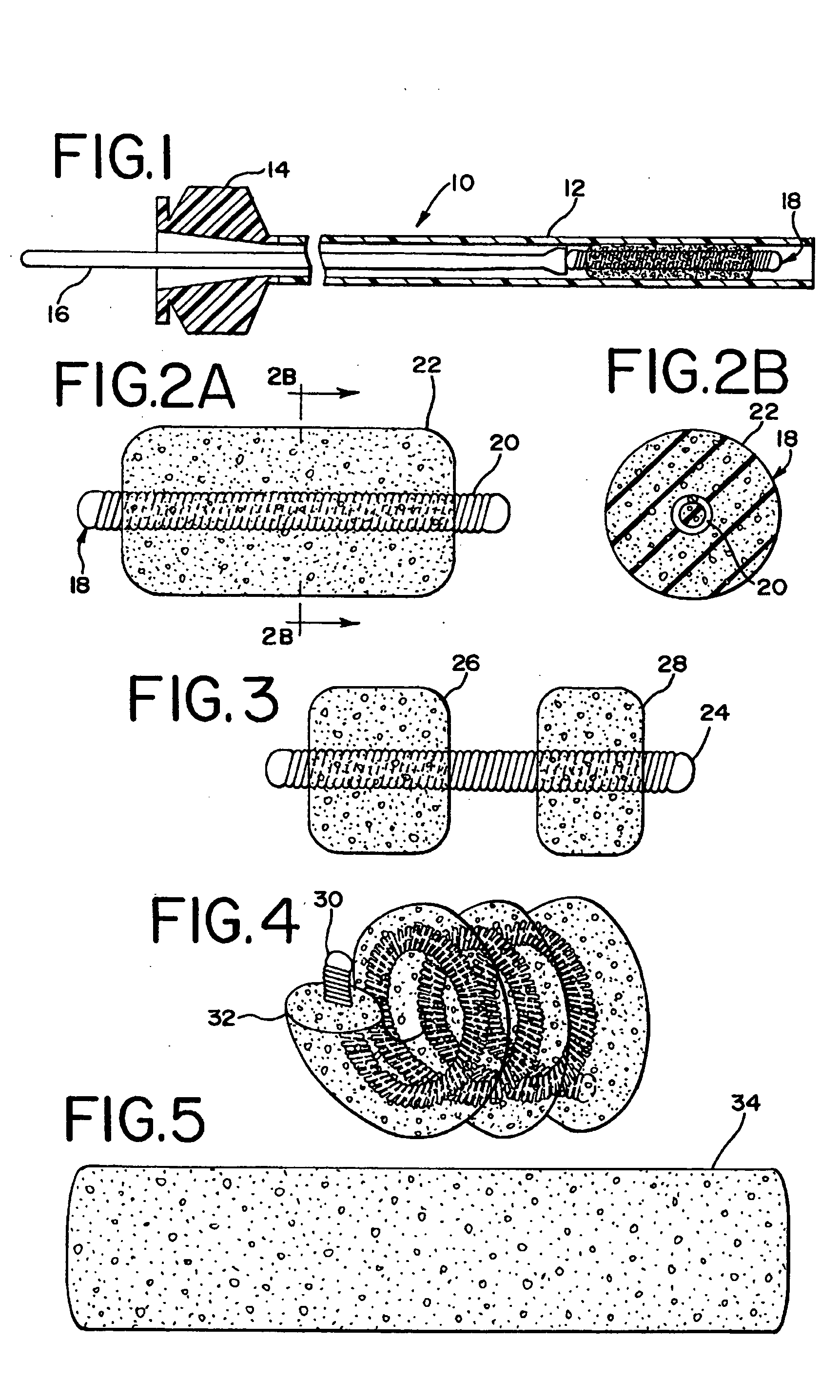
[0093] A 10-cm embolic coil was inserted in a polytetrafluoroethylene (PTFE) tubing (0.015″ ID, ZEUS Industrial products, Inc. Orangeburg, S.C.) that was 2 centimeters longer that the coil. The tube served as a mold. The tube mold containing the coil was filled with the 1% w / w solution of the epsilon-polycaprolactone / glycolide (PCL / PGA) copolymer in 1,4-dioxane described in Example 1, using a glass syringe with a 30 G needle attached. The tube mold containing the coil and solution was sonicated for approximately 5 minutes in a sonication bath (BRANSON, Model 3210R-DTH, Danbury, Conn.) to ensure the solution got into the inter-diameter of the coil, between the coils and into the coil lumen. Bubbles generated during sonication were replaced with fresh polymer solution. Then, the tube mold was frozen in liquid nitrogen for about five minutes, and placed on a pre-cooled (−17° C.) shelf of a freeze-dryer (lyophilization unit) (FTS...

example 3

Fibronectin Absorption Experiment

[0094] A 4-cm foam-embedded coil from Example 2 was cut into about 4-mm segments. Coil segments were then placed into Siliconized Eppendorf tubes. The coil segments were wet with 0.5 milliliters of 70% ethanol, and then washed twice with 1 milliliter of phosphate buffered saline (PBS). The coil segments were incubated with 100 microliters of fibronectin probe solution. The solution was purchased from Sigma Chemical Co. (St. Louis, Mo.). and was radiolabeled by iodine 125 at Lofstrand Labs Limited (Gaithersburg, Md.). The incubation was for 1 hour at 37° C. Afterwards, the incubated coil segments were washed three times with 1 milliliter 0.1% Triton in PBS. The coil segments were transferred to a scintillation vial, and the radioactivity of the samples was counted. Absorption of fibronectin on foamed coils of Example 2 was about three fold higher than on the bare coils under similar treatment.

[0095] As can be appreciated, numerous variations of the ...


PUM

PropertyMeasurementUnit
Pore sizeaaaaaaaaaa
Pore sizeaaaaaaaaaa
Pore sizeaaaaaaaaaa

Abstract

The present invention relates to a medical device for placement at a predetermined location within a passageway of the human body, and more particularly, relates to a flexible embolization device which may be delivered by a catheter to a pre-selected position within a blood vessel to thereby embolize a blood vessel or a blood vessel defect, such as an aneurysm or fistula. Specifically, the embolization device comprises an elongated coil having a lumen, the coil and lumen being at least partially embedded in an elongated foam member comprising, in some embodiments, a flexible, biodegradable, water insoluble, open, interconnecting-cell foam material having embolic characteristics, and capable of allowing cell proliferation into the open cell foam interior.

Description

CROSS-REFERENCE TO RELATED APPLICATION(S) [0001] This patent application is a continuation in part of U.S. patent application Ser. No. 10 / 775,398, filed Feb. 10, 2004, which is a continuation of U.S. patent application Ser. No. 09 / 663,768 (Attorney Docket No. CRD 0869), filed on Sep. 18, 2000, entitled, “Foam Matrix Embolization Device”, now U.S. Pat. No. 6,723,108.BACKGROUND OF THE INVENTION [0002] 1. Field of the Invention [0003] The present invention relates to a medical device for placement at a predetermined location within a passageway of the human body, and more particularly, relates to a flexible embolization device which may be delivered by a catheter to a pre-selected position within a blood vessel to thereby embolize a blood vessel or a defect in a blood vessel, such as an aneurysm or fistula. [0004] Elongated coil embolization devices are well known in their use to embolize a blood vessel or a defect in a blood vessel such as an aneurysm or fistula, for example a brain a...

Claims


Application Information

Patent Timeline
IPC IPC(8): A61M29/00A61F2/95
CPCA61B17/12022A61B17/12145A61B17/1215A61B17/12163A61B17/12181A61B17/1219A61L2430/36A61L31/10A61L31/146A61B2017/1205A61B2017/0053C08L67/04
Inventor DO, HIEP Q.DAVIS, RICHARD CHAMPION IIIJONES, DONALD K.LORENZO, JUAN A.MITELBERG, VLADIMIRSHERMAN, DARREN R.VYAKARNAM, MURTY
Owner CORDIS CORP
Why Eureka
Try Eureka