

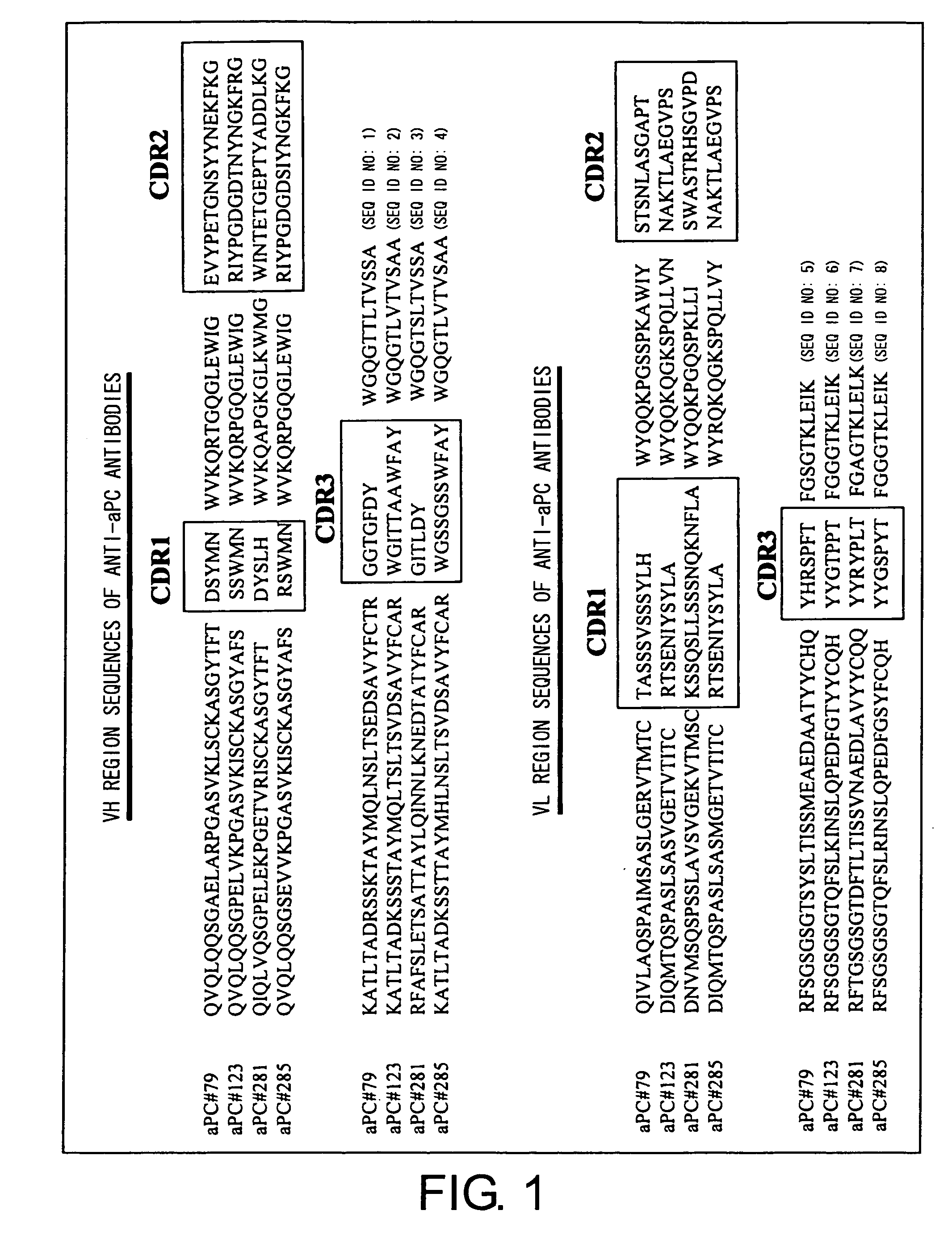
Apc non-neutralizing anti-body
a non-neutralizing, anti-activated technology, applied in the direction of antibacterial agents, antibody medical ingredients, extracellular fluid disorders, etc., can solve the problems of limiting the combined use of other drugs, affecting the effect of apc, and causing bleeding between the two drugs, so as to suppress the inactivation of apc, the effect of potentiating the activity of apc and the effect of endogenous ap
- Summary
- Abstract
- Description
- Claims
- Application Information
AI Technical Summary
Benefits of technology
Problems solved by technology
Method used
Image

Examples
example 1
Preparation of Anti-Human aPC Monoclonal Antibodies
[0140] BALB / c mice were immunized by subcutaneous injection of aPC (Sigma P-2200) as an antigen into their abdominal areas. After elevation of the antibody titers in the sera was confirmed, the final immunization was carried out by injecting the antigen to the caudal vein at a dose of 20 μg / mouse. Three days after the final immunization, the spleen was excised and spleen cells were prepared. The cells were then fused with P3U1 cells. The fused cells were prepared in 2648 wells.
[0141] The day of fusion is defined as “Day 0”. Using HAT medium, the culture medium was changed on Day 1, 2, 3, and 5, to select hybridomas using the HAT medium (which contained RPMI1640, 10% FCS, 0.1% penicillin-streptomycin, 2% BM-Condimed H1, and HAT). The culture supernatant was collected on Day 8, and the first screening was carried out using ELISA.
example 2
First Screening
[0142] The first screening was carried out with ELISA using aPC (SIGMA P-2200) as an antigen. After aPC was diluted to 0.5 μg / mL with a coating buffer (100 mmol / L NaHCO3 (pH 9.6) and 0.02 w / v % NaN3), 100 μL of the solution was aliquoted into each well of 96-well ELISA plates (Nunc, Maxisorp) and immobilized. The plates were washed with rinse buffer (PBS(−) and 0.05% Tween 20) using amicro plate washer (Bio-Rad, Model 1550). A 200-μL aliquot of the diluent buffer (1 w / v % BSA, 50 mmol / L Tris-HCl (pH 8.1), 150 mmol / L NaCl, 1 mmol / L MgCl2, 0.05% Tween 20, and 0.02 w / v % NaN3) was added to each well, and the plates were allowed to stand at room temperature for one hour. After the diluent buffer was removed, a 100-μL aliquot of culture supernatant of hybridomas was added to each well. The plates were incubated at room temperature for one hour. After the plates were washed with the rinse buffer, a 100-μL aliquot of a solution of alkaline phosphatase-conjugated anti-mouse ...
example 3
Second Screening
[0144] aPC has anticoagulant activity and thus extends blood plasma coagulation time. Longer incubation of aPC with blood plasma attenuates this effect because aPC is inactivated in blood plasma over time. If an antibody comprises the activity of suppressing aPC inactivation, the anticoagulant activity of aPC can be maintained by adding the antibody to aPC prior to incubation with blood plasma. Conversely, if the antibody is an aPC-neutralizing antibody, the anticoagulant activity of aPC will be lost. In this Example, hybridoma culture supernatants were tested for the activity of suppressing aPC inactivation. APTT (activated partial thromboplastin time) was used as an indicator of coagulation time. 10 μL of 10 μg / mL aPC (SIGMA, P-2200) solution was combined with 40 μL of hybridoma culture supernatant (cultured under an atmosphere of 5% CO2 at 37° C. for three days) or P3U1 cell culture supernatant. The resulting mixture was incubated at room temperature for 60 minut...
PUM
| Property | Measurement | Unit |
|---|---|---|
| mean molecular weight | aaaaa | aaaaa |
| total volume | aaaaa | aaaaa |
| pH | aaaaa | aaaaa |
Abstract
Description
Claims
Application Information