Coumarin-based cyanine dyes for non-specific protein binding
a cyanine dye and non-specific technology, applied in the field of cyanine dyes for non-specific protein binding, can solve the problems of time-consuming, labor-intensive or incomplete procedures of this kind, and achieve the effect of measurable fluorescence and effective use in protein detection and quantification
- Summary
- Abstract
- Description
- Claims
- Application Information
AI Technical Summary
Benefits of technology
Problems solved by technology
Method used
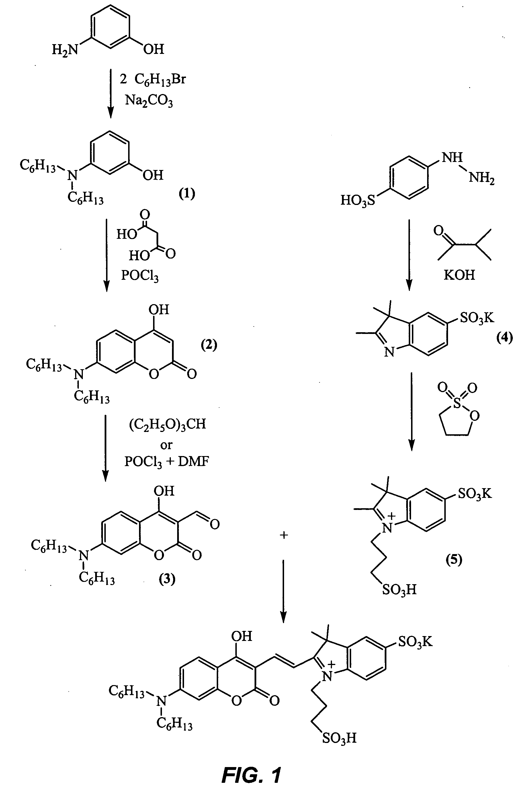
Image
Examples
example 1
Effect of Protein on Fluorescence of Coumarin-Based Cyanine Dyes
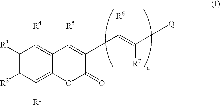
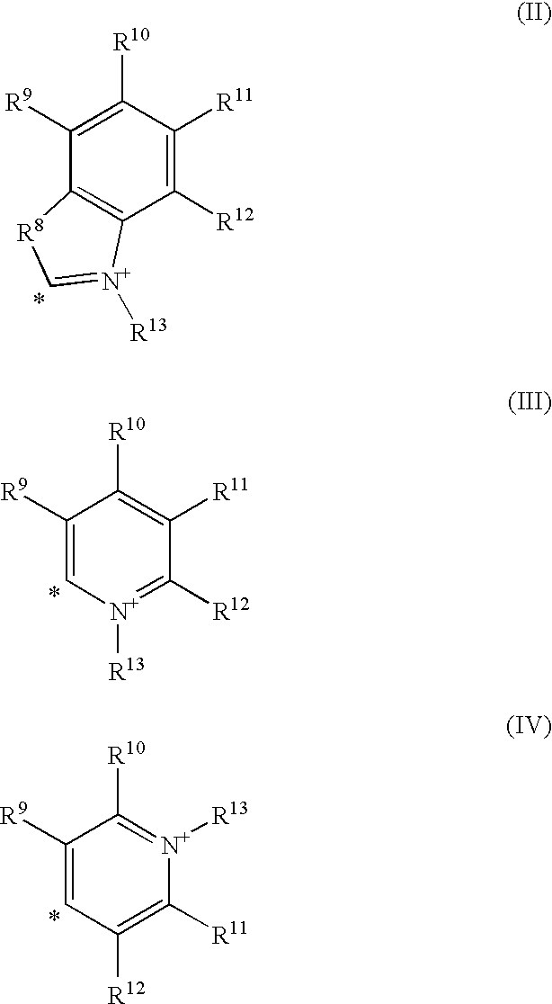
[0023] A series of coumarin-based cyanine dyes within the scope of Formula I above were tested by the following procedure. A 20 mg / mL solution of denatured protein (yeast alcohol dehydrogenase or bovine carbonic anhydrase in 8 M urea) was diluted 100-fold into 50 mM sodium formate pH 4.0 to a final concentration of 0.2 mg / mL. Dye was added from a DMSO stock solution (0.4-1 mM) to a final concentration of 200 nM. The resulting mixture was incubated at room temperature for at least 15 minutes and fluorescence readings were taken on an Aminco Bowman Series 2 Luminescence spectrometer with the excitation and emission monochrometers set at the absorbance and emission maxima, respectively, for each dye. The spectrometer slit width was set at 4 nm. Fluorescence readings at the same wavelengths were also taken of each dye diluted identically into buffer without added protein.
[0024] The peak fluorescence intensities measured i...
example 2
Use of Coumarin-Based Cyanine Dye for Staining 1-D SDS-PAGE Gel
[0025] An 18-well 4-20% Tris-Ci gel (Criterion® System of Bio-Rad Laboratories, Inc., Hercules, Calif., U.S.A.) was loaded with serial dilutions of broad range SDS-PAGE standards (Bio-Rad Laboratories, Inc.) and subjected to electrophoresis according to the manufacturer's instructions. The dilutions and the lanes in the gel in which each dilution was d were as listed below:
Lane No.Load (ng of each protein)1(blank)296034804240512066073081598104112121130.5140.25150.125
[0026] The gel was fixed for 16.5 hours in 300 mL of 40% (volume / volume) ethanol and 10% (volume / volume) acetic acid. Following the fixing step, the gel was stsained in 125 mL of a solution consisting of 0.2 μM of Compound 8 of Example 1 in 30% (volume / volume) methanol and 1% (volume / volume) oxalic acid. After 2 hours and without washing of the gel after the application of Compound No. 8, the gel was scanned on an FX fluorescence scanner (Bio-Rad Laborator...
example 3
Use of Coumarin-Based Cyanine Dye for Staining 2-D Gel
[0027] Immobilized pH gradient strips (ReadyStrip® IPG strips, 11 cm pH 3-10 NL, Bio-Rad Laboratories, Inc.) were loaded with 40 μg of E. coli protein in 8 M urea, 2% CHAPS, 40 mM dithiothreitol, 0.2% (weight / volume) Bio-Lyte ampholyte pH 3-10 (Bio-Rad Laboratories, Inc.). First-dimension isoelectric focusing followed by equilibration and second-dimension of SDS-PAGE were performed as described in the ReadyStrip® instruction manual. Criterion® 8-16% Tris-Cl gels (Bio-Rad Laboratories, INc.) were used for the second dimension. The resulting 2-dimensional gels were fixed for 1 hour in 170 mL per gel of 40% (volume / volume) ethanol and 10% (volume / volume) acetic acid, and stained with 200 mL of a solution consisting of 0.2 μM of Compound 8 of Example 1 in 30% (volume / volume) methanol and 7% (volume / volume) acetic acid. After 2 hours and without washing of the gel after the application of Compound No. 8, the gel was scanned on an FX ...
PUM
 Abstract
Description
Claims
Application Information
 - R&D
- Intellectual Property
- Life Sciences
- Materials
- Tech Scout
- Unparalleled Data Quality
- Higher Quality Content
- 60% Fewer Hallucinations
Browse by: Latest US Patents, China's latest patents, Technical Efficacy Thesaurus, Application Domain, Technology Topic, Popular Technical Reports.
© 2025 PatSnap. All rights reserved.Legal|Privacy policy|Modern Slavery Act Transparency Statement|Sitemap|About US| Contact US: help@patsnap.com


