Microdissection-based methods for determining genomic features of single chromosomes

a single chromosome and genomic feature technology, applied in the field of microdissection-based methods for determining genomic features of single chromosomes, can solve the problems of complicated, difficult to determine the genomic origin of cancer chromosome abnormalities, and limited strategy, and achieve the effect of quick determination of snp haplotypes and facilitate both individual- and population-based genetic association studies

Inactive Publication Date: 2006-09-21
WISCONSIN ALUMNI RES FOUND
View PDF2 Cites 95 Cited by
  • Summary
  • Abstract
  • Description
  • Claims
  • Application Information

AI Technical Summary

Benefits of technology

[0011] The present invention provides a chromosome microdissection-based strategy for detailed molecular analysis of any cytogenetically visible chromosome regions from a single cell. It is a straightforward “what you see is what you get” approach that possesses several unique advantages. It allows the direct isolation of any normal or abnormal chromosomes or chromosome regions from targeted single cells without the interference of the surrounding cells. The isolated chromosomes or chromosome regions are efficiently amplified for high-resolution genomic analysis, such as determination of the breakpoints, gene content, DNA content and genetic variations of the target regions. This strategy provides a unique tool to analyze clonal acquired abnormalities in cancer cells and mosaic constitutional abnormalities observed in a variety of genetic disorders. In addition, this strategy is particularly useful for unambiguously determining haplotypes of any genomic regions without the requirement of family pedigrees and genotypes, LD and population genomic information, or complicated statistical analysis and computing. For example, it can quickly determine SNP haplotypes of any single or multiple haplotype blocks, chromosome regions or chromosomes regardless their genomic locations and sizes, as well as the frequency of LD in the target regions. Furthermore, this strategy has a strong potential to be used for genome-wide and population-based haplotype analysis when it can be applied with large scale SNP array technologies. Amplified single chromosome DNA samples may be analyzed using SNP arrays or, alternatively, subjected to genotyping by primer extension and DNA sequencing. For the genome-wide analysis, one may separate and amplify haploid chromosome sets of an individual, and directly determine the haplotypes of each haploid set using appropriate SNP arrays. This new strategy may largely facilitate both individual- and population-based genetic association studies. In addition, this strategy may also be applied to haplotype analysis of species other than human.

Problems solved by technology

However, this strategy has limitations.
Due to limited resolution and often poor cancer chromosome morphology, determining the genomic origins of cancer chromosome abnormalities can be particularly difficult.
It is often further complicated when the aberrations are only detected in a small number of cancer cells that are mixed with a large population of cells with an apparently normal karyotype.
Because of this limitation, the genomic origins of many observed cancer chromosome abnormalities cannot be identified for further analysis.
Second, the cytogenetics-based positional cloning is not a straightforward, high-throughput strategy; the analytical process of this strategy is cumbersome and labor-intensive.
In addition, such an analysis usually requires a significant amount of cancer specimens, but that are not always available.
However, this method is more commonly used for studying constitutional Mendelian diseases and rare familial cancer cases; its application seems to be limited in genetic analysis of sporadic cancers, perhaps due to the complexity of the cancer genetics and genomics.
However, these techniques also have limitations.
They usually cannot detect balanced chromosomal rearrangements that often result in cancer-causing gene fusion and / or breaking apart, such as the Bcr / Abl fusion gene in CML and the MLL gene split in various types of leukemia.
In addition, the capability of these techniques to detect DNA copy number imbalance can be complicated or impaired in mixed cell populations.
Unfortunately, most cancer specimens are contaminated with more or less normal cells, and genomic changes in most cancers are heterogeneous; cancer tissues are often mixed with multiple cell lines with multiple clonal and / or non-clonal (random) genomic abnormalities.
Furthermore, these techniques may not directly reveal the dosage of individual genes involved in the unbalanced genomic regions.
], which is better than that of the cytogenetic analysis, but is not enough to determine the dosage alterations in individual genes.
As of yet, however, most current technical strategies do not have the ability to directly characterize detectable chromosome aberrations.
In addition, none of the techniques described above can directly uncover the genomic features, such as DNA sequence variations, of the abnormal chromosome regions.
However, unambiguously determining haplotypes of SNP blocks, especially large blocks, is particularly challenging due to technical limitations.
The former is expensive, time-consuming and requires DNA samples from several generations, which are not always available.
In addition, accurately assigning SNP phase using family-based methods becomes increasingly difficult as more loci are considered.
Meanwhile, the latter currently relies on enrichment of DNA of single-chromosome origin using complicated methods, such as somatic cell hybrid and multi-step allele-specific PCR, which is also time-consuming and expensive.
These limitations make it difficult to apply unambiguous SNP haplotype analysis to either individual- or population-based genetic association studies.
Taken together, effectively identifying disease-causing and disease-risk genes as well as their genomic alterations / variations, particularly from cancer-associated clonal chromosome aberrations and constitutional mosaicism from mixed cell populations, remains a great challenge in genetic research.

Method used


Image

  • Microdissection-based methods for determining genomic features of single chromosomes
  • Microdissection-based methods for determining genomic features of single chromosomes
  • Microdissection-based methods for determining genomic features of single chromosomes

Examples


example 1

Determining Single Nucleotide Polymorphism (SNP) Haplotypes Using Dissected Single Chromosomes

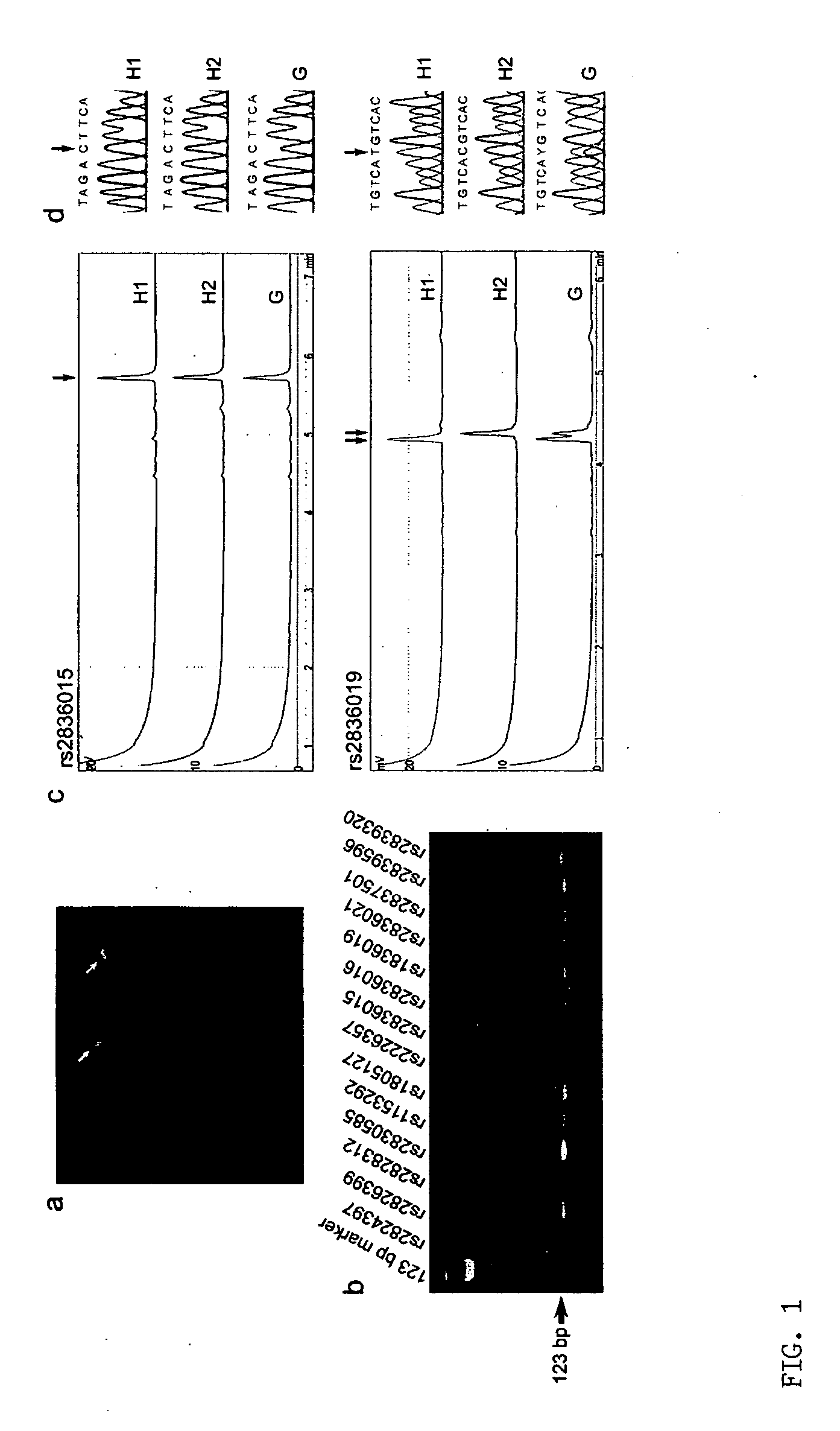
[0035] This example describes the unambiguous determination of SNP haplotypes of a selected chromosome region using a technique according to the present invention. The disclosed technique includes the steps of single chromosome microdissection, universal DNA modification, and direct analysis of amplified single-chromosome DNA by primer extension.

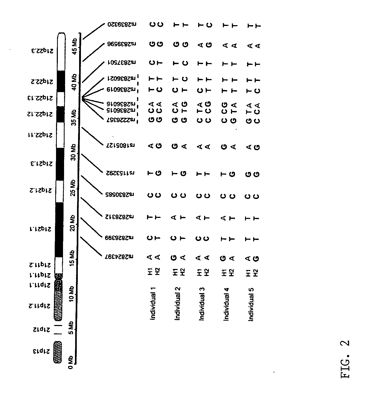
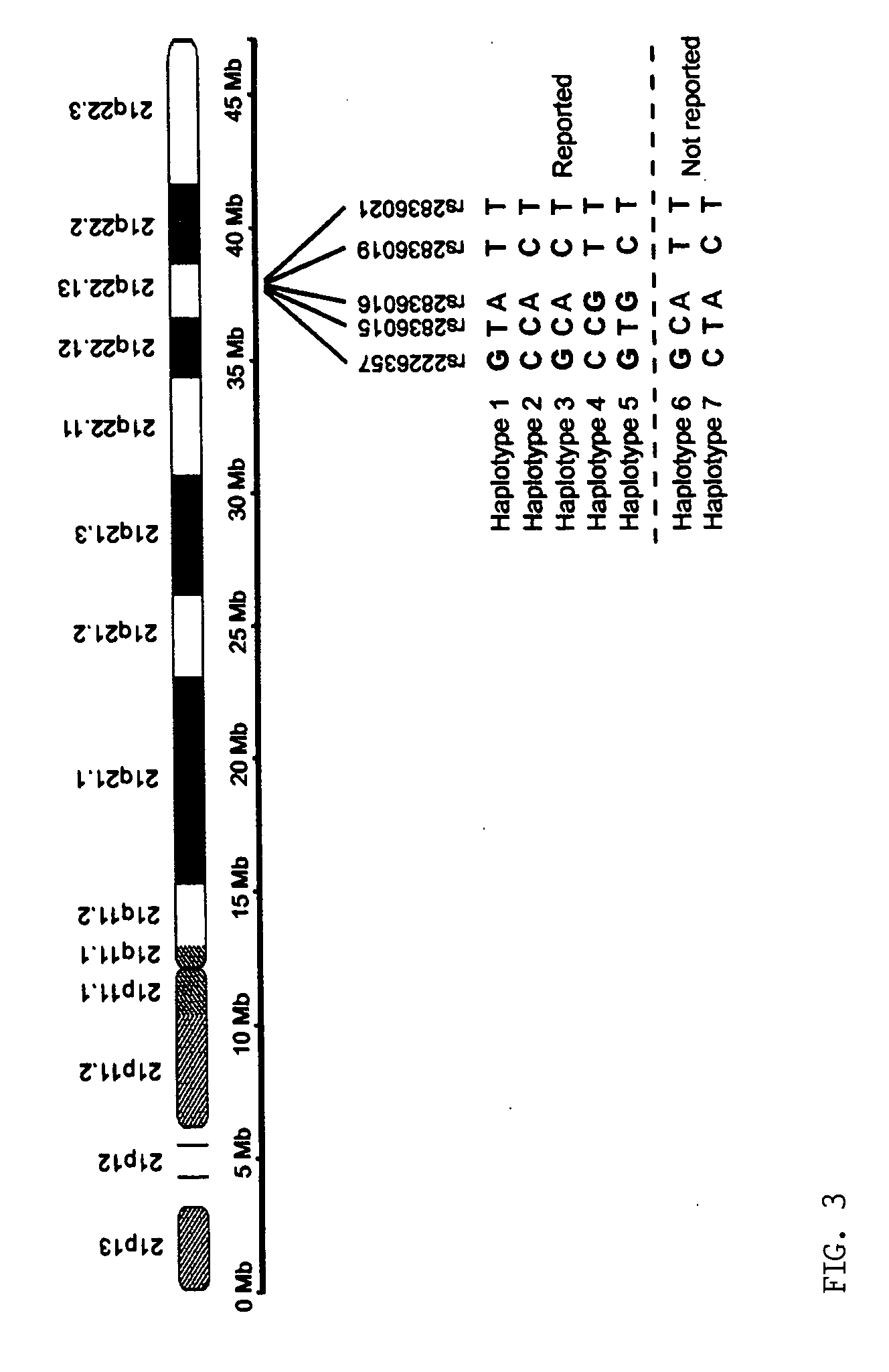
[0036] The inventors analyzed the haplotypes of 14 SNP loci across the long arm of chromosome 21 (21q) in five normal Caucasians, including three unrelated individuals, a 5 mother and her son. Briefly, the inventors isolated 10 single-copy 21qs from each individual in five cultured metaphase peripheral blood cells by chromosome microdissection and amplified each individual 21q using modified degenerate oligonucleotide primed PCR (DOP-PCR). The quality of the dissection and amplification was evaluated by fluorescence in situ hybridization (FISH), i...

example 2

Characterization of Visible Regions of Normal Chromosome 21

[0046] This example illustrates a method according to the invention for haplotype determination of a human chromosome, specifically, human chromosome 21 [Hattori, et al., Nature 405: 311-321 (2000)]. The strategy involves several technical steps, including microdissection of chromosome regions, universal amplification of dissected DNA, reverse FISH for identification of the genomic origins of the chromosome regions, region-specific genomic DNA / gene array and genome-wide SNP array analysis. Single copies of dissected chromosome 21 may be obtained from a transformed normal lymphoblast cell line. A suitable cell line is GM 03657 (Coriell Institute; normal karyotype of 46,XY).

[0047] Metaphase cells are spread on clean 25×50 mm coverslips and G-banded for microdissection, as described in our previous studies [Kao, et al., Proc. Natl. Acad. Sci. U.S.A. 88: 1844-1848 (1991)]. The chromosome and the chromosome region to be dissect...

example 3

Characterization of Lymphoma-Specific Chromosome Abnormalities

[0054] Lymphomas are a group of heterogeneous hematological malignancies which often show chromosome aberrations. Certain lymphoma-specific chromosome abnormalities have been identified, such as t(8;14)(q24;q32) and t(11;14)(q13;q32) translocations in Burkitt lymphoma and Mantle cell lymphoma, respectively. However, the genomic features of many other clonal chromosome aberrations that are frequently seen in lymphomas, particularly marker chromosomes, remain unknown. Such aberrations can be readily characterized by the invention. In this example, the inventors characterized two cytogenetically undistinguishable chromosome deletions in only two cells from two unrelated pediatric patients, respectively, to demonstrate the proof of principle. This example involves several technical steps, including microdissection of chromosome regions, universal amplification of dissected DNA, reverse FISH for identification of the genomic ...


PUM

PropertyMeasurementUnit
temperatureaaaaaaaaaa
sizeaaaaaaaaaa
reverse fluorescence inaaaaaaaaaa

Abstract

The present provides a microdissection-based method for identifying a genomic feature present within a visible chromosome region. The method includes steps of: (a) micro-dissecting a single copy of a chromosome to obtain a visible chromosome region; (b) amplifying the visible chromosome region to obtain amplified single chromosome DNA; and (c) subjecting the amplified single chromosome DNA to micro-array analysis whereby such analysis identifies at least one genomic feature present within the visible chromosome region. The method is applicable to determining genomic features including, but not limited to, genomic DNA size, gene content, DNA breakpoint, or DNA polymorphism (e.g., single nucleotide polymorphisms).

Description

FIELD OF THE INVENTION [0001] This invention relates generally to the identification and characterization of genomic features of chromosomes. More specifically, the present invention is directed to microdissection-based methods that facilitate direct determination of genomic features of either normal or abnormal single chromosomes. BACKGROUND OF THE INVENTION [0002] Chromosome aberration is a hallmark of cancer, which provides valuable clues to pinpoint the candidates for cancer-related genes. Cancer-specific chromosome aberrations often result in alterations in the structure and / or the dosage of cancer-causing genes [Balmain, et al. Nat. Genet. 33 Suppl: 238-244 (2003)]. Examples include Bcr / Abl fusion gene caused by the translocation between chromosomes 9 and 22 (Philadelphia chromosome) in chronic myeloid leukemia (CML), and loss of a copy of the Rb gene caused by the deletion of the proximal region of the long arm of chromosome 13 in retinoblastoma. Chromosome rearrangement in c...

Claims


Application Information

Patent Timeline
Patent Type & Authority Applications(United States)
IPC IPC(8): C12Q1/68C12P19/34
CPCC12Q1/6806C12Q1/6841C12Q2565/519
Inventor YU, JINGWEIQI, ZHONGXIA
Owner WISCONSIN ALUMNI RES FOUND
Why Eureka
Try Eureka