Methods and compositions for growing corneal endothelial and related cells on biopolymers and creation of artifical corneal transplants
a technology of biopolymerization and composition, which is applied in the direction of drug composition, peptide/protein ingredient, prosthesis, etc., can solve the problem of not being able to grow antibodies to the polymer
- Summary
- Abstract
- Description
- Claims
- Application Information
AI Technical Summary
Benefits of technology
Problems solved by technology
Method used
Image
Examples
example 1
Non-Enzymatic Dissection of Primary Human Corneal Endothelial Cells
[0040] The corneal rims from human donors (after the central portion has been removed for transplantation) or whole donor corneas will be rinsed in a large volume (50 ml) of phosphate buffered saline (PBS). They will then be placed in endothelial side up on a holder. The trabecular meshwork and remnants of iris will be removed carefully by micro-dissection. By using sharp pointed jeweler's forceps, the endothelial cell layers and the Descemet's membrane will be peeled off very carefully with great care taken not to include any underlying stromal tissue. This step can be confirmed by viewing the dissected Descemet's membrane under an inverted microscope to make sure it only carries the corneal endothelial cells on one side and nothing on the other side. The piece of tissue will be placed onto an ECM coated 35 mm tissue culture dish or similarly suitable container, filled with approximately 0.5 ml of culture medium (D...
example 2
Culture of Human Corneal Endothelial Cells at High Split Ratio
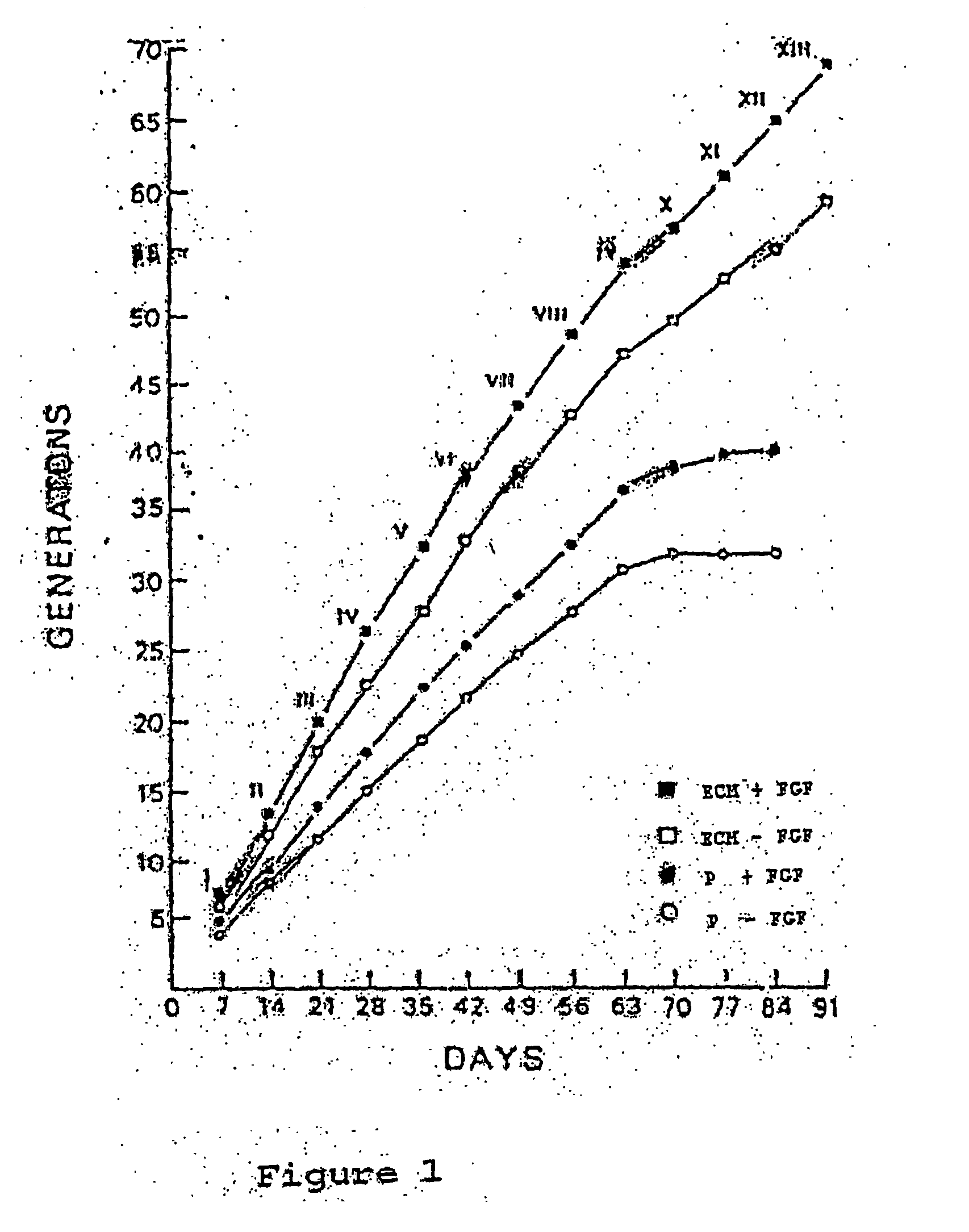
[0041] When the primary cell count from the tissue sample outgrowth reaches a number of 200 to 500, the cells will be released from the dish with STV solution (0.05% trypsin, 0.02% EDTA in normal saline). The STV solution will be removed when the cells round up but are still attached to the culture dish. No centrifugation step is necessary since the remaining STV will be inactivated by the growth media containing 15% fetal calf serum. The corneal cells will be placed onto a 60-mm ECM-coated dish (about 500 cells per dish). The medium will be changed every other day and b-FGF at a concentration of 250 ng / ml will be added at the time of medium change. At confluence (about 7 to 10 days after plating), the cells will be passaged again at the same split ratio (1:16 to 1:64) or will be frozen in 10% DMSO, 15% FCS at a density of 106 cells / ml per ampoule and stored in liquid nitrogen for future use. The passaging can be carried...
example 3
Denudation of Corneal Button
[0043] Human donor corneal buttons are obtained from the Eye Bank. These corneal buttons are deemed unsuitable for transplantation-due to inadequate endothelial cell counts, but otherwise are healthy and disease free and obtained under eye banking guidelines.
[0044] The corneal button will be placed endothelial side up in a holder, and rinsed three times with PBS. Then a solution of ammonium hydroxide at a concentration ranging from 10 mM to 2200 mM will be added carefully into the corneal button without spilling over the top. The cornea will be kept at temperatures of about 10° C. to 25° C. for a period of 5 minutes up to 2 hours. Then the ammonium hydroxide will be removed, and the inside of the cornea button rinsed approximately 10 times with PBS. A cotton swab will be slid gently across the endothelial surface to remove any residual cell skeletons or debris. The corneal button is rinsed again three times with PBS, punched with an 11 mm trephine, and ...
PUM
| Property | Measurement | Unit |
|---|---|---|
| Temperature | aaaaa | aaaaa |
| Length | aaaaa | aaaaa |
| Mass | aaaaa | aaaaa |
Abstract
Description
Claims
Application Information
 - R&D
- Intellectual Property
- Life Sciences
- Materials
- Tech Scout
- Unparalleled Data Quality
- Higher Quality Content
- 60% Fewer Hallucinations
Browse by: Latest US Patents, China's latest patents, Technical Efficacy Thesaurus, Application Domain, Technology Topic, Popular Technical Reports.
© 2025 PatSnap. All rights reserved.Legal|Privacy policy|Modern Slavery Act Transparency Statement|Sitemap|About US| Contact US: help@patsnap.com


