Optical fingerprinting of nucleic acid sequences

Inactive Publication Date: 2007-06-28
WISCONSIN ALUMNI RES FOUND
View PDF3 Cites 11 Cited by
  • Summary
  • Abstract
  • Description
  • Claims
  • Application Information

AI Technical Summary

Benefits of technology

[0015] The present invention provides methods for the optical fingerprinting of nucleic acid molecules. In certain embodiments, the invention provides methods labeling nucleic acid molecules using site-specific nucleic acid binding partners that bind to nucleic acids without cleaving the molecule. The nucleic acid binding partners can be labeled directly with a fluorophore, such as a quantum dot (QD), or indirectly by recombinantly adding a biotin moiety to the binding molecule and using an avidin conjugated fluorophore to bind to the biotin moiety using standard biotin-avidin chemistry. Examples of suitable binding molecules include cut-deficient restriction endonucleases (cdREs), transcription factors, the binding domains of nucleic acid polymerases, antibodies and the like.

Problems solved by technology

However, such large-scale sequencing is limited by the comparative slowness of preparing individual genomes or genome libraries for large-scale nucleotide sequencing.
For example, the YAC vector can accommodate inserts of up to 4 megabases (Mb) while BAC vectors can only accommodate inserts of up to 300 kb, a limit imposed by decreased transfection efficiency.
For sequencing purposes, YAC vectors are comparatively unstable and prone to chimerism of the insert.
Although high-quality maps can be created with these methods, they share significant technical limitations that reduce both information content and throughput.
Too many or too few fragments create problems with resolution sensitivity and/or information content.
Second, fingerprints obtained in this manner are unordered.
Merging different maps entails significant additional effort requiring either fragment hybridization or end sequencing.
Even despite these technical issues, no matter which method is implemented, substantial human involvement is required and remains the rate-limiting step in fingerprint contig assembly.
Unlike traditional agarose gel fingerprinting, the ability to determine the size of the BAC insert is no longer possible.
Further, the presence of chromosomal DNA in the preparation often produces extraneous fragments, a sensitivity issue not apparent in agarose separations.
Thus, extremely pure DNA is required for fluorescent fingerprinting, as erroneous chromosomal fragments can impair automated contig assembly.
These improvements will inevitably lead to additional projects aimed at genome-level sequencing.
However, it is doubtful that the preparation of sequence-ready BAC libraries can keep pace with current sequencing technologie

Method used


Image

  • Optical fingerprinting of nucleic acid sequences
  • Optical fingerprinting of nucleic acid sequences
  • Optical fingerprinting of nucleic acid sequences

Examples


Example

Example 1

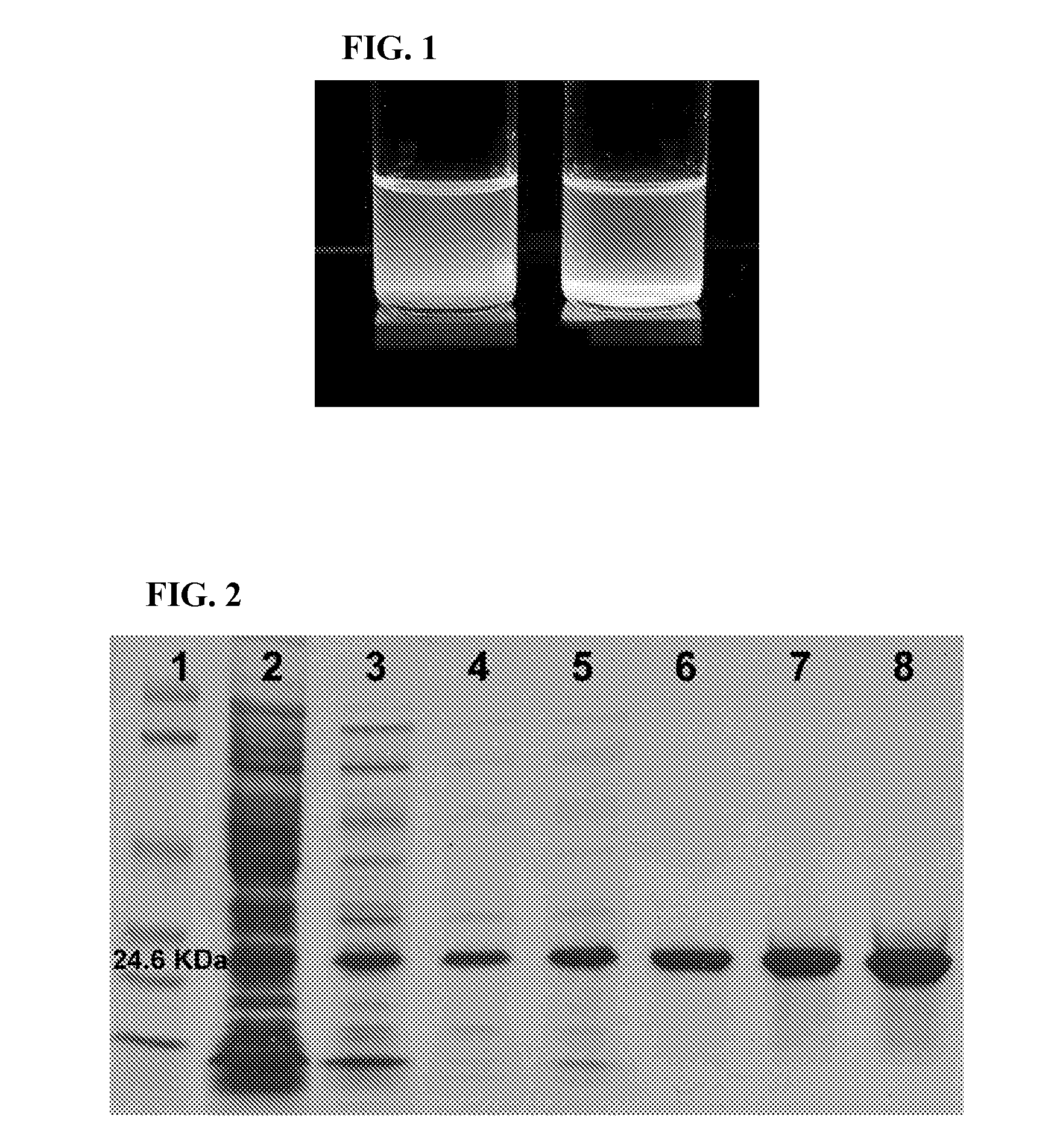
Purification of Recombinant Cut-Deficient BamHI

[0090] A histidine tag was fused to the N-terminal end of the D94N sequence and it was ligated into the T7 expression vector pIVEX (Roche Diagnostics, Indianapolis, Ind.). This construct was electroporated into a MDS42 E. coli strain (Scarab Genomics, Madison, Wis.) containing T7 polymerase regulated by the auto-inducible rhamnose operon. This procedure was required as it was discovered that even tiny amounts of expression (e.g. from a “leaky” promoter) prior to induction in a standard host cell (such as, for example JM109) failed to produce measurable amounts of D94N and even induced numerous mutations in the D94N sequence. This problem was solved using MDS42 cells as an expression host. Ni-NTA purification of the expression product typically yields more than 100 mg protein per 250 ml culture. This product is easily purified and washed using imidazole as shown in FIG. 2.

Example

Example 2

Bioconjugation of QDs to cdRE

[0091] Recombinant D94N molecules were used in an electrophoretic mobility shift assay using 1.5% agarose. FIGS. 5A and 5B show the results of mobility shift assays with the D94N protein. FIG. 5A EtBr stained gel of BpmI digested pGEM3Z with unlabelled D94N: Lane 1, marker; lane 2, pGEM3Z; lane 3, pGEM3Z+D94N cell lysate; lane 4, pGEM3Z+purified 6×His-D94N. FIG. 5B QD-conjugated D94N: lane 1, marker; lane 2, pGEM3Z; lane 3, pGEM3Z+QD labeled 6×His-D94N. The QD-labeled D94N proteins caused an enhanced mobility shift of BpmI-linearized pGEM-3Z (compare FIG. 5B, lane 3 with FIG. 5A, lane 4). The results demonstrate that D94N binds stably to the BamHI site in pGEM-3Z (FIG. 5A), but does not cleave the DNA. This shows that the D94N protein, even with the HIS tag attached, behaves as the native D94N. Similar mobility shifts are observed with D94N / QD bioconjugates where the QDs have surface-capping layers consisting of DTT, cysteine, or mercaptoaceti...

Example

Example 3

Quantum Dot Labeling of Cut-Deficient Restriction Enzymes: Site-Specific Biotinylation of D94N

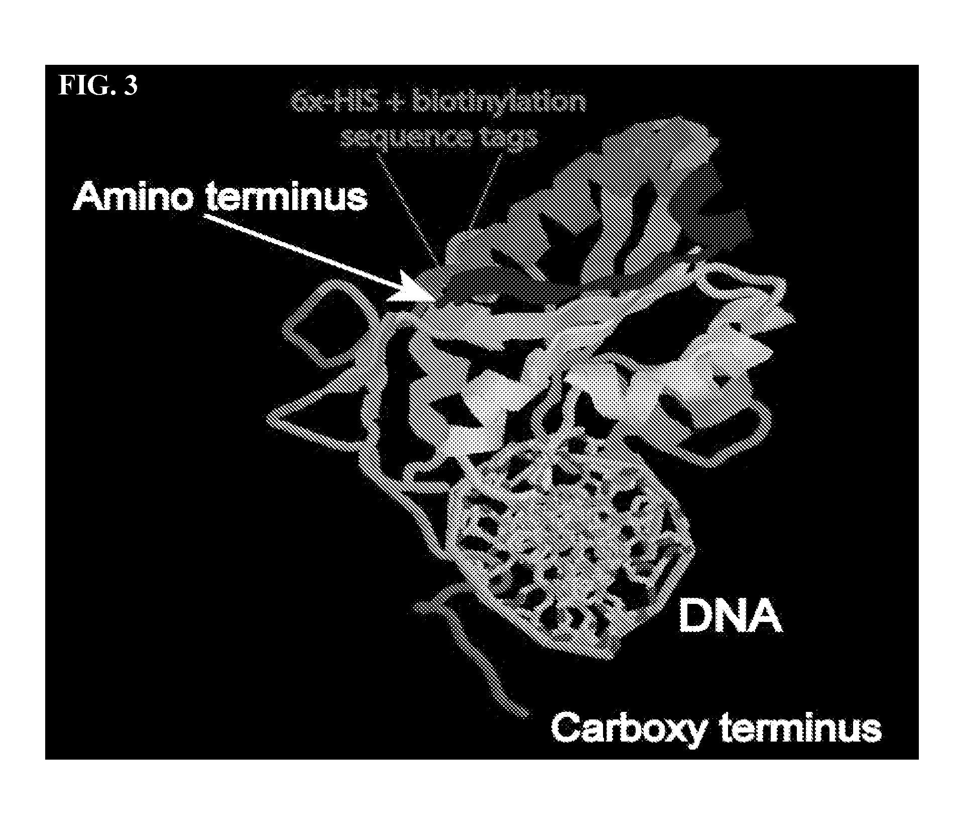
[0092] To correct the problem of binding-site occlusion a new D94N protein containing a biotinylation tag, a 15 amino acid sequence that is specifically biotinylated by the E. coli birA gene was constructed. The N-terminal placement of the tag is almost completely opposite to the DNA binding site (FIG. 3) and is freely available for interaction with streptavidin, to which QDs directly can be directly bioconjugated.

[0093] In vivo biotinylation may be more efficient than in vitro methods. Therefore, the D94N gene containing the biotin target peptide sequence and the E. coli biotinylation gene birA were co-transformed into MDS42. In this investigation the inventors chose to put the D94N and birA constructs are on separate T7-promoter vectors containing ampicillin and chloramphenicol markers respectively. The unmodified birA gene was placed into a CAM-selectable single-copy vector (...


PUM

PropertyMeasurementUnit
Elongationaaaaaaaaaa

Abstract

The present invention provides methods and apparatus for the optical fingerprinting of nucleic acid molecules. In certain embodiments, the invention provides methods for labeling nucleic acid molecules using-site-specific nucleic acid binding partners that bind to nucleic acids without cleaving the molecule. The nucleic acid binding partners can be labeled directly with a fluorophore, such as a quantum dot (QD), or indirectly. Examples of suitable binding partners include cut-deficient restriction endonucleases (cdREs), transcription factors, the binding domains of nucleic acid polymerases, antibodies and the like. The methods disclosed make the assembly of fingerprint contigs easier and allows for digitizing the results so as to provide for easier computer manipulation and assembly. The invention also includes a microarray, the microarray designed to provide for easy deposition and high-throughput fingerprinting. The invention also provides for methods of using the microarrays. The invention also provides kits for the use of the invention.

Description

CROSS REFERENCE TO RELATED APPLICATIONS [0001] This application claims the benefit of U.S. Provisional Application 60 / 753,564 filed on Dec. 23, 2005, incorporated herein by reference in its entirety for all purposes.GOVERNMENT SUPPORT [0002] Development of this invention was supported in part by grants from the National Institutes of Health Grant HG002530. The Government of the United States of America may have certain rights in this invention.FIELD OF THE INVENTION [0003] This invention is generally directed to methods of fingerprinting nucleic acids. More particularly, this invention is directed to methods of optically fingerprinting nucleic acid molecules using site-specific recognition molecules. BACKGROUND OF THE INVENTION [0004] The technological explosion accompanying the growth in molecular biology has made possible the large-scale sequencing of complete genomes. However, such large-scale sequencing is limited by the comparative slowness of preparing individual genomes or ge...

Claims


Application Information

Patent Timeline
IPC IPC(8): C12Q1/68C12M3/00
CPCC12Q1/6816C12Q1/6837C12Q1/6869G01N33/5308C12Q2565/601C12Q2565/513C12Q2522/101
Inventor BERRES, MARK E.FRISCH, DAVID A.BLATTNER, FREDRICK R.
Owner WISCONSIN ALUMNI RES FOUND
Why Eureka
Try Eureka