
Apparatus and method of removing occlusions using ultrasonic medical device operating in a transverse mode

a transverse mode and medical device technology, applied in the field of medical devices, can solve the problems of occlusion, myocardial infarction, stroke, death, etc., and achieve the effect of enhancing the performance of angioplasty procedures and reducing the likelihood of subsequent clot formation

Inactive Publication Date: 2007-09-27
CYBERSONICS
View PDF15 Cites 47 Cited by
  • Summary
  • Abstract
  • Description
  • Claims
  • Application Information

AI Technical Summary

Benefits of technology

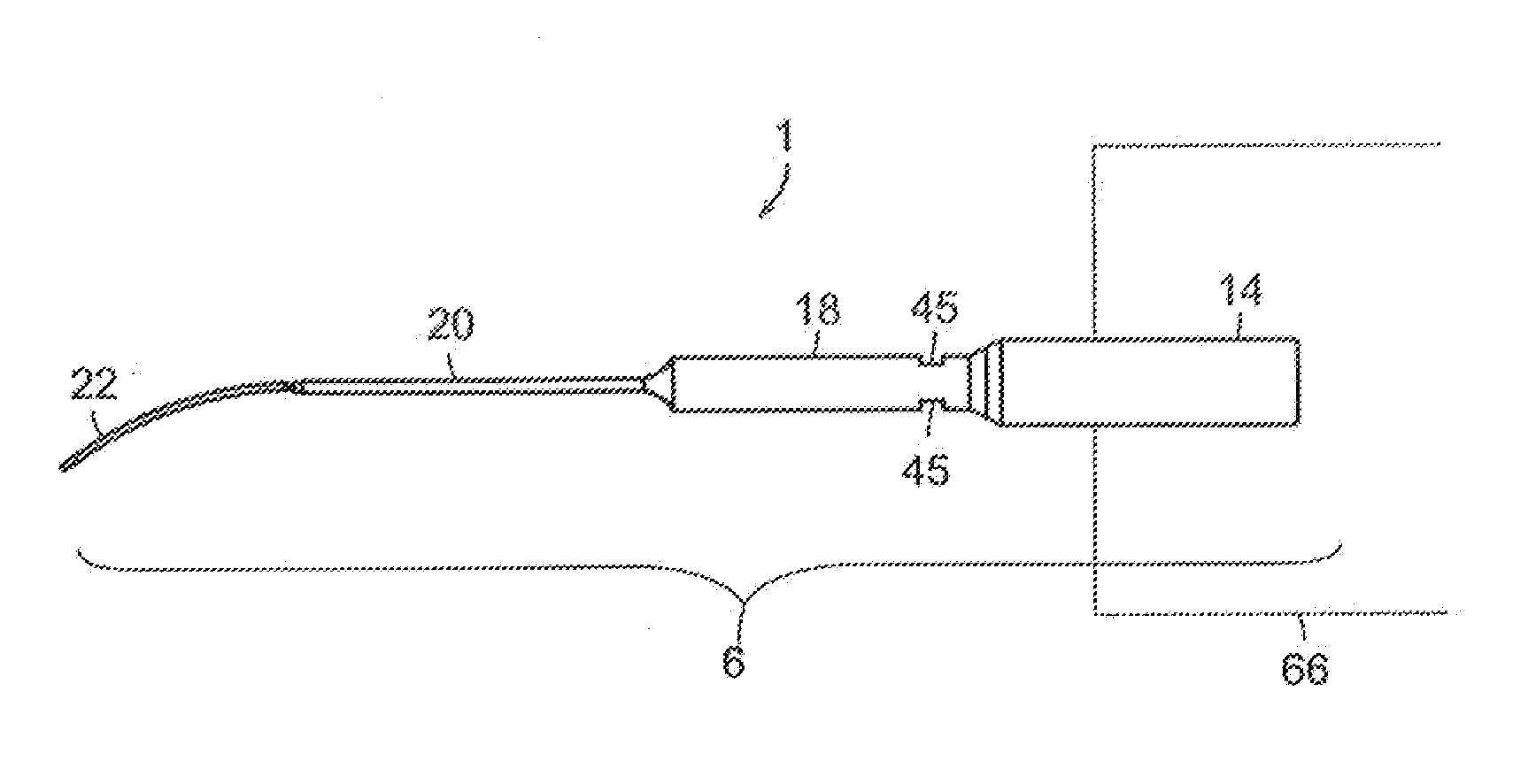
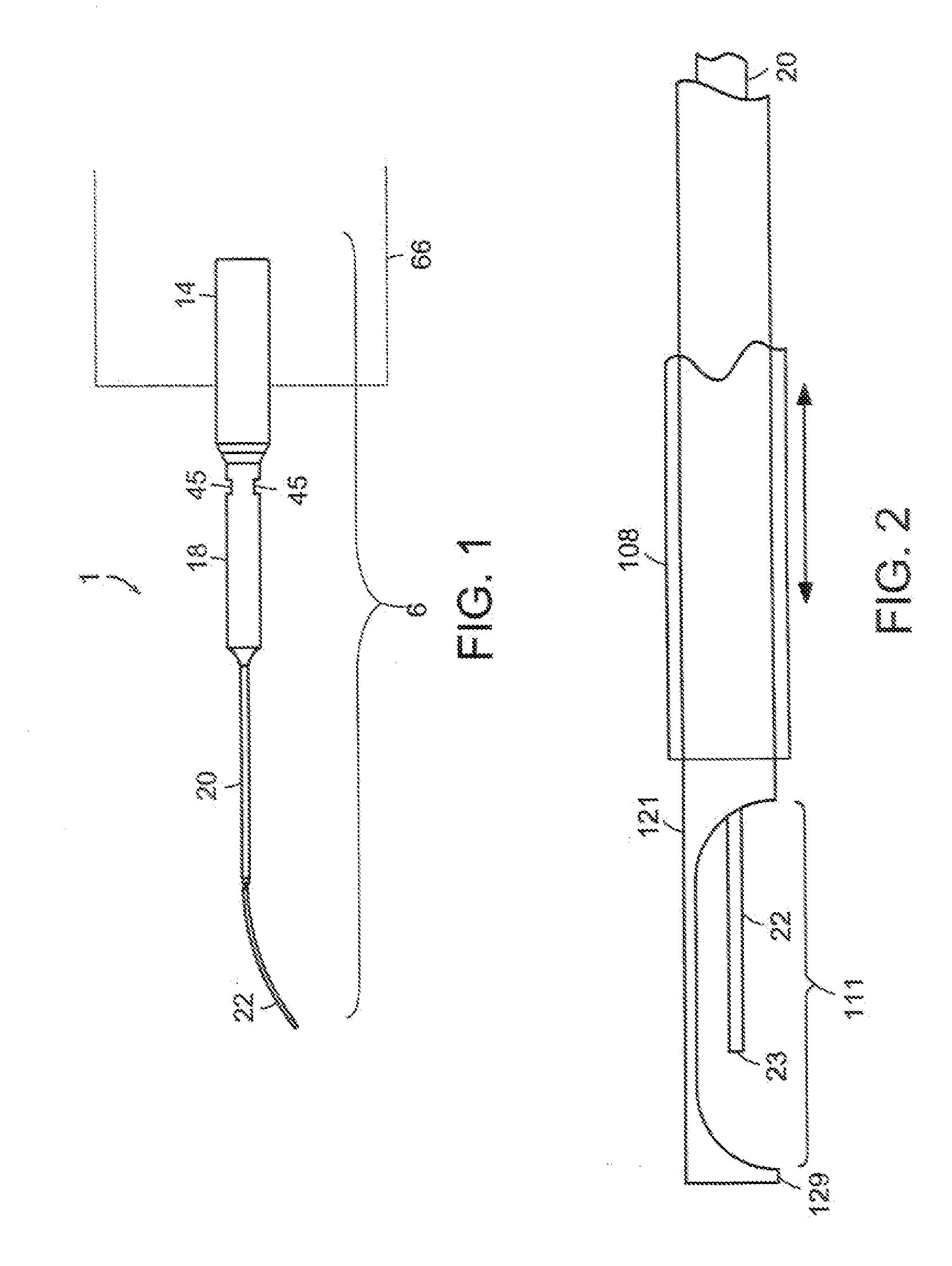
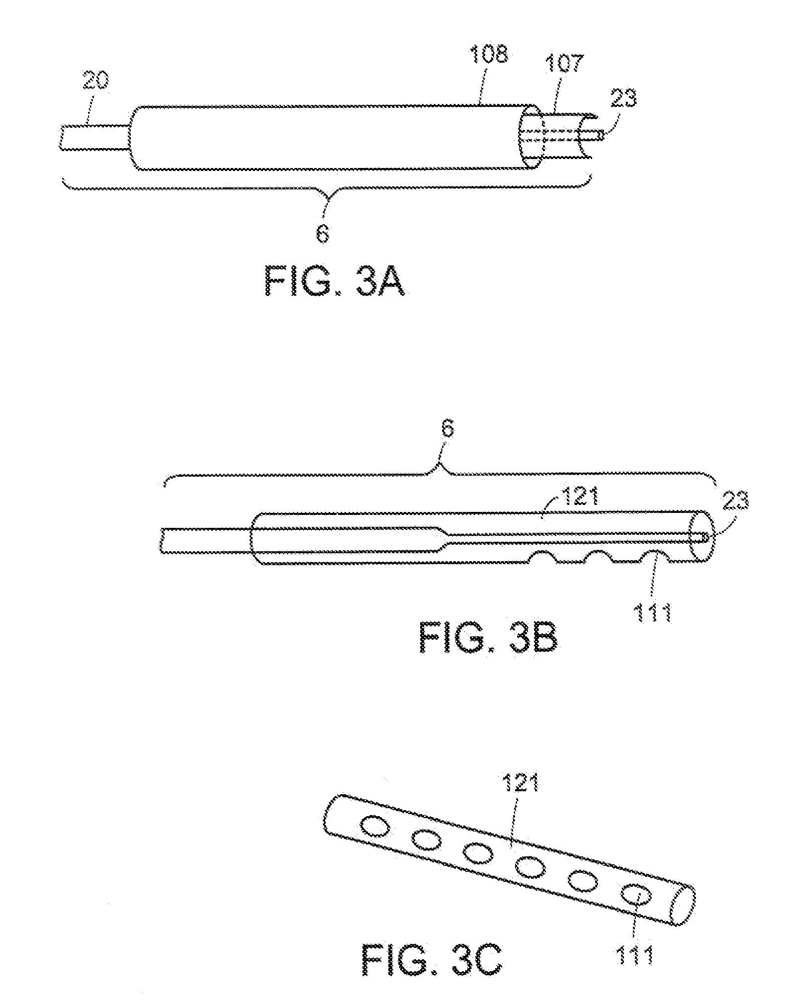
[0010] In one aspect, the invention relates to one or more sheaths which can be adapted to the probe tip, thereby providing a means of containing, focusing, and transmitting energy generated along the length of the probe to one or more defined locations. Sheaths for use with an ultrasonic medical device are described in the Applicant's co-pending utility application U.S. Ser. No. 09 / 618,352, now U.S. Pat. No. 6,551,337, the entirety of which is hereby incorporated by reference. The sheaths of the present invention also provide the user with a means of protecting regions of tissue from physical contact with the probe tip. In one embodiment of the invention the sheaths also comprise a means for aspiration and irrigation of the region of probe activity. In another embodiment of the invention, a plurality of sheaths are used in combination to provide another level of precision control over the direction of cavitation energy to a tissue in the vicinity of the probe. In one embodiment of the invention, the sheath encloses a means of introducing fluid into the site of the procedure, and a means of aspirating fluid and tissue debris from the site of the procedure. In a further embodiment, the probe tip can be moved within the sheath. In yet another embodiment, the irrigation and aspiration means, and the probe tip, can all be manipulated and repositioned relative to one another within the sheath. In another embodiment, the sheath is shaped in such a way that it may capture or grasp sections of tissue which can be ablated with the probe. In yet another embodiment, the sheath provides a guide for the probe tip, protecting tissues from accidental puncture by the sharp, narrow diameter tip, or from destruction by energy emitted radially from the probe during introduction of the probe to the site. The sheath may be applied to the probe tip prior to insertion of the probe into the patient, or the sheath can be inserted into the patient prior to the insertion of the probe. The sheath of the present invention can be used to fix the location of one or more shapes relative to the nodes or anti-nodes of a probe acting in transverse action. The location of the reflective shapes can amplify the acoustical wave thereby magnifying the energy. This allows for the use of very small diameter probes which themselves would not have the requisite structural integrity to apply and translate acoustical energy into sufficient mechanical energy to enable ablation of tissues. The reflective shapes can also focus or redirect the energy, effectively converting a transverse probe emitting cavitation energy along its length, to a directed, side fire ultrasonic device.
[0011] In another embodiment, the probe, which may or may not contain a probe sheath, is used in conjunction with an expandable balloon dilatation catheter, providing a means of resolving the occlusion without imparting stress, or inflicting stress injury to a vessel. The balloon catheter acts as a carrier means for guiding the probe wire to the desired site, and acts as a means to position the wire within the lumen of the vessel. With the balloon inserted within the confines of an occlusion, inflation of the balloon provides a means of continuous contact with the potentially irregularly shaped vessel lumen. Introduction of ultrasonic energy into the balloon by the transversely vibrating probe wire thereby results in uniform communication of energy to the regions of the occluded vessel in contact with the balloon. Since the balloon is inflated to much lower pressures than in traditional balloon angioplasty procedure, neither the occlusion or the vessel is compressed, thereby eliminating the problems of stress injury to the vessel. Likewise, as the ultrasound energy fragments the occlusion, the vessel is cleared of the problematic material, rather than simply compressing it into the vessel.

Problems solved by technology

Occlusions result in oxygen deprivation (“ischemia”) of tissues supplied by these blood vessels.
Prolonged ischemia results in permanent damage of tissues which can lead to myocardial infarction, stroke, or death.
However, many thrombolytic drugs are associated with side effects such as severe bleeding which can result in cerebral hemorrhage.
Mechanical methods of thrombolysis include balloon angioplasty, which can result in ruptures in a blood vessel, and is generally limited to larger blood vessels.
Another common problem is secondary vasoconstriction (classic recoil), a process by which spasms or abrupt closure of the vessel occurs.
These problems are common in treatments employing interventional devices.
However, these extreme forces result in the application of extreme stresses to the vessel, potentially rupturing the vessel, or weaking it thereby increasing the chance of post-operative aneurysm, or creating vasoconstrictive or restenotic conditions.
Other mechanical devices that drill through and attempt to remove an occlusion have also been used, and create the same danger of physical damage to blood vessels.
This process is called cavitation and results in shock waves running outward from the collapsed bubbles which can wear away or destroy material such as surrounding tissue in the vicinity of the probe.
Another drawback of existing ultrasonic medical probes is that they typically remove tissue slowly in comparison to instruments which excise tissue by mechanical cutting.
Because the tip of the probe is vibrated in a direction in line with the longitudinal axis of the probe, a tissue-destroying effect is only generated at the tip of the probe.
Thus, while destruction of tissue in proximity to the tip of the probe is more efficient, tissue destruction is still predominantly limited to the area in the immediate vicinity at the tip of the probe.
Although tissue destruction at the tip of the device is more efficient, the tissue destroying effect of the probe is still limited to the tip of the probe.

Method used


Image

  • Apparatus and method of removing occlusions using ultrasonic medical device operating in a transverse mode
  • Apparatus and method of removing occlusions using ultrasonic medical device operating in a transverse mode
  • Apparatus and method of removing occlusions using ultrasonic medical device operating in a transverse mode

Examples


example 1

Removing Occlusions Using An Ultrasonic Medical Device and A Balloon Catheter

[0055] In one embodiment of the invention, the transverse mode ultrasonic medical device, is used in a procedure to remove an occlusion from a small diameter vessel (e.g., a native vessel, or a grafted vessel). In one embodiment, device is used in a method to reduce or eliminate an occlusion of a saphenous vein graft (e.g., such as used in a coronary bypass procedure).

[0056] A transverse mode ultrasonic probe is selected by the surgeon who will perform the procedure. The probe of the present invention further comprises a plurality of sheaths adapted to the probe, and a balloon catheter operably attached to one of the sheaths, all incorporated within a sharps container, and the container further sealed inside a sterile package, for example, a plastic bag. The user removes the container from the package and attaches the probe to the ultrasonic medical device by applying the threaded end of the probe to the ...

example 2

Clearing Occlusions From a Hemodialysis Graft

[0059] In another embodiment, the invention can be used to clear occlusions from and restore the patency of a hemodialysis graft. The graft will not require shielding from ultrasonic energy, or the use of a balloon catheter as in example 1. A probe is selected and affixed to the ultrasonic transducer in the manner previously described, through the use of the container. The probe is withdrawn from the container, and inserted into the lumen of the hemodialysis graft. In one embodiment, the probe is directly introduced into the hemodialysis graft. In another embodiment, the probe is inserted using a trocar or other vascular insertion device, such as for example, the insertion device of Applicant's utility application 09 / 618,352, now U.S. Pat. No. 6,551,337. Application of ultrasonic energy causes the probe to vibrate transversely along its longitude. Occlusive materials, such as for example a thrombus, are fragmented by the action of the pr...


PUM


Abstract

A method for removing an occlusion is provided comprising introducing a transverse mode ultrasonic probe into a blood vessel, positioning the probe in proximity to the occlusion, and transmitting ultrasonic energy to the probe, until the occlusion is removed. The probe has a small cross-sectional lumen and is articulable for navigating in a tortuous vessel path. The probe can be used with acoustic and / or aspirations sheaths to enhance destruction and removal of an occlusion. The probe can also be used with a balloon catheter. The probe, sheaths, and catheter can be provided in a sharps container which further provides a means of affixing and detaching the probe from an ultrasonic medical device.

Description

RELATED APPLICATIONS [0001] This application is a continuation of application Ser. No. 10 / 665,445, filed Sep. 19, 2003, which is a continuation of application Ser. No. 09 / 776,015, filed Feb. 2, 2001, now U.S. Pat. No. 6,652,547, which is a continuation-in-part of application Ser. No. 09 / 618,352, filed Jul. 19, 2000, now U.S. Pat. No. 6,551,337, which claims the benefit of Provisional Application Ser. No. 60 / 178,901, filed Jan. 28, 2000, and claims the benefit of Provisional Application Ser. No. 60 / 157,824, filed Oct. 5, 1999, the entirety of all these applications are hereby incorporated herein by reference.FIELD OF THE INVENTION [0002] The present invention relates generally to medical devices, and more particularly to an apparatus and method for using an ultrasonic medical device operating in a transverse mode to remove occlusions from a blood vessel. The invention also relates to an apparatus and method of using balloon catheters emitting ultrasonic energy in transverse mode, to ...

Claims


Application Information

Patent Timeline
Patent Type & Authority Applications(United States)
IPC IPC(8): A61N7/00A61BA61B17/00A61B17/22A61B17/32A61B18/00A61FA61M1/00A61N7/02
CPCA61B17/22012A61B2017/22081A61B2017/00137A61B2017/00274A61B2017/22007A61B2017/22008A61B2017/22015A61B2017/22018A61B2017/22051A61B2017/320084A61B2018/00547A61B2018/00982A61B2217/005A61B2217/007A61M1/0084A61N7/022A61B17/22004A61B2017/22028A61B17/320068A61B2017/32007A61B2017/320089A61M1/85A61B2017/00778
Inventor RABINER, ROBERTHARE, BRADLEY A.
Owner CYBERSONICS
Learn More