Composite vascular prosthesis

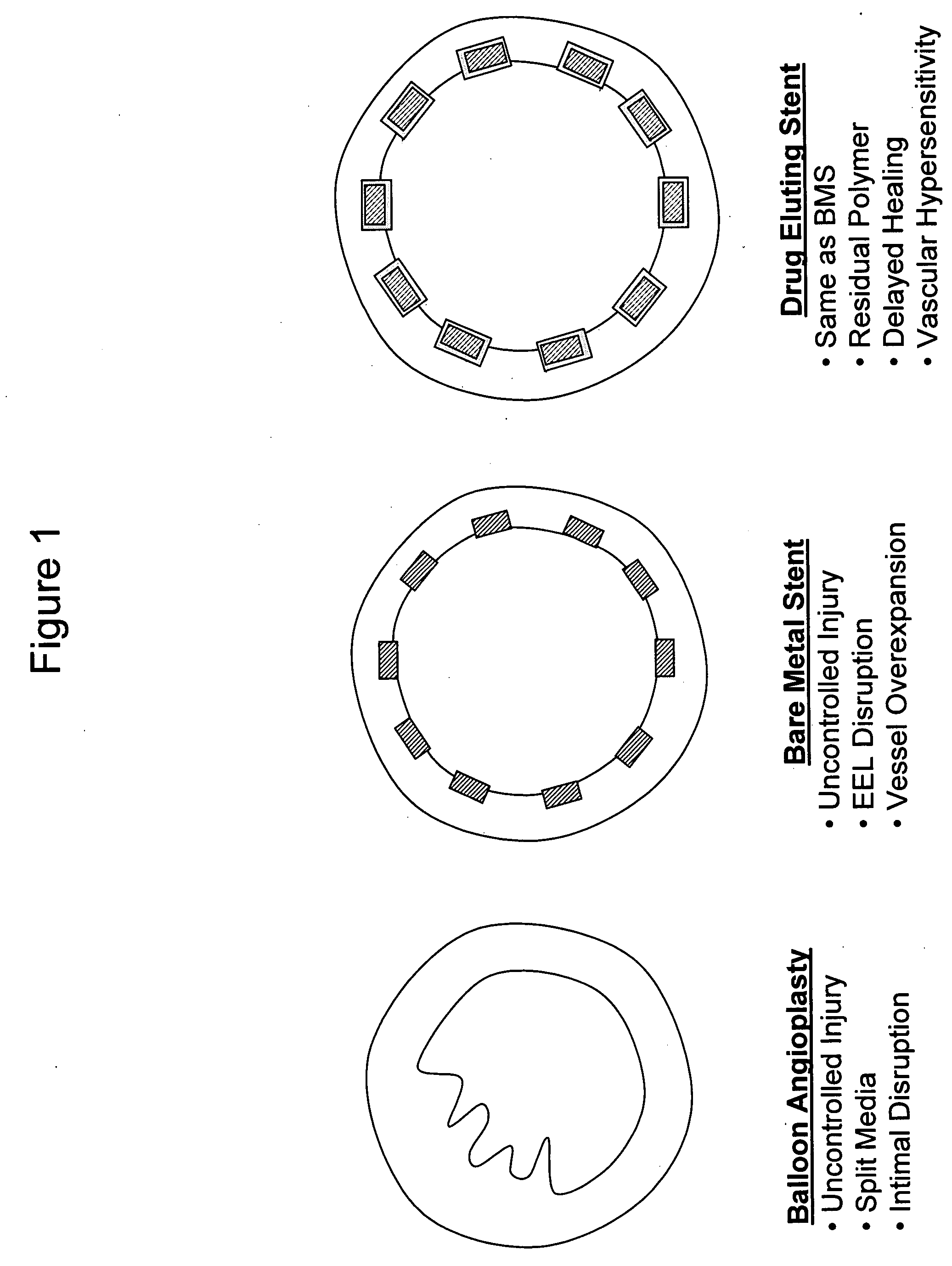
a vascular prosthesis and composite technology, applied in the field of composite vascular prosthesis, can solve the problems of high restnosis rate, high recoil of the vessel, abrupt closure, etc., and achieve the effect of increasing the injury rate of the vessel

Inactive Publication Date: 2007-09-27
GRANADA JUAN +1
View PDF60 Cites 50 Cited by
  • Summary
  • Abstract
  • Description
  • Claims
  • Application Information

AI Technical Summary

Benefits of technology

[0004]Most of the existing vascular scaffoldings constructed today are based on metals. Self-Expanding (SE) stents are typically constructed from nickel-titanium alloys, fabricated either from laser cut and electro-polished tubing or welded wire braids, coils or other wire mesh forms that allow for a small unexpanded profile to reach distal lesions in tortuous vessels which can be deployed and expanded in place when released from a captive sheath. SE stents are not currently used for coronary applications and typically require both pre and...

Problems solved by technology

In its simplest form, balloon angioplasty treats vascular obstructions by applying high dilatation forces that split the vessel wall structure resulting in vessel recoil, abrupt closure and high restenosis rates.
These metallic structures are deployed using balloon delivery systems that deliver the device at higher deployment pressures disrupting at different levels the integrity of the elastic structures of the vessel wall architecture.
Although effective in reducing the accumulation of scar tissue, current evidence suggest that hypersensitivity and allergic reaction to the polymer retained into the vessel wall occurs after drug eluting stent implantation and that this biological effect may be associated to lethal late thrombotic events.
In summary, as shown in FIG. 1, balloon angioplasty is associated with uncontrolled injury, split media and intimal disruption, use of bare metal stents is associated with uncontrolled injury, EEL disruption and vessel overexpansion, and use of drug eluting stents is associated with not only the problems of bare metal stents but also issues of residual polymer, delayed healing and vascular h...

Method used


Image

  • Composite vascular prosthesis
  • Composite vascular prosthesis
  • Composite vascular prosthesis

Examples


example 1

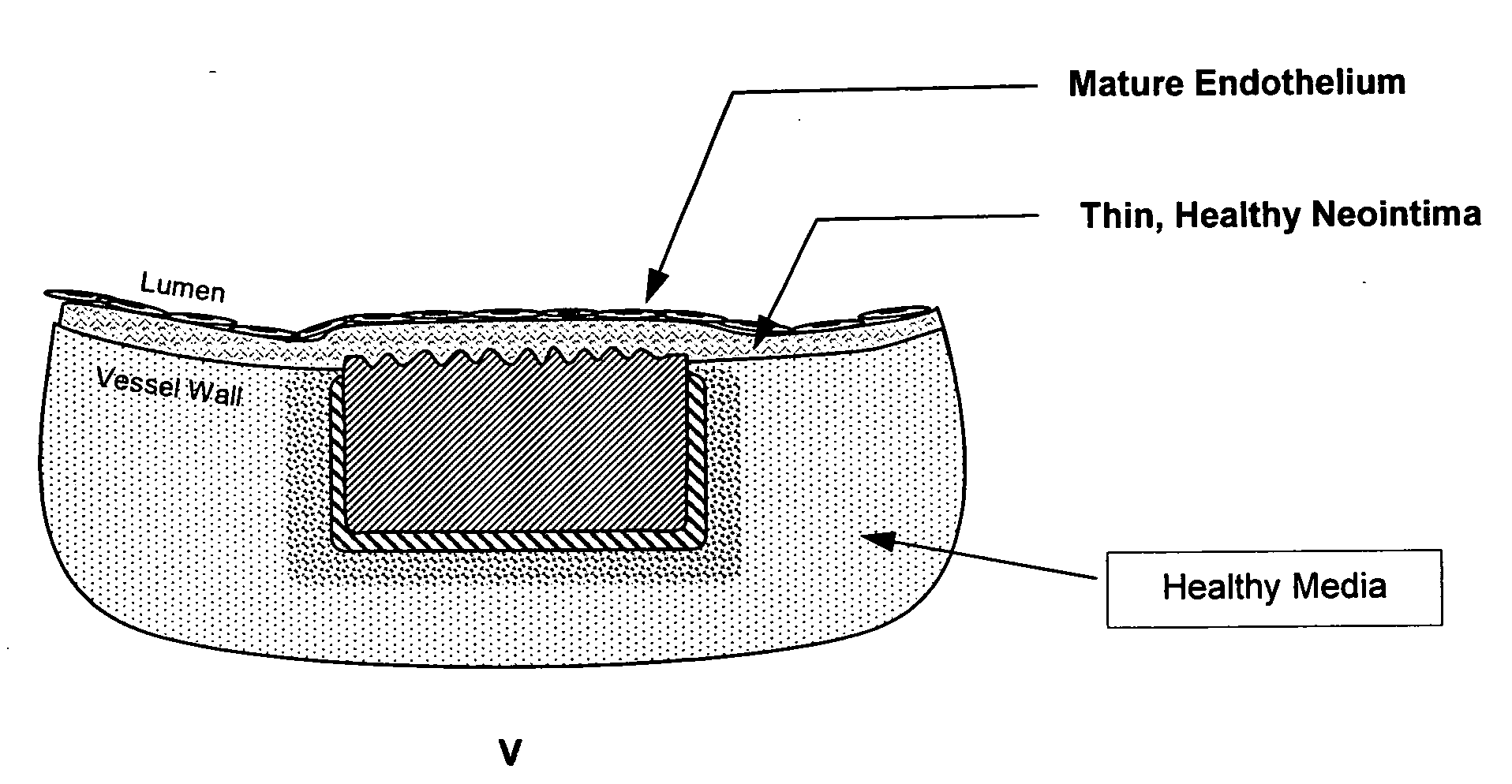
[0053]In one embodiment, the matrix can include a structural material, a bioadhesive component and a bioactive component. The structural material is composed of a metallic alloy or a durable or bioabsorbable polymer that has very thin strut thickness and width is highly flexible and conforms to the vessel wall. The bioadhesive component is composed of one or several natural proteins resembling extracellular matrix proteins, mainly collagen or collagen derivates. This component is preferentially located on the outer abluminal surface of the device. The bioactive component is achieved through direct modification of the interior adluminal surface of the prosthesis. In a preferred embodiment, the adluminal surface modification is the application of an etched surface topography tailored for improved endothelial cell migration, growth, adhesion and maturation. In an alternate embodiment, the adluminal layer is a deposited surface coating for achieving the same purpose. Other combinations ...

example 2

[0054]In another embodiment, the bioadhesive layer can act as the structural layer of the device. In this embodiment, the mixture of proteins must provide the structural support for the device. Blends of proteins such as collagen and elastin can be coupled with other compounds. These proteins can be assembled together to form tubes or preformed sheets that can be apposed to the vessel in-situ by an expandable delivery system such as a low-pressure balloon catheter. The bioadhesive component is deposited onto the external abluminal surface of the device to allow anchoring and apposition of the prosthesis to the vessel wall. This component will be preferentially located in the outer surface of the device but could be located throughout the entire surface of it

[0055]In a further derivation from the embodiments described above, the bioadhesive component incorporates molecules to allow bioactivation via secondary mechanism. These molecules could be incorporated via nanoliposomes, nanopar...

example 3

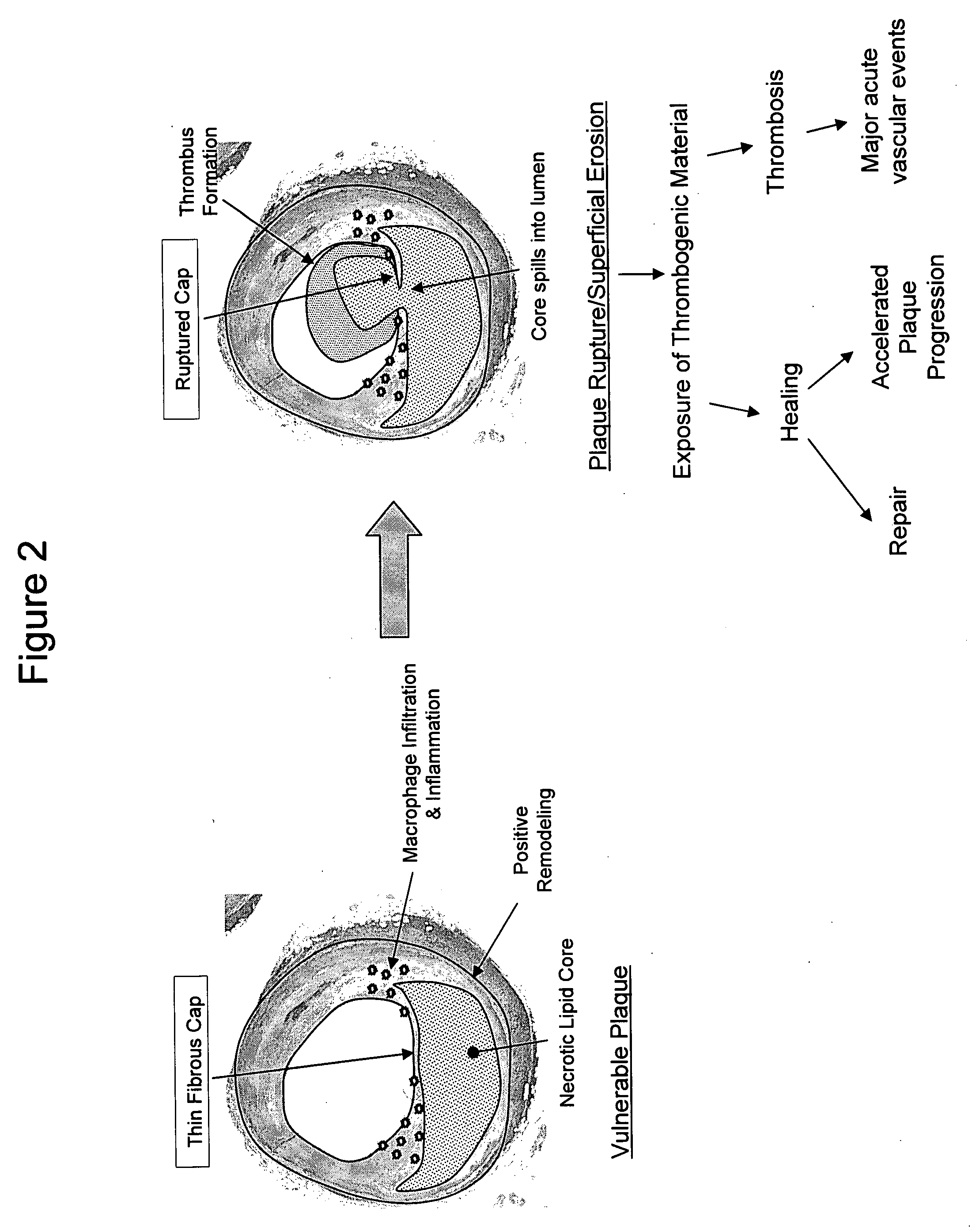
[0092]The structural layer or skeleton may be an ultra-thin self-expandable Nitinol alloy with a specific configuration in which the skeleton is covered by a thin bioadhesive component. In a preferred embodiment, this bioadhesive component includes or is composed of collagen. The layer may have a thickness of from 400 nm to 120 microns (enough to reinforce the thickness of the thinned fibrous cap). The average fiber size may be 100 to 800 nm and the average porosity size is preferably from 1 to 20 microns, enough to allow cell seeding and protein incorporation. The collagen layer may have a degradation time of less than 2 weeks, reaching 50% degradation in less than 4 days. In this embodiment, the coating may be disposed around the struts (all surfaces) or can cover only the abluminal side of the prosthesis. In a preferred variation, the inner surface of the device is modified to allow endothelial cell adhesion and colonization. This biological process is achieved by either directly...


PUM


Abstract

A novel treatment for atherosclerotic vascular disease is described utilizing the implantation of a thin, conformable biocompatible prosthesis constructed from a composite of various structural and therapeutic scaffolds in combination with one or more bioactive agents. This prosthesis can be delivered into position over a lesion in order to passivate atherosclerotic plaques with minimal remodeling of the artery, or alternatively can be applied with a balloon to passivate the remodeled site. The composite prosthesis itself provides mild structural reinforcement of the vessel wall and an evenly distributed platform for the introduction of bioactive therapeutic agents.

Description

[0001]This application claims the benefit of U.S. provisional patent application Ser. Nos. 60 / 785,579 filed Mar. 24, 2006 and 60 / 582,643 filed Oct. 19, 2006, each of which is incorporated by reference herein in its entirety.FIELD OF THE INVENTION[0002]The invention relates generally to a composite vascular prosthesis and more particularly to a highly conformable and biologically active endovascular system for treating vascular disease by promoting the regeneration of vascular tissue after implantation of the prosthesis.BACKGROUND OF INVENTION[0003]The field of percutaneous vascular intervention has been exclusively focused in the treatment of obstructive and symptomatic obstructive vascular disease. In fact, endovascular therapy is exclusively reserved for the patient presenting with symptoms related to an obstruction of the lumen of the vessel. In its simplest form, balloon angioplasty treats vascular obstructions by applying high dilatation forces that split the vessel wall struct...

Claims


Application Information

Patent Timeline
IPC IPC(8): A61F2/82
CPCA61F2/91A61F2250/003A61F2220/0075A61F2210/0076A61F2220/005A61F2250/0067
Inventor GRANADA, JUANFURNISH, SIMON M.
Owner GRANADA JUAN
Why Eureka
Try Eureka