Biological Tissue Sheet, Method Of Forming The Same And Transplantation Method By Using The Sheet

a technology of biological tissue and tissue plate, which is applied in the field of biological tissue plate, can solve the problems of corneal intransparency, limited collection size, and long time required to generate epithelium, and achieve excellent culture substrate, excellent proliferation of epidermal keratinocytes, and good cell migration ability.

Inactive Publication Date: 2008-02-14
KOUJI HASHIMOTO +1
View PDF0 Cites 61 Cited by
  • Summary
  • Abstract
  • Description
  • Claims
  • Application Information

AI Technical Summary

Benefits of technology

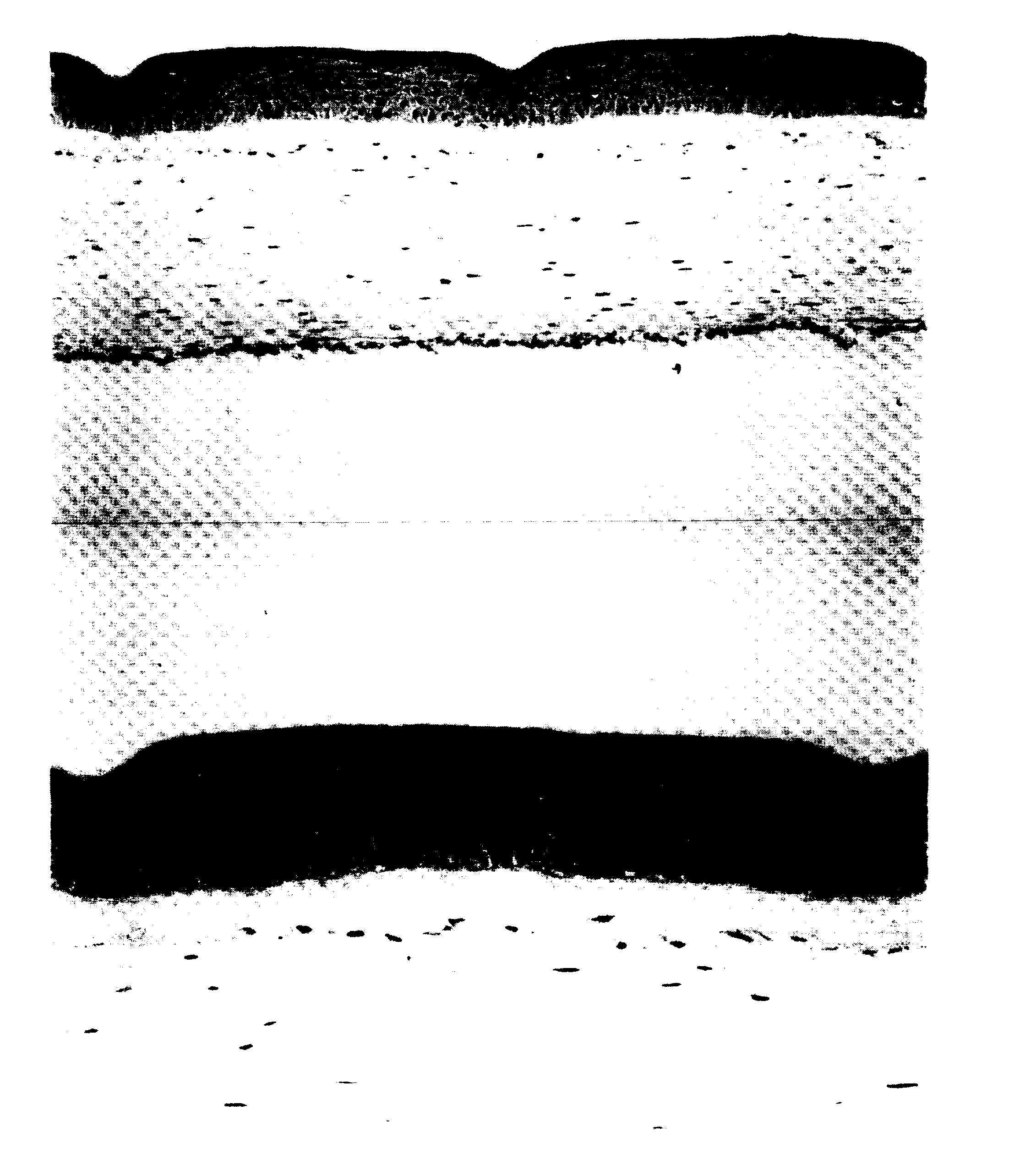
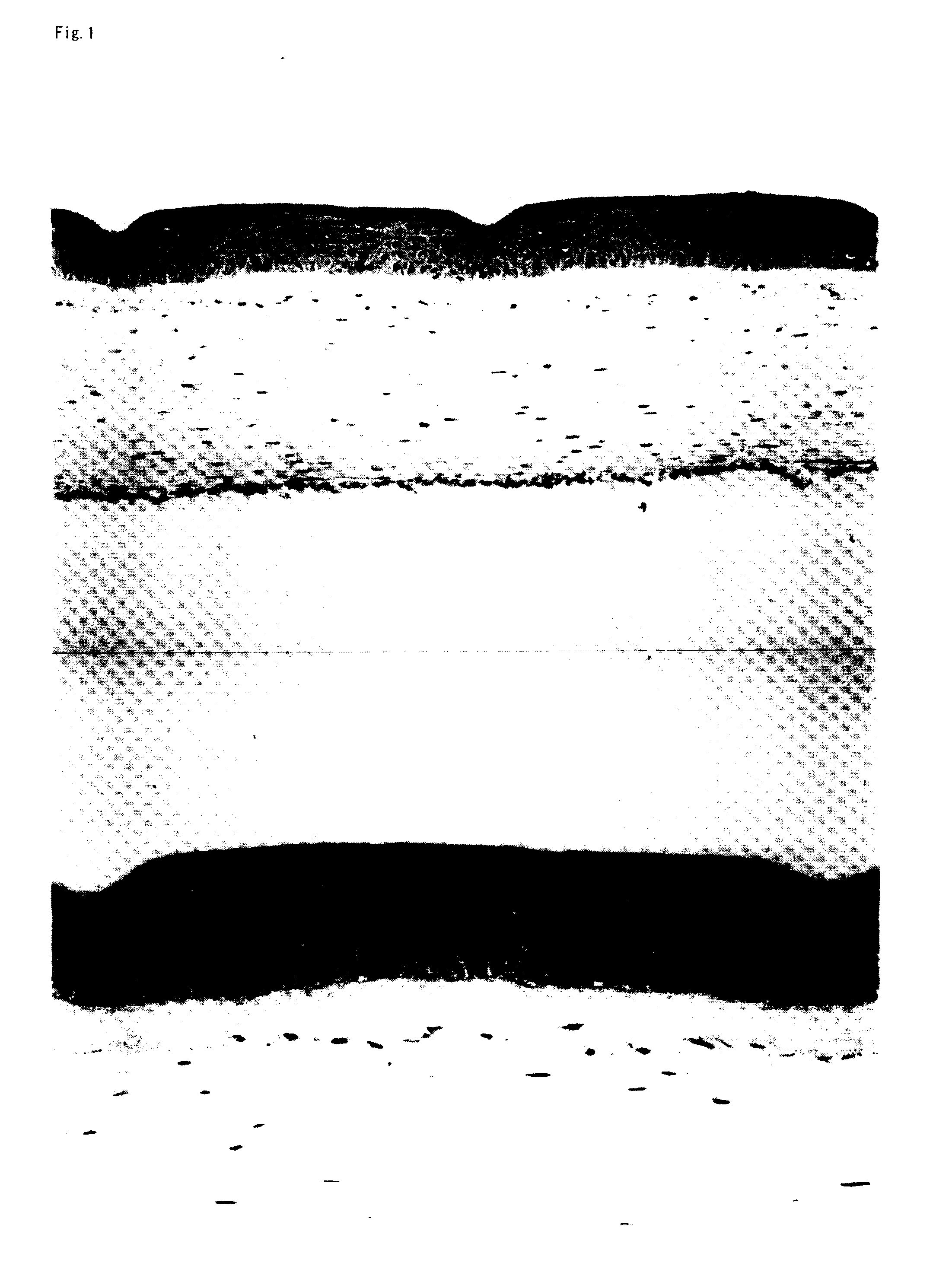
[0022] In view of the above-mentioned circumstances and problems, the present invention was made. The objective of the present invention is to provide a biological tissue sheet by which high therapeutic effect can be expected and which offers high degree of safety when transplantation is carried out. In order to achieve such a objective, the present inventors firstly have attempted to produce a cultured epidermal sheet. Specifically, in view of safety, under the conditions where cells (feeder cells) derived from xenogeneic animals are not used when epithelial cells are cultured, as a developed system of a conventional cultured epidermal sheet auto-transplantation, three-dimensional cultured skin was produced and then immunohistological and electron-microscopic investigation was carried out with respect to a basal membrane constituting component, cell adhesive molecule, and differentiated antigen. As a result, as compared with a conventional cultured epidermal sheet, the formation of strong horny cell layer is found, and the cell adhesion molecule and differentiation marker are sufficiently expressed similar to vivo epidermis. As to the basal membrane component, hemidesmosomes were well formed and pemphigoid antigen and β4 integrin were sufficiently expressed.
[0023] The survival of the cultured epidermal sheet is affected by the formation of the component of the basal membrane. That is to say, the cultured epidermal sheet is cultured in a state in which it is brought into close contact with the bottom surface of a plastic petri dish and it is necessary that the cultured epidermal sheet is peeled off from the bottom surface of the petri dish when the sheet is produced. This peeling is carried out by using dispase or collagenase and these enzymes decomposes the component of the basal membrane. According to reports to date, due to dispase, pemphigoid antigen cannot be detected by western blotting, and in fluorescent antibody technique, laminin 5 recognized by GB3 monoclonal antibody is degraded. Furthermore, it is reported that collagenase reduces the expression of anchoring fiber or IV type collagen, VII type collagen, but does not affect α6β4 integrin. On the contrary, a three-dimensional cultured skin keeps a structure which is similar to in vivo epidermis and the formation of component of a basal membrane is confirmed. The investigation by the present inventor demonstrated that β1 integrin, β4 integrin and pemphigoid antigen are formed relatively favorably. Also from the findings of electron microscopy, it was confirmed that hemidesmosome is formed favorably.
[0025] As mentioned above, the present inventors have succeeded in producing safe and practical biological tissue sheet without using cells derived from xenogeneic animals at all. Furthermore, the present inventors have found that a biological tissue sheet can be produced even under the conditions of serum free culture.
[0029] In one embodiment of the present invention, in vivo-derived cells are proliferated in a state in which the amniotic membrane is placed on a collagen gel containing human fibroblasts. Thus, the improvement in the proliferation rate of the in vivo-derived cells is to be achieved.

Problems solved by technology

However, in a burn injury in which a wide range of epidermis is lost at one time or in highly refractory ulcer from which hair follicle is lost, it takes a long time to generate epithelium only by regeneration of the epidermis from the vicinity.
However, there is a limitation to the size of collected cite.
The cornea may become not transparent by conditions such as keratitis, cornea ulcer, punch, and the like, and the transparent may be lost.
Although such cornea transplantation offers an effective treatment effect, there are some diseases that cannot be treated only by transplantation of the cornea.
However, it has come to be reported that in the above-mentioned conditions, the stem cell tissue for regenerating the cornea has become impaired.
In the above-mentioned pathologic conditions, it is thought that this stem cell tissue itself undergoes some impairment and become deficient.
Thus, the transparency is lost, resulting in extreme deterioration of the visual acuity.
In this way, in the above-mentioned pathologic conditions, since the corneal limbus is deleted, even if only the cornea is transplanted, the transplanted cornea cannot be maintained for a long term.

Method used


Image

  • Biological Tissue Sheet, Method Of Forming The Same And Transplantation Method By Using The Sheet
  • Biological Tissue Sheet, Method Of Forming The Same And Transplantation Method By Using The Sheet
  • Biological Tissue Sheet, Method Of Forming The Same And Transplantation Method By Using The Sheet

Examples


example 1

Production of Three-Dimensional Cultured Skin Sheet (Cultured Epidermal Sheet) Using Amniotic Membrane

1. Preparation of Amniotic Membrane

[0091] 1-1. Collection of Amniotic Membrane

[0092] After giving a pregnant woman who does not have a systemic complication and would undergo cesarean section sufficient informed consent together with an obstetrician in advance, amniotic membrane was obtained at the time of the cesarean section in the operation room. The operation was carried out cleanly. In accordance with the operation work, the operators washed hands, and then wore a special gown. Before delivery, a clean vat for obtaining amniotic membrane and physiologic saline for washing were prepared. After delivery, the placenta tissue was transferred to the vat and amniotic membrane tissue was manually detached from the placenta. A portion where amniotic membrane and placenta were strongly attached to each other was separated from each other with scissors.

[0093] 1-2. Treatment of Amniot...

example 2

Production of Three-Dimensional Cultured Skin Sheet

Cultured Epidermal Sheet

In the Case Where Collagen Gel is not Used

1. Collection of Amniotic Membrane and Epidermal Keratinocyte

[0114] Amniotic membrane and epidermal keratinocytes were prepared by the same procedure as described in Example 1.

2. Seeding of Keratinocytes

[0115] Preserved amniotic membrane is washed with PBS twice and further washed with a culture solution for keratinocytes once. The amniotic membrane is attached to the bottom surface of a culture insert with the side of substantial cells facing downward. The keratinocytes that have been prepared in advance are detached from the dish and collected by using trypsin-EDTA. The keratinocytes are subjected to centrifugation at 1000 rpm for five minutes to remove the supernatant. The cells are suspended so that the concentration becomes 200 million cells / 0.25 ml. The cell suspension (0.25 ml) is seeded to the amniotic membrane inside the culture insert, transferred to a...

example 3

Production of Three-Dimensional Cultured Corneal Epithelial Sheet Using Amniotic Membrane

1. Preparation of Amniotic Membrane

[0118] Amniotic membrane was prepared by the same procedure as described in Example 1.

2. Preparation of Corneal Epithelial Cell

[0119] 2-1. Procurement of Cornea

[0120] Donor corneas were purchased from Northwest Lions Eye Bank (Seattle, USA).

[0121] 2-2. Serum Free Culture Method of Corneal Epithelial Cells

[0122] Cornea is transferred to a petri dish containing Dulbecco's phosphate buffer (PBS) and the limbus is cut into strip with the size of 2 to 3 mm×2 to 3 mm by using a surgical knife under stereoscopic microscope. The limbus strip is washed with PBS several times and was sterilized by dipping it into 70% ethanol for one minute. The strip is washed with PBS, dipped in Dispase solution (Dispase II, Goudou Shusei, 250 units / ml, Dulbecco's Modified MEM medium; DMEM) and stood still overnight (18 to 24 hours) at 4° C. On the following day, by using forceps...


PUM

PropertyMeasurementUnit
turnover timeaaaaaaaaaa
sizeaaaaaaaaaa
temperatureaaaaaaaaaa

Abstract

A biological tissue sheet which is expected as exerting a favorable therapeutic effect and a high safety in transplantation. The biological tissue sheet formed by (a) preparing in vivo-derived cells; (b) sowing the in vivo-derived cells on amniotic membrane; and (c) culturing and proliferating the in vivo-derived cells in the absence of any xenogeneic animal cells. As the cells of a biological origin, for example, cells originating in corneal epithelium, conjunctival epithelium, skin epidermis, hair follicle epithelium, oral mucosa, respiratory tract mucosa, or intestinal tract mucosa.

Description

TECHNICAL FIELD [0001] The present invention relates to a biological tissue sheet. More specifically, the present invention relates to a biological tissue sheet produced by culturing and proliferating in vivo-derived cells such as corneal and conjunctival epithelial cells, epidermal cells, hair follicle epithelial cells, oral mucosa epithelial cells, respiratory tract mucosa epithelial cells, intestinal tract mucosa epithelial cells, and the like, on amniotic membrane without using cells derived from xenogeneic animals as feeder cells, a method of forming the sheet and use (a transplantation method, and the like) of the sheet. BACKGROUND ART [0002] The skin is an organ that covers the outermost layer of organism and forms a kind of barrier for protecting organism from the outside. The skin is made up of epidermis, dermis and subcutaneous tissue. The epidermis mainly consists of keratinocytes and includes a small amount of pigment cells, Langerhans cells, and the like. Cells constitu...

Claims


Application Information

Patent Timeline
Patent Type & Authority Applications(United States)
IPC IPC(8): A61F2/10A01N1/00A61P43/00A61L27/00A61L27/38C12N5/071
CPCA61L27/3604A61L27/3804A61L27/3869C12N5/0621C12N2533/92C12N5/0698C12N2502/094C12N2502/1323C12N5/0629C12M25/02A61P43/00C12N11/16A61L27/00C12N5/0602
Inventor HASHIMOTO, KOUJISHIRAKATA, YUUJIOHASHI, YUUICHIHAMURO, JUNJI
Owner KOUJI HASHIMOTO
Why Eureka
Try Eureka