
System and method for securing an implantable interface to a mammal

a technology of implantable interface and mammalian body, which is applied in the field of medical devices, can solve the problems of reducing lifespan, traditional weight loss techniques, diet, drugs, exercise, etc., and reducing quality of li

Active Publication Date: 2008-04-24
NUVASIVE SPECIALIZED ORTHOPEDICS INC
View PDF35 Cites 164 Cited by
  • Summary
  • Abstract
  • Description
  • Claims
  • Application Information

AI Technical Summary

Benefits of technology

[0034]In still another embodiment of the invention, a method for laparoscopically placing a gastric restriction device around the stomach of a patient includes inserting a multi-lumen trocar into the patient's abdominal cavity and affixing the gastric restriction device in an encircling manner around the stomach under laparoscope guidance from a laparoscope placed in one of the lumens of the multi-lumen trocar. A tool is then inserted in one of the lumens of the multi-lumen trocar, the tool including a releasable clip having first and second gripping portions. The first gripping portion is secured to a first portion of the stomach and the second gripping portion is secured to a second portion of the stomach. The clip is then released from the tool.
[0035]In yet another embodiment, an implant system includes an actuator configured for implantation within a mammal and a first resonant structure configured for implantation within the mammal and configured to resonate at a first natural frequency, the first resonant structure being operatively coupled to the actuator. The system further includes an external activator configured to apply energy at a one or more frequencies from a location outside the mammal, wherein the application of the energy at a frequency substantially the same as the first natural frequency causes the first resonant structure to substantially resonate and to effectuate movement of the actuator.

Problems solved by technology

Since traditional weight loss techniques, such as diet, drugs, exercise, etc., are frequently ineffective with many of these patients, surgery is often the only viable alternative.
Surgical treatments for obesity continue to be a strong focus of research due to their high level of effectiveness although no treatment is considered ideal.
It is well-established in the medical literature that obesity adversely affects general health, and can result in reduced quality of life and reduced lifespan.
Open surgery typically requires a ten day hospitalization and a prolonged recovery period with a commensurate loss of productivity.
This operation, therefore, is both a restrictive and malabsorptive procedure, because it limits the amount of food that one can eat and the amount of calories and nutrition that are absorbed or digested by the body.
Some of the major risks associated with the Roux-en-Y Gastric Bypass procedure include bleeding, infection, pulmonary embolus, anastomotic stricture or leak, anemia, ulcer, hernia, gastric distention, bowel obstruction and death.
Following this operation, many patients have reported feeling full but not satisfied after eating a small amount of food.
These practices can result in vomiting, tearing of the staple line, or simply reduced weight loss.
Major risks associated with VBG include: unsatisfactory weight loss or weight regain, vomiting, band erosion, band slippage, breakdown of staple line, anastomotic leak, and intestinal obstruction.
It is a modification of the biliopancreatic diversion or “Scopinaro procedure.” While this procedure is considered by many to be the most powerful weight loss operation currently available, it is also accompanied by significant long-term nutritional deficiencies in some patients.
Many surgeons have stopped performing this procedure due to the serious associated nutritional risks.
This irreversible procedure, therefore, is both restrictive (the capacity of the stomach is greatly reduced) and malabsorptive (the digestive tract is shortened, severely limiting absorption of calories and nutrition).
The major risks associated with the Duodenal Switch are: bleeding, infection, pulmonary embolus, loss of too much weight, vitamin deficiency, protein malnutrition, anastomotic leak or stricture, bowel obstruction, hernia, nausea / vomiting, heartburn, food intolerances, kidney stone or gallstone formation, severe diarrhea and death.
A great disadvantage of repeatedly injecting liquid via the injection port is the increased risk of the patient getting an infection in the body area surrounding the injection port.
Moreover, such an infection might be spread along the tube interconnecting the injection port and the band to the stomach, causing even more serious complications.
Thus, the stomach might be infected where it is in contact with the band, which might result in the band migrating (eroding) through the wall of the stomach.
Also, it is uncomfortable for the patient when the necessary, often many, post-operation adjustments of the stoma opening are carried out using a relatively large injection needle penetrating the skin of the patient into the injection port.
It may happen that the patient swallows pieces of food too large to pass through the restricted stoma opening.
Again, these measures require the use of an injection needle penetrating the skin of the patient, which is painful and uncomfortable for the patient, and can sometimes be the cause of infection, thus risking the long-term viability of the implant.
For example, if some air is inadvertently injected with the liquid (sterile saline), it can cause some compressibility to the pressurization media and take away some of the “one-to-one” feel when pressurizing and depressurizing.
As the band inflates, the size of the stoma shrinks, thus further limiting the rate at which food can pass from the upper stomach pouch to the lower part of the stomach.
The esophagus, unlike the stomach, is not capable of handling highly acidic contents so the condition results in the symptoms of heartburn, chest pain, cough, difficulty swallowing, or regurgitation.
These episodes can ultimately lead to injury of the esophagus, oral cavity, the trachea, and other pulmonary structures.
The open surgical repair procedure, most commonly a Nissen fundoplication, is effective but entails a substantial insult to the abdominal tissues, a risk of anesthesia-related iatrogenic injury, a 7 to 10 day hospital stay, and a 6 to 12 week recovery time, at home.
However, this procedure still requires surgery, which is more invasive than if an endogastric transluminal procedure were performed through the lumen of the esophagus or stomach, such as via the mouth.
However, despite the advantages provided by gastric banding methods, they nonetheless suffer from drawbacks that limit the realization of the full potential of this therapeutic approach.
For example, slippage may occur if a gastric band is adjusted too tight, or too loose, depending on the situation and the type of slippage.
During slippage, the size of the upper pouch may grow, causing the patient to be able to consume a larger amount of food before feeling full, thus lowering the effectiveness of the gastric band.
On the other hand, erosion may occur if the gastric band is adjusted or secured too tightly.
Furthermore, current methods of adjusting gastric bands and restriction devices require invasive procedures.
However, the use of X-ray procedures in a significant number of patients is highly undesirable.
In addition, while fluoroscopy can monitor flow of a radio-opaque material such as barium sulfate, it is not particularly well suited to provide accurate information about the size of the band aperture, the size of the lumen in the alimentary canal where the band is placed, or whether the band is causing secondary problems such as erosion of the gastric wall.

Method used


Image

  • System and method for securing an implantable interface to a mammal
  • System and method for securing an implantable interface to a mammal
  • System and method for securing an implantable interface to a mammal

Examples


Embodiment Construction

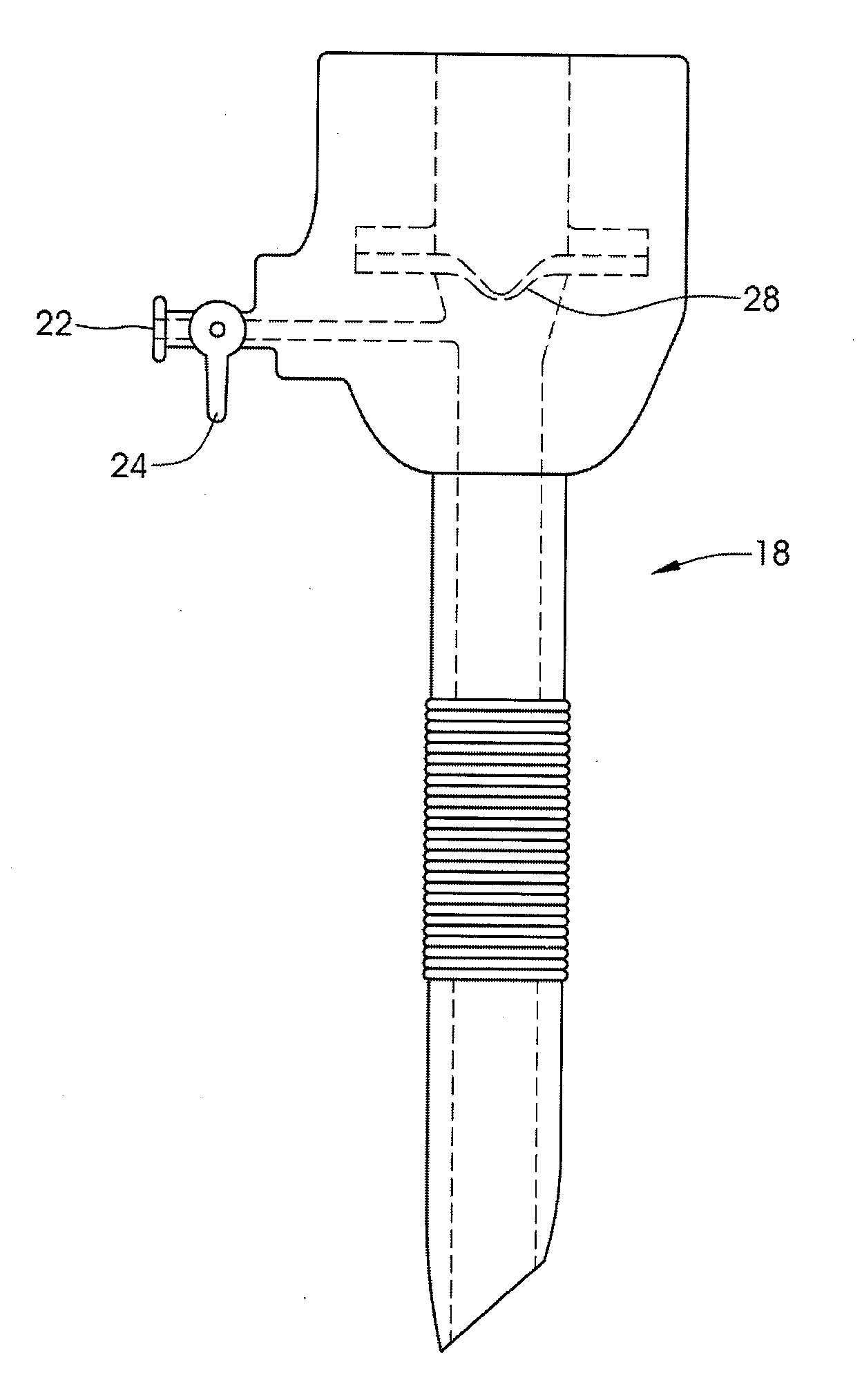
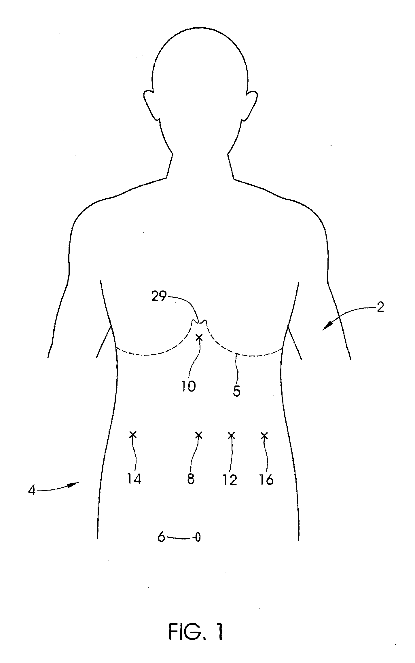
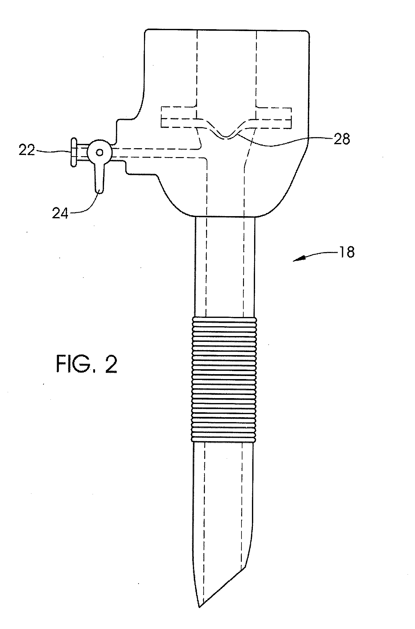
[0133]FIG. 1 illustrates the abdomen 4 of a patient 2. The navel 6 and the ribline 5 are shown for reference. In typical laparoscopic surgeries for placement of gastric restriction systems, a 12 mm trocar (or a larger trocar) is placed at first site 8. FIGS. 2 and 3 illustrate a trocar 18 of this type. This trocar 18 is placed prior to insufflation (inflation of the abdominal cavity by pressurized gas, such as carbon dioxide), so for safety purposes, often a trocar with an optically clear tip 20 is used. A scope (such as a 5 mm laparoscope) is inserted inside the tip 20 and can view the separation of tissue layers and the safe entrance into the abdominal cavity. Alternatively, instead of using the trocar tip 20 to separate the tissue, an incision can first be made in the skin followed by finger dissection into the abdominal cavity. The trocar 18 is then placed through the tract made by the finger dissection. After insertion of the trocar 18, pressurized CO2 is injected into the abdo...


PUM


Abstract

A system for securing an implantable apparatus to a mammal includes a mount including a base portion having a plurality of holes dimensioned to receive rotationally-driven fasteners, each fastener comprising a helical portion having a tip configured for tissue penetration, the mount configured to secure the implantable apparatus relative to tissue of the mammal upon driving the fasteners into the tissue. The system further includes a fastening tool configured to rotationally drive the helical portion of the fasteners into the tissue. The mount may be secured to the fascia covering the sternum via a subcutaneous securement method, or it may be attached to the intra-abdominal wall, behind the sternum, or it may be attached to the sternum directly via bone screws or the like.

Description

RELATED APPLICATIONS [0001]This Application claims priority to U.S. Provisional Patent Application Nos. 60 / 853,105 filed on Oct. 20, 2006, 60 / 854,574 filed on Oct. 25, 2006, and 60 / 904,625 filed on Mar. 1, 2007. Priority is claimed pursuant to 35 U.S.C. §§119, 120. The '105, '574, and '625 Provisional Patent Applications are incorporated by reference as if set forth fully herein.FIELD OF THE INVENTION [0002]The field of the invention generally relates to medical devices for accessing and controlling dimensions of body lumens and cavities along a mammalian alimentary canal, including methods and devices for treating obesity and gastroesophageal reflux disease (GERD).BACKGROUND OF THE INVENTION [0003]Obesity is a common disease of unknown etiology. It is a chronic, multifactorial disease that develops from an integration of genetic, environmental, social, behavioral, physiological, metabolic, neuron-endocrine and psychological elements. This disease is considered a cause or co-morbidi...

Claims


Application Information

Patent Timeline
Patent Type & Authority Applications(United States)
IPC IPC(8): A61B17/122
CPCA61B17/064A61B17/3417A61B17/3462A61B17/3474A61F5/0053A61B2017/0649A61B2017/3484A61F5/003A61B2017/00876
Inventor CHANG, ARVINPOOL, SCOTTWALKER, BLAIRTRAN, PETER P.
Owner NUVASIVE SPECIALIZED ORTHOPEDICS INC
Learn More