
Prion test

a prion and test technology, applied in the field of prion test, can solve the problems of not being able to discern the difference between prion strains, no diagnostic method is available to discriminate between bse and scrapie in sheep at present, and the risk of scoring is reduced. , the effect of reducing the risk of scoring

Inactive Publication Date: 2008-06-26
ID LELYSTAD INSTITUUT VOOR DIERHOUDERIJ EN DIERGEZONDHEID BV
View PDF4 Cites 0 Cited by
  • Summary
  • Abstract
  • Description
  • Claims
  • Application Information

AI Technical Summary

Benefits of technology

The patent describes a method for reducing false-positive results when testing samples from mammals for the presence of aberrant prion protein. The method involves using guanidine thiocyanate or a functional equivalent thereof to reduce the signal arising from normal prion protein. This allows for a more accurate and reliable method of testing for TSE, which is both sensitive and simple. The method can be used in various applications, such as mass screening of samples from ruminants or experimental animals. It is also a faster and more efficient method than other similar methods.

Problems solved by technology

There is also concern that the BSE strain that seems to be transmissible to humans may have infected sheep, where it could produce a disease hardly distinguishable from scrapie.
When its ominous strain-specific properties are maintained across the species barrier, sheep BSE may be a threat to human health, although scrapie by itself does not seem to transmit to humans.
With the exception of a bioassay in mice, no diagnostic method is available to discriminate between BSE and scrapie in sheep at present.
However, except for one case (Korth et al., 1997), there have been no antibodies developed that can discriminate between native forms of PrPC and PrPSc, and such antibodies cannot likely discern the difference between prion strains.
However, in BSE-infected cattle, PrPSc could not be detected outside the central nervous system, not even when clinical symptoms were present.
Moreover, human blood and blood products may form a health threat on account of possible contamination with blood of CJD patients and the recent occurrence of the nvCJD.
Bioassays for PrPSc in which different doses of the analyte are administered to target animals are generally regarded a gold standard but otherwise are cumbersome and costly.
Moreover, their quantitative character is limited by a high variation.
However, and in general, histological or cytological methods are not quantitative and hardly applicable on a large scale.
(1997) state that their method could be a basis for a sensitive screening method for PrPSc in crude tissue extracts, their extraction and purification steps are impracticable and time consuming (over 22 hours).
However, the practical conditions under which PrPSc, in order to be detected as an antigen, is brought into an unfolded state by chaotropic agents like 3-4 M gdnSCN, are not compatible with the immunochemistry of a sandwich type of ELISA.
This means that sampling of brain tissue could lead to false negatives due to regional variations in PrPSc content.
The test, however, is still far from lending itself to large-scale use in view of too much labor and long incubation times.
In spite of the high sensitivity of this method, sample processing is time consuming (approximately 24 hours) and cumbersome since PrPSc from brain stem has to be concentrated and purified through steps like ultracentrifugation and HPLC.
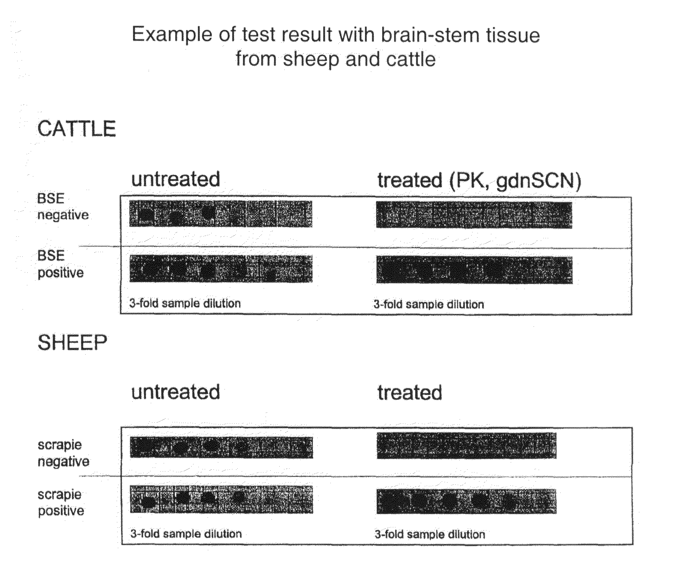
However, these two commercial tests, although claiming high sensitivity in detecting the aberrant protein, and thus claiming to have a low number of false-negative results, suffer from the low specificity associated with the claimed high sensitivity.
When using the above tests, one, therefore, runs an increased risk of falsely identifying a negative sample as false positive, thereby falsely identifying an animal as positive.
A false-negative diagnosis thus results in missing positive cases.
This causes a wrong prognosis being established and wrong treatment being given, until a second test is done.
Such meat and meat products will be traded and eaten and can thus be a source for further infection, notably of humans who even falsely trust that the animal has been tested well and the meat or meat product bears no risk.
For humans, “false positive” means that a false diagnosis of TSE is made, here again resulting in false prognosis and in faulty treatment.
For animals, “false positive” means that a false diagnosis of TSE is made, however, since TSEs are notifiable diseases that in general are met with strict eradication measures, the animal shall, at least in most Western countries, be killed and destroyed.
Furthermore, the herd from which the animal originated runs the same risk of being destroyed when the diagnosis is not corrected.
For the slaughterhouse, it might mean that special laborious decontamination actions have to be implemented, which mean temporary interference of use of the facilities and thus considerable loss of productivity.
It goes without saying that, especially when the country has no (present) reported cases of TSE, such a false-positive diagnosis is highly detrimental for the country's position on foreign markets for animal products.
Understanding the above risks associated with false-negative or false-positive diagnoses becomes even more complicated when one understands that, in general, the level of false positives scored by a diagnostic method or test is inversely related to the number of false negatives scored by the same test.

Method used


Image

  • Prion test
  • Prion test
  • Prion test

Examples


Embodiment Construction

Materials and Methods

[0065]Phosphate buffered saline (PBS), pH 7.2 contained 136.89 mM NaCl, 2.68 mM KCl, 8.10 mM Na2HPO4 and 2.79 mM KH2PO4 in water.

[0066]PBTS: 0.2% (w / v) Tween-20 in PBS.

[0067]Two extraction buffers were used:[0068](a) 10 mM phosphate buffer, pH 7.0, 0.15 M NaCl and 0.25 M sucrose, used by Pan et al. (1992) to prepare microsomal fractions;[0069](b) lysis buffer (Collinge et al., 1996) consisted of 0.5% (w / v) Tergitol (type NP-40, nonylphenoxy polyethoxy ethanol, Sigma NP-40) and 0.5% (w / v) deoxycholic acid, Na-salt (Merck) in PBS, pH 7.2.

[0070]Guanidine thiocyanate (gdnSCN, purity >99%; Sigma G 9277) solutions of 4 M were made up in water (pH 5.8).

[0071]Alkaline phosphatase-conjugated goat anti-rabbit IgG (GAR / AP) was from Southern Biotechnology As. (ITK, Diagnostics B.V., Uithoorn).

[0072]Substrate for alkaline phosphatase was 5-bromo-4-chloro-3-indolyl phosphate / nitro blue tetrazolium (BCIP / NBT; tablets; Sigma B5655).

[0073]Usually, after PrP extraction, protease ...


PUM

PropertyMeasurementUnit
pHaaaaaaaaaa
pHaaaaaaaaaa
pHaaaaaaaaaa

Abstract

The invention is related to diagnostic methods for detecting transmissible spongiform encephalopathies (TSEs) such as BSE and scrapie and related disease in humans. The invention provides use of guanidine thiocyanate (gdnSCN), or a functional equivalent thereof, for treating at least one sample derived from a mammal, including humans, for reducing the risk of scoring a false-positive test result in testing the sample for the presence or absence of aberrant prion protein.

Description

CROSS-REFERENCE TO RELATED APPLICATIONS[0001]This application is a continuation of U.S. patent application Ser. No. 09 / 913,345, filed Mar. 25, 2002, U.S. Pat. No. ______, which application was a national phase entry under 35 U.S.C. § 371 of International Patent Application No. PCT / NL01 / 00079, filed Feb. 9, 2000, published in English as International Patent Publication WO 00 / 48003 on Aug. 17, 2000, which claims priority to European Patent Application No. EP 99200391.3, filed Feb. 11, 1999, the contents of each of which are incorporated herein by this reference.TECHNICAL FIELD[0002]The invention is related to diagnostic methods for detecting transmissible spongiform encephalopathies (TSEs) such as BSE, scrapie and related diseases in animals and humans.BACKGROUND[0003]Bovine spongiform encephalopathy (BSE or mad cow disease) of cattle and scrapie of sheep are fatal, non-inflammatory neurodegenerative diseases caused by prions and are characterized by a long incubation period. In human...

Claims


Application Information

Patent Timeline
Patent Type & Authority Applications(United States)
IPC IPC(8): G01N33/00G01N33/53C12Q1/37G01N33/68
CPCG01N33/6896G01N2800/2828G01N2333/47
Inventor GARSSEN, GERRIT JANJACOBS, JORG GUNTHERLANGEVELD, JOANNES PIETER M.SMITS, MARINUS ADRIANUSVAN KEULEN, LUCIEN JOHANNES M.SCHREUDER, BRAM EDWARD C.BOSSERS, ALEXANDER
Owner ID LELYSTAD INSTITUUT VOOR DIERHOUDERIJ EN DIERGEZONDHEID BV
Learn More