

Preserved Viable Cartilage, Method for Its Preservation, and System and Devices Used Therefor
a cartilage and cryogenic technology, applied in the field of cryogenic preservation of cartilagecontaining tissue, can solve the problems of long time-consuming and laborious, low self-repair ability, and inapplicability of the process to larger lesions, and achieves easy surgical technique, uniform surface area, and large damage
- Summary
- Abstract
- Description
- Claims
- Application Information
AI Technical Summary
Benefits of technology
Problems solved by technology
Method used
Image

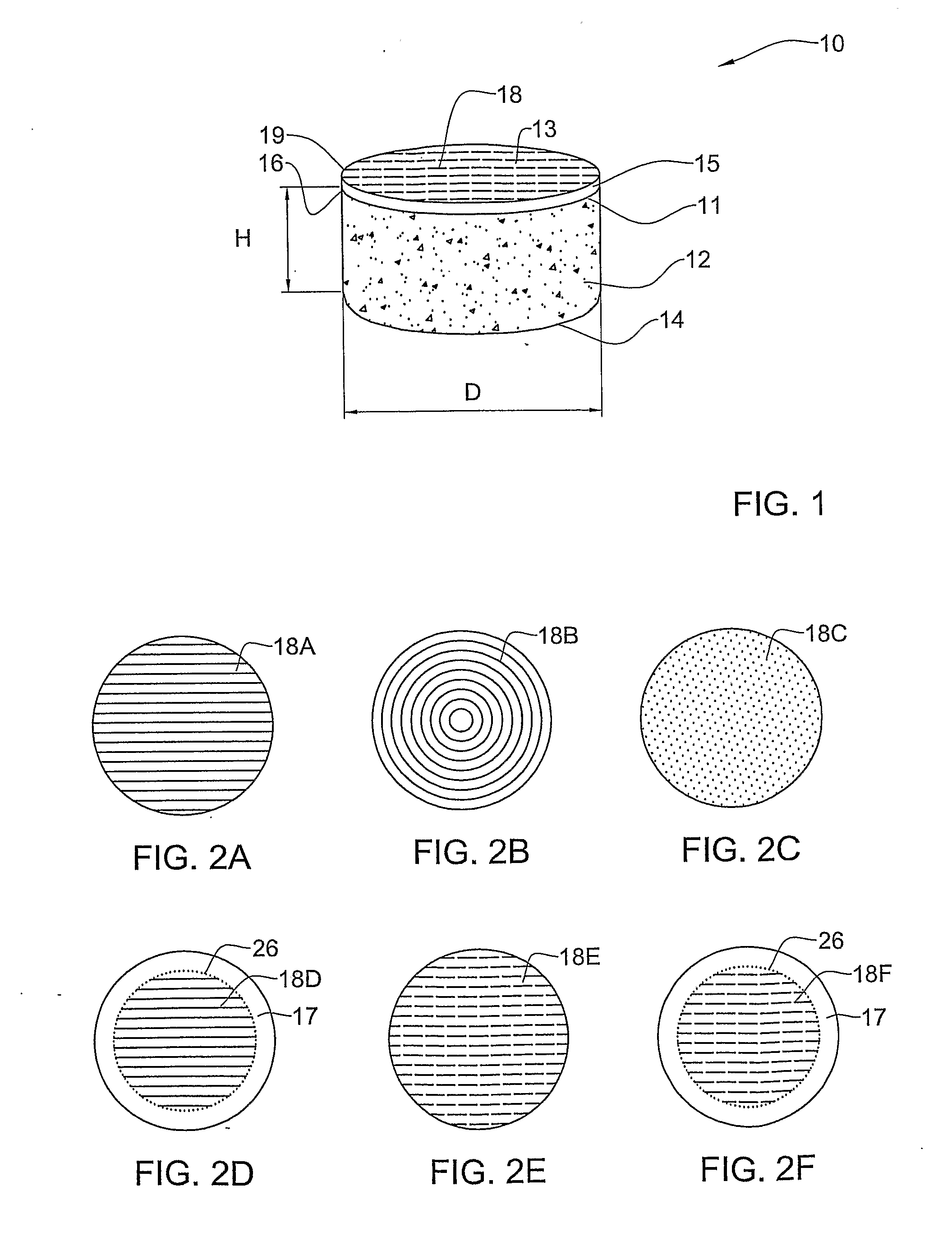
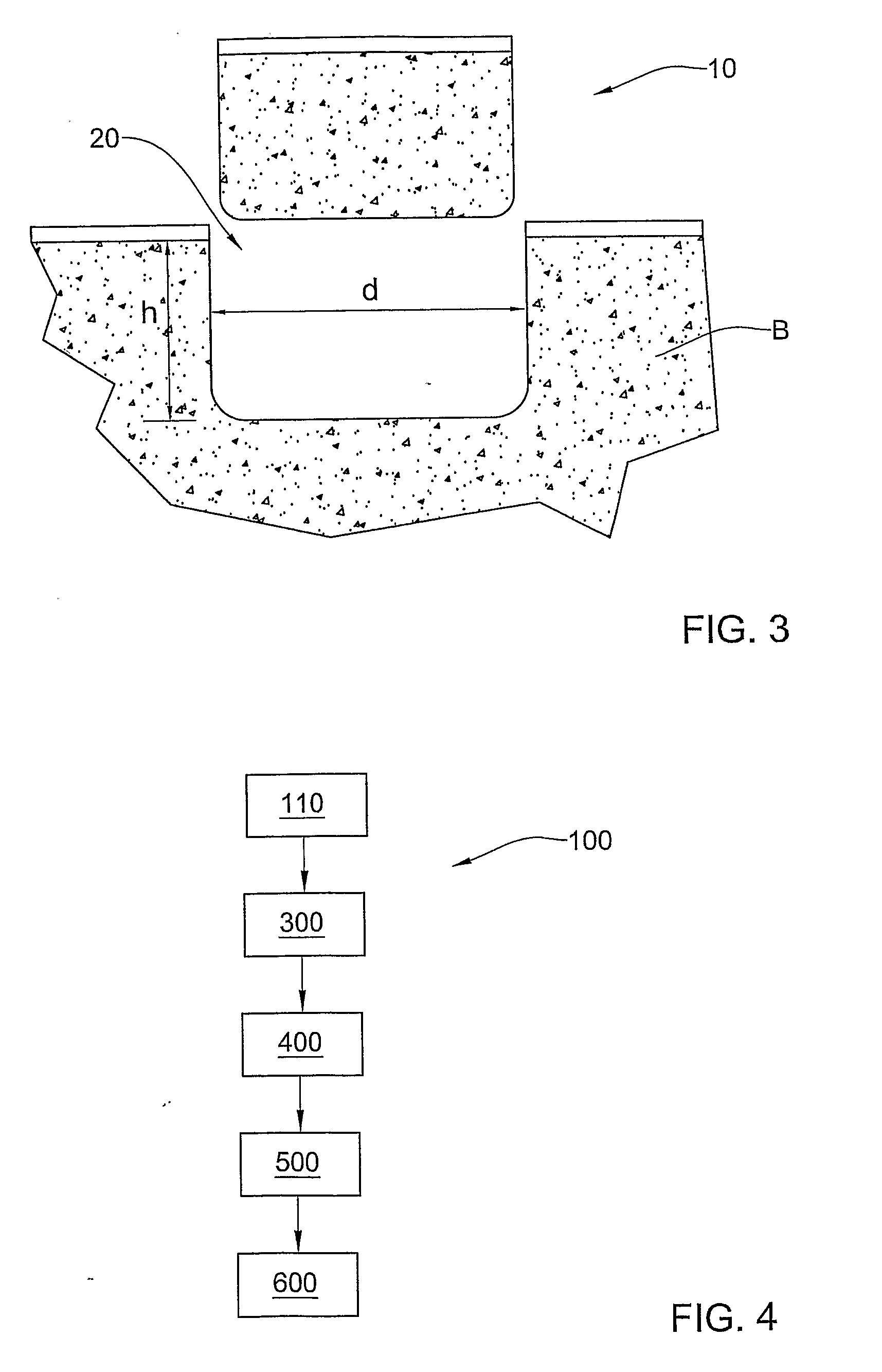
Examples
examples
Cartilage Preparation and Protocols
Materials
[0167]Unless specifically said otherwise, materials were obtained as follows: Sucrose S-5016 and Ethylene Glycol E9129 / L (Sigma, Israel) F12 medium-01-095-1A, PBS and Penicillin-Streptomycin-Nystatin solution 03-032-1B (Biological Industries, Israel). Viability was tested using live / dead fluorescent dyes (SYTO-13 / Propidium Iodide (PI), Molecular probe, USA, according to the manufacturer's manual).
Handling and Receipt of Human Knee Joint
[0168]Human knee joints were provided from cadaver donors by DIZG German Institute for Cell and Tissue Replacement, Berlin, Germany, after being tested for HIV (Human Immunodeficiency Virus), HBV (Hepatitis B Virus) and HCV (Hepatitis C Virus). The knee joints were packaged in RPMI 1640 storage medium (Biological Industries, Israel Cat#01-104-1, [Moore, G. E., Gerner R. E. and Franklin, H. A. (1967) Culture of Normal Human Leucocytes. JAMA 199, 519-524]) containing antibiotics and antimycotics and shipped in...
PUM
 Abstract
Description
Claims
Application Information