Compositions and methods of sphingosine kinase inhibitors in radiation therapy of various cancers
- Summary
- Abstract
- Description
- Claims
- Application Information
AI Technical Summary
Benefits of technology
Problems solved by technology
Method used
Image
Examples
example 1
Immunohistochemistry Staining for SPK Expression
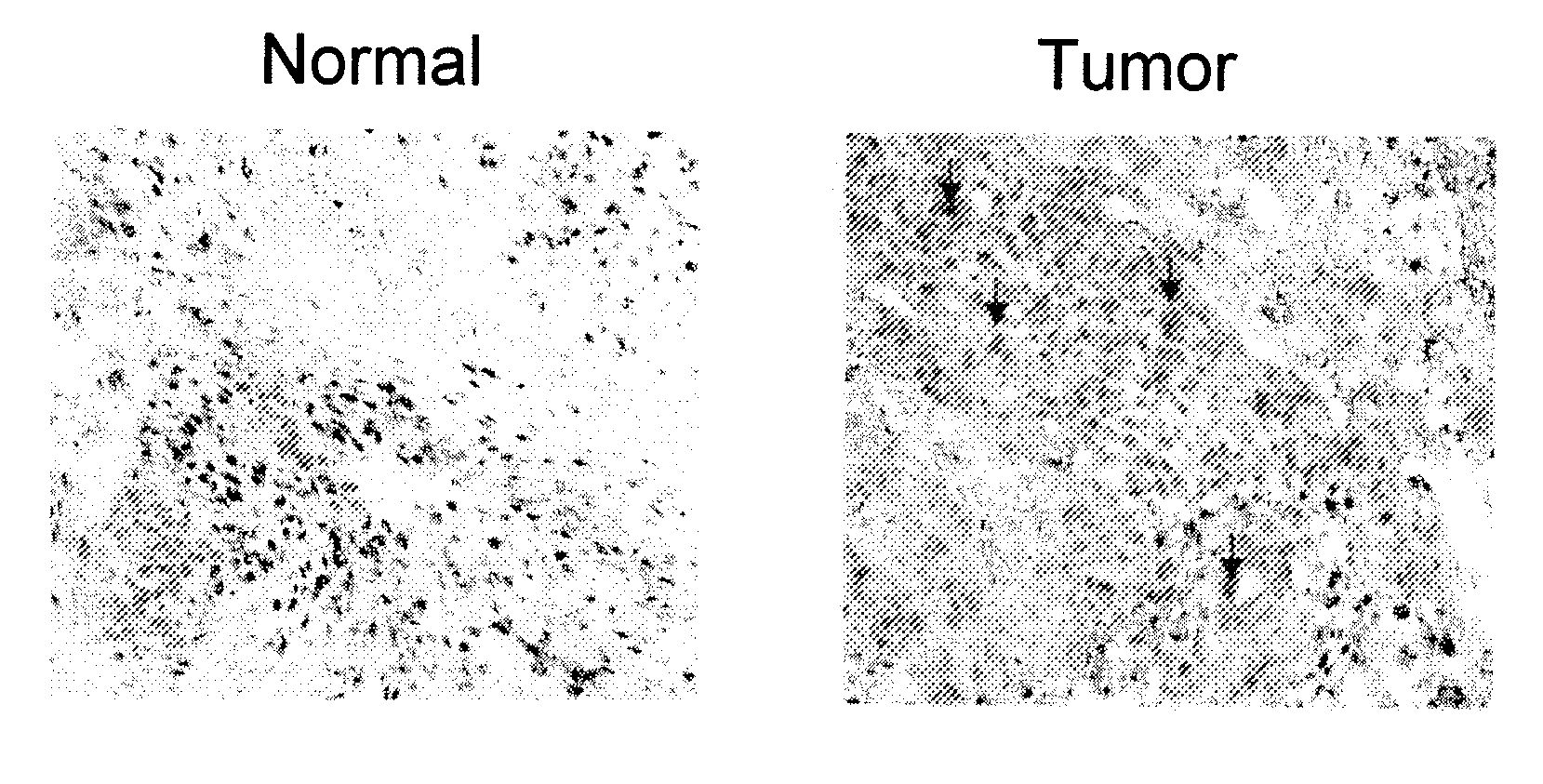
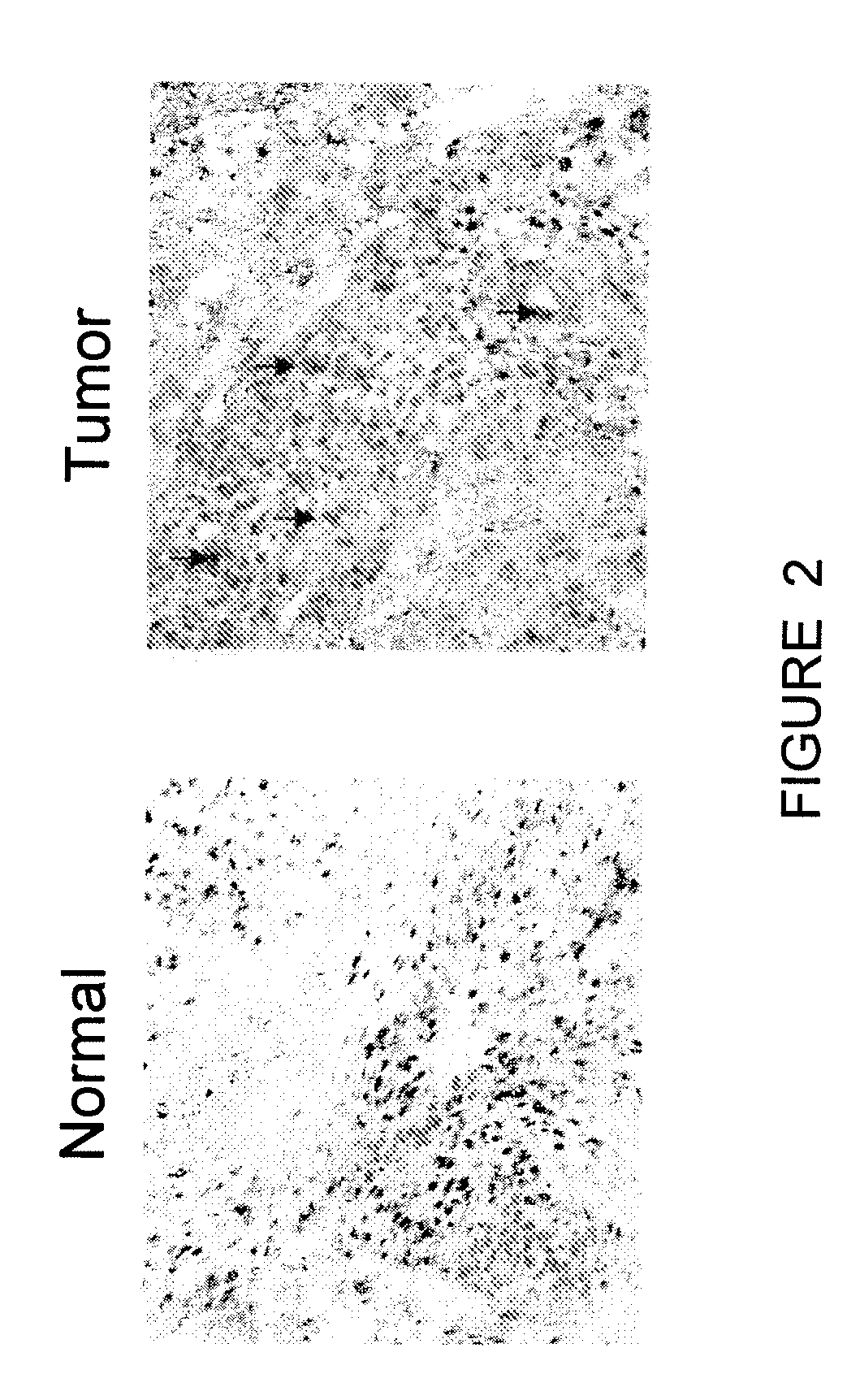
[0058]Head and neck tumor biopsy samples and adjacent normal tissue were paraffin-embedded according to the methods of Atkins et. al., who demonstrated a preferred method for detection of protein with specific antibody (21). Sections were studied by immunohistochemistry within one week of cutting the sections. Cut sections were placed on slides, rinsed twice in PBS and preincubated with blocking buffer (0.2% Triton-X100, 1% BSA in PBS) for 20 min, followed by incubation at 4° C. for 16 hr with SPK as a primary antibody that was added to the blocking buffer. After washing three times in PBS, sections were incubated with HRP-conjugated secondary antibody for 1 hr at 25° C. Peroxidase activity was revealed by the diaminobenzidine (Sigma) cytochemical reaction. Sections were counterstained with 0.12% methylene blue or H&E and mounted in IMMU-MOUNT (Shandon, Astmoor UK). SPK was expressed in the tumor regions only, as there was no SPK stain...
example 2
Western Blotting for SPK Expression in Tumor Tissue
[0059]Western blots of tissue from primary tumor, lymph node metastases, and normal tissue were carried out to determine the relative levels of SPK expression in those sites. Normal tissues adjacent to tumors, tumor, and lymph nodes were collected from several patients. SPK expression was observed in each of the tumor samples. Similarly, all tumor-positive lymph nodes showed SPK expression that was equal to or slightly greater than the primary tumor (FIG. 3). No or minimal expression was observed in the normal tissue that was a adjacent to the tumor. Quantitative analysis by stage and lymph node status showed that SPK levels were substantially higher in stage III / IV compared to stage I / II.
[0060]Western blotting was performed by adding tissues to 0.5 ml of cold lysis buffer (50 mM Tris, pH 8, 150 mM NaCl, 1% Triton X-100, 0.5 mM EDTA, containing Halt Protease Inhibitor cocktail [Pierce, Rockford Ill.]) and homogenizing them on ice us...
example 3
Western Blotting for SPK-1 Expression in siRNA Treated Cells
[0061]SCC cell lines (SCC-04, SCC-15, SCC-25 and SCC-71) from ATCC were seeded on six well plates. Lipofectamine™ 2000 was used according to the manufacturer's instructions to introduce siRNA (10-100 nM of HNB-001) into the SCC cells. Control SCC cells were transfected with lipofectamine only. Four hours post-transfection, the cells were returned to growth media (DMEM / 10% FCS). Three days after transfection, the cells were harvested and washed once with cold PBS. The cell pellet was lysed in lysis buffer and the extracted protein was tested for SPK expression by Western Blotting. SPK expression in SCC cells treated with siRNA was reduced by 95% as compared to the control (FIG. 4, left image). This clearly demonstrates that, the siRNA inhibitor has silenced or knocked out the gene responsible for SPK expression. In addition, the sphingolipid rheostat teaches that the concentration of S1P is decreased while the concentration ...
PUM
| Property | Measurement | Unit |
|---|---|---|
| Composition | aaaaa | aaaaa |
| Therapeutic | aaaaa | aaaaa |
| Antisense | aaaaa | aaaaa |
Abstract
Description
Claims
Application Information
 - R&D
- Intellectual Property
- Life Sciences
- Materials
- Tech Scout
- Unparalleled Data Quality
- Higher Quality Content
- 60% Fewer Hallucinations
Browse by: Latest US Patents, China's latest patents, Technical Efficacy Thesaurus, Application Domain, Technology Topic, Popular Technical Reports.
© 2025 PatSnap. All rights reserved.Legal|Privacy policy|Modern Slavery Act Transparency Statement|Sitemap|About US| Contact US: help@patsnap.com


