

Methods of using miRNA for detection of in vivo cell death
a cell death and mirna technology, applied in the field of mirna for in vivo cell death detection, can solve the problems of limited number of such techniques, inflammation and secondary damage to surrounding cells, catastrophic necrosis, etc., and achieve the effect of reducing the degradation of nucleic acid in said urine sampl
- Summary
- Abstract
- Description
- Claims
- Application Information
AI Technical Summary
Benefits of technology
Problems solved by technology
Method used
Image

Examples
example 1
Extraction of miRNA from Urine
[0078]Urine collection: For these experiments, urine specimens from patients or volunteers were collected in a sterile 110 ml urine collection cup and were immediately supplemented with EDTA up to final concentration between 10 and 150 mM, preferably 50 mM. Specimens were stored in 10-50 ml aliquots at −80° C. Optional filtration of urine was carried out on Stericup™ (Millipore, Vacuum Driven Filtration System, 0.45μ Durapore™ filter) immediately after specimen collection before the EDTA was added.
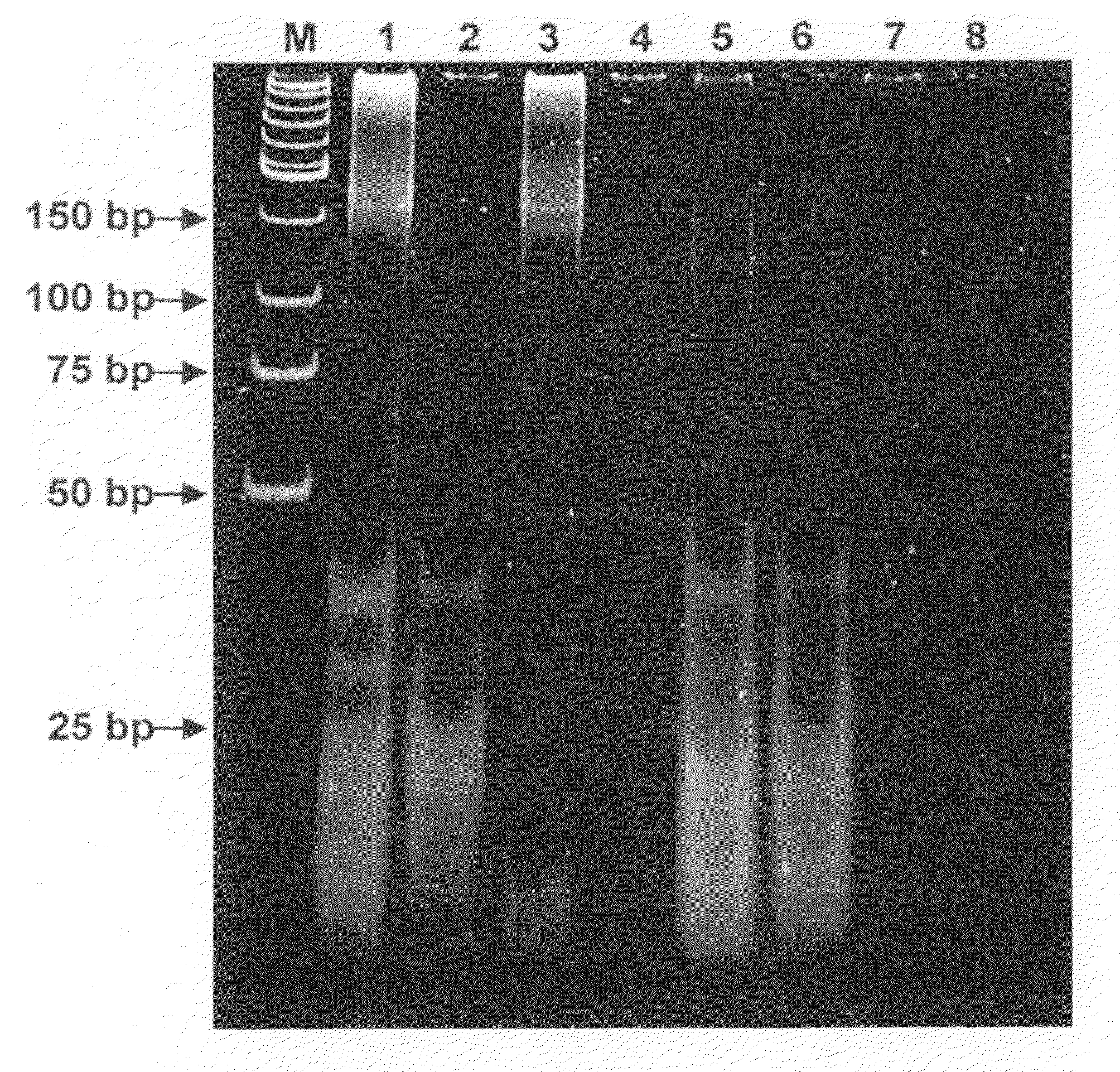
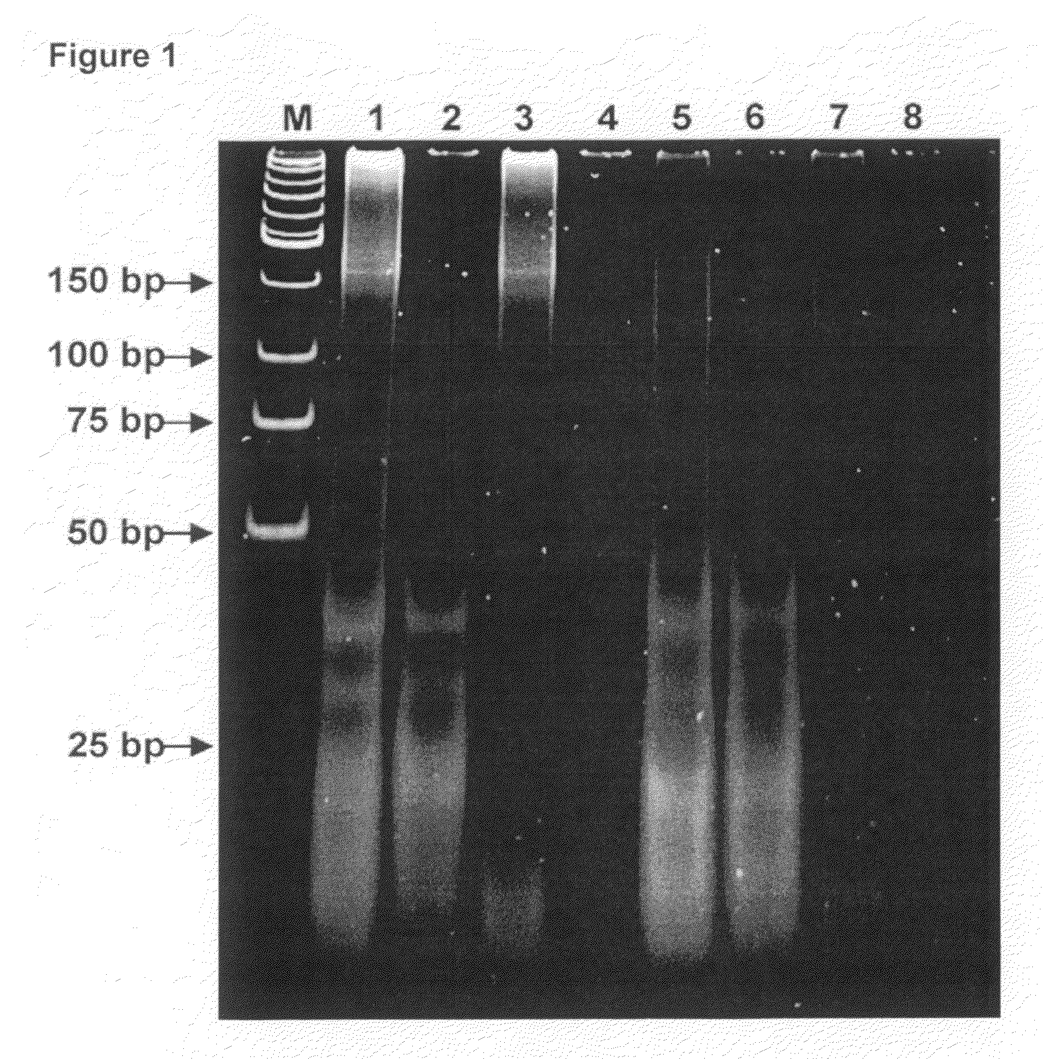
[0079]Q Binding: In a 50 ml tube 20 mL of urine was diluted with equal volume of 50 mM EDTA (pH 8.0) and 50 mM Tris-HCl (pH 8.0) which was then supplemented with 1-2 ml of Q-Sepharose™ (GE Healthcare; 17-0510-10) slurry and rigorously mixed 10-30 min at room temperature. The resin, with bound nucleic acids, was collected by centrifugation at 2000 g for 5 minutes at room temperature in a table top clinical centrifuge using a swing bucket rotor. All but ˜500 μl ...
example 2
[0087]This Example demonstrates that miRNA, from dying cells, cross the kidney barrier and may be detected in the urine of a patient.
Detection of Human miRNA Molecules in Urine
[0088]Micro RNA species that were analyzed in this example can be grouped in three distinct types, namely ubiquitous miRNAs, which are expressed in all or multiple tissues, tissue-specific miRNAs, and miRNAs in which expression is significantly altered in a particular tissue or cell type. As shown by Table 1, 20 different miRNAs were obtained from urine of 16 healthy volunteers and enrolled donors and later detected by real time RT-PCR using commercially available miRNA expression analysis kit (ABI). Corresponding synthetic miRNA oligonucleotides were used as standards. Reactions were carried out strictly as recommended by the supplier.
TABLE 1Detected miRNASEQIDNO:IDSequenceExpression1hsa-miR-127UCGGAUCCGUCUGAGCUUGGCUBrain overexpressed2hsa-miR-153UUGCAUAGUCACAAAAGUGABrain overexpressed3hsa-miR-129CUUUUUGCGGUC...
example 3
[0090]The Example demonstrates that miRNA from human nasopharyngeal carcinoma (NPC) cells can cross the patient's kidney barrier and can be detected in patient's urine by real time RT-PCR.
Virus-Derived miRNA in Urine
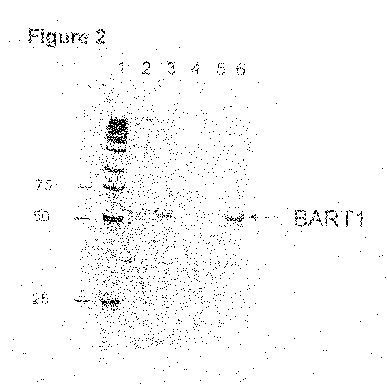
[0091]It is known that some viruses also encode and produce miRNAs. Since Epstein-Barr virus (EBV) is involved in development of nasopharyngeal carcinoma (NPC), the instant system was used to find out if viral miRNA from NPC cells can reach a patient's urine and be detected there. Urine samples from NPC patients were collected and stored according to the procedures described in the Example 1 of this application. EBV infection was confirmed by the detection of virus-specific DNA sequences in urine. Urine collected from healthy donor was negative for EBV specific DNA sequences. Two EBV-specific miRNAs BART3-3p and BART1-3p were analyzed in this study:
SEQ ID NO:21BART3-3PCGC ACC ACU AGU CAC CAG GUG USEQ ID NO:22BART1-3PUAG CAC CGC UAU CCA CUA UGU CU
[0092]Reverse transcripti...
PUM
| Property | Measurement | Unit |
|---|---|---|
| Time | aaaaa | aaaaa |
| Cell death | aaaaa | aaaaa |
| Degradation properties | aaaaa | aaaaa |
Abstract
Description
Claims
Application Information