Real-Time Ultrasound Monitoring of Heat-Induced Tissue Interactions

a real-time ultrasound and tissue technology, applied in the field of thermal therapy, can solve the problems of inability to provide an accurate temperature read-out, method that cannot work, and limited technique to skin applications,

Inactive Publication Date: 2009-04-23
BOARD OF RGT THE UNIV OF TEXAS SYST
View PDF27 Cites 80 Cited by
  • Summary
  • Abstract
  • Description
  • Claims
  • Application Information

AI Technical Summary

Benefits of technology

[0010]In one embodiment, the present invention is an apparatus to monitor and control radiation therapy that includes a radiative source that emits energy that enters a tissue and is absorbed at or a near a target site in the tissue to heat the tissue; an ultrasound transmitter directed at the target site, wherein the ultrasound transmitter emits ultrasound signals to the tissue that has been heated by the radiative source; an ultrasound receiver directed at the target site, wherein the ultrasound receiver receives ultrasound signals emitted from the ultrasound transmitter and reflected from the tissue that may or may not have been heated by the radiative source; and a...

Problems solved by technology

This technique is limited to just skin applications as it relies on knowing the order of placement of tissues, i.e. skin, followed by fat.
However, the laser therapy on the skin is essentially performed without pre-treatment monitoring of tissue composition and without real-time imaging of therapy progression during the treatment.
However, if the tissue composition (e.g., fat vs. water) is not known, this method will not work or provide an erroneous temperature read-out.
In addition, ultrasound frames are compared before and after therapy which is not realistic as therapy is...

Method used


Image

  • Real-Time Ultrasound Monitoring of Heat-Induced Tissue Interactions
  • Real-Time Ultrasound Monitoring of Heat-Induced Tissue Interactions
  • Real-Time Ultrasound Monitoring of Heat-Induced Tissue Interactions

Examples


example 1

Ultrasound-Based Thermal and Elasticity Imaging to Assist Photothermal Cancer Therapy

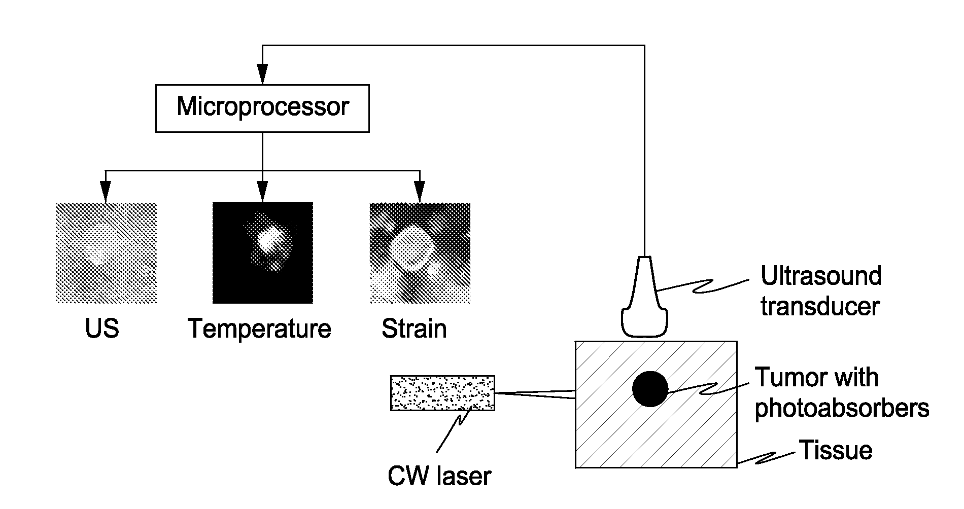
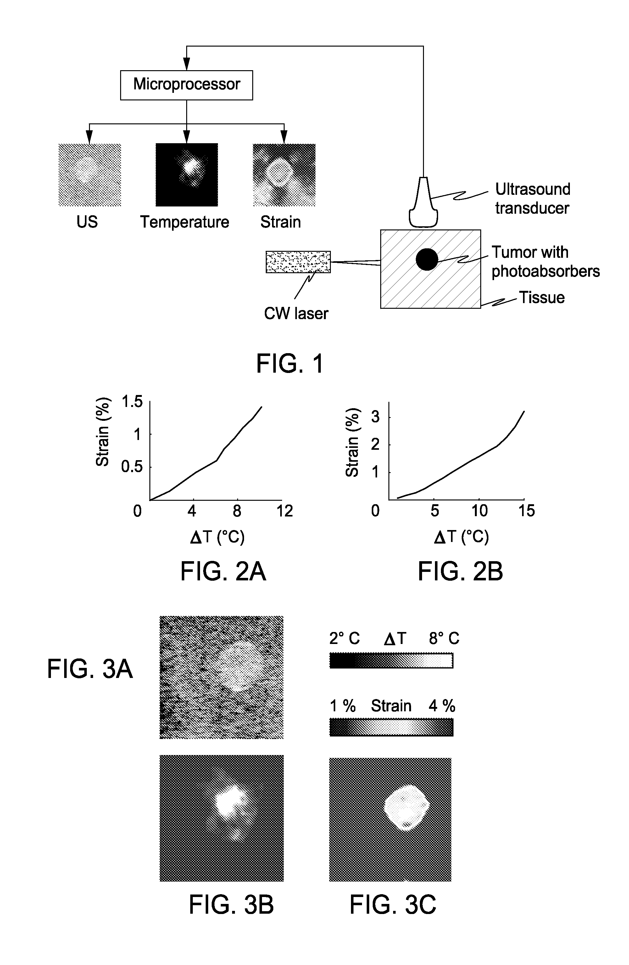
[0042]Photothermal therapy is a targeted, non-invasive thermal treatment of cancer. Up to 40° C. temperature increase is obtained in a small volume of malignant cells by using appropriate photoabsorbers and irradiating the tissue with a continuous wave laser. However, in order to ensure successful outcome of photothermal therapy, the tumor needs to be imaged before therapy, the temperature needs to be monitored during therapy and, finally, the tumor needs to be evaluated for necrosis during and after therapy. We investigated the feasibility of ultrasound imaging to track temperature changes during photothermal therapy and elasticity imaging to monitor tumor necrosis after treatment. The image-guided therapy was demonstrated on tissue mimicking phantoms and ex-vivo animal tissue with gold nanoparticles as photoabsorbers. Ultrasound-based thermal imaging effectively generates temperature scans during ...

example 2

Ultrasound Imaging to Monitor Photothermal Therapy Augmented by Plasmonic Nanoparticles

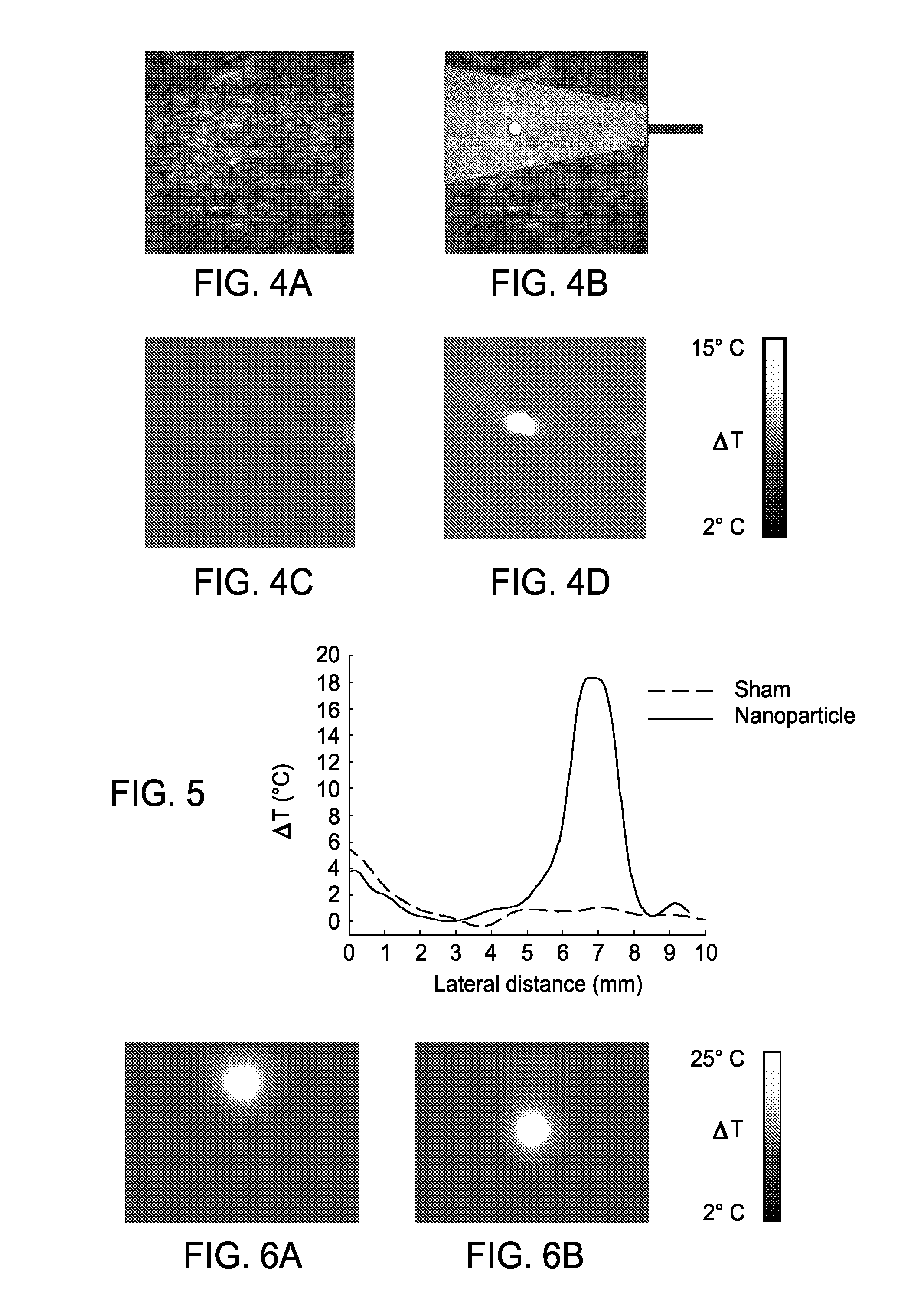
[0065]Metal nanoparticles are often used during photothermal therapy to efficiently convert light energy to thermal energy causing selective cancer destruction. This study investigates the feasibility of ultrasound imaging to monitor temperature changes during photothermal treatment. A continuous wave laser was used to perform photothermal therapy on tissue mimicking phantoms with embedded gold nanoparticles acting as photoabsorbers. Photothermal therapy studies were also carried out on ex-vivo tissue specimen with gold nanoparticles injected at a specific site. Prior to therapy, the structural features of the phantoms and tissue were assessed by ultrasound imaging. Thermal mapping, performed by measuring thermally induced motion of ultrasound signals, showed that temperature elevation obtained during therapy was localized to the region of embedded or injected nanoparticles. The results of our stu...

example 3

Ultrasound Guidance and Monitoring of Laser-Based Fat Removal

[0088]The present example used ultrasound imaging to guide laser removal of subcutaneous fat. Ultrasound imaging was used to identify the tissue composition and to monitor the temperature increase in response to laser irradiation. Laser heating was performed on ex-vivo porcine subcutaneous fat through the overlying skin using a continuous wave laser operating at 1210 nm optical wavelength. Ultrasound images were recorded using a 10 MHz linear array-based ultrasound imaging system. Ultrasound imaging was utilized to differentiate between water-based and lipid-based regions within the porcine tissue and to identify the dermis-fat junction. Temperature maps during the laser exposure in the skin and fatty tissue layers were computed. This example demonstrates the use of ultrasound imaging to guide laser fat removal.

[0089]Liposuction, also known as lipoplasty, is an invasive procedure for subcutaneous fat removal and body resha...


PUM


Abstract

The present invention includes an apparatus, method and system for monitoring and controlling radiation therapy, the system including a radiative source that emits energy that enters a tissue and is absorbed at or a near a target site in the tissue to heat the tissue; an ultrasound transmitter directed at the target site, wherein the ultrasound transmitter emits ultrasound signals to the tissue that has been heated by the radiative source; an ultrasound receiver directed at the target site, wherein the ultrasound receiver receives ultrasound signals emitted from the ultrasound transmitter and reflected from the tissue that has been heated by the radiative source; and a signal processor that processes the received ultrasound signal to calculate a tissue composition scan or tissue temperature scan.

Description

CROSS-REFERENCE TO RELATED APPLICATIONS[0001]This application claims priority to U.S. Provisional Application Ser. No. 60 / 976,994, filed Oct. 2, 2007, the entire contents of which are incorporated herein by reference.STATEMENT OF FEDERALLY FUNDED RESEARCH[0002]This invention was made with U.S. Government support awarded by the National Institutes of Health under grant EB 004963. The government has certain rights in this invention.TECHNICAL FIELD OF THE INVENTION[0003]The present invention relates in general to the field of thermal-therapy, and more particularly, to methods and systems for the scanning, mapping, monitoring and treatment of a tissue target.BACKGROUND OF THE INVENTION[0004]Without limiting the scope of the invention, its background is described in connection with tissue ablation methods and systems.[0005]U.S. Pat. No. 6,524,250 teaches a fat layer thickness mapping system to guide liposuction surgery. This patent uses ultrasound signals to identify fat layer under the ...

Claims


Application Information

Patent Timeline
IPC IPC(8): A61B18/04A61B8/00
CPCA61B5/01A61B5/4869A61B8/00A61B8/5223A61N5/062A61B2017/00084A61B2017/00106A61B2019/5276A61N5/0601A61B18/20A61B2090/378
Inventor EMELIANOV, STANISLAVMILNER, THOMASSHAH, JIGNESH
Owner BOARD OF RGT THE UNIV OF TEXAS SYST
Why Eureka
Try Eureka