Artificial cartilage containing chondrocytes obtained from costal cartilage and preparation process thereof

a technology of chondrocytes and costal cartilage, which is applied in the field of artificial cartilage containing chondrocytes obtained from costal cartilage, can solve the problems of premature degeneration, limited ability of adult cartilage to regenerate, and damage to articular cartilage, so as to confirm the ability of use and achieve effective articular regeneration

Active Publication Date: 2009-09-10
BIO SOLUTION CO LTD +2
View PDF2 Cites 69 Cited by
  • Summary
  • Abstract
  • Description
  • Claims
  • Application Information

AI Technical Summary

Benefits of technology

[0015]As a result, they discovered that chondrocytes obtained from costal cartilage are better than those from articular cartilage as donor cell source for cartilage repair. Also, they discovered that dedifferentiated chondrocytes during the passage show MSC properties to confirm their ability for use as cell therapeutic agent as well as artificial cartilage, by dedifferentiating them into desired cells in the differentiation inducible medium. In addition, the present inventors confirmed that chondrocytes-loaded chitosan-based scaffolds when transplanted to articular defects show effective articular regeneration, to complete the present invention.

Problems solved by technology

Damage to articular cartilage is an exceedingly common problem affecting the joints of millions of people.
The ability of adult cartilage to regenerate is limited due to avascularity and the absence of stem cell in this tissue.
Although defects that extend to subchondral bone provoke the formation of a fibrous or fibrocartilagious tissue, it undergoes premature degeneration because the repair tissue is biochemically and biomechanically different from hyaline cartilage.
A variety of clinical procedures have been developed to repair articular cartilage defects, but success has been limited.
However, these methods have disadvantages that the repair fibrocartilage lacks such biochemical and biomechanical property as normal hyaline cartilage has.
Further, their effects have not been proved yet for the large area defects, osteoarthritis and the older patient.
Osteochondral / chondral graft is simple and effective for small size defect, but if the tissue is moved from low weight part to high weight part, it may cause a harmful compression due to non-physiological bearing in the transplanted position.
Periosteal and perichondrial graft has a potential advantage to introduce new cell assembly which is able to construct cartilage, but has also disadvantage that hyaline-like repair tissue tends to undergo calcification through osteogenesis in cartilage.
However, the progenitor cells capacity to construct articular chondrocytes is limited, and the age of patient is directly related to clinical results.
Autologous articular chondrocyte transplantation (ACT) has been clinically applied in small defects of articular cartilage, but only limited success has been reported.
Even though articular chondrocytes can be easily isolated from mature articular cartilage by enzyme digestion, it requires two step-procedures for harvesting and grafting which are invasive to joint and considerably expensive.
Such dedifferentiation is a main problem which limits cell expansion in vitro and ACT application.
However, so far, only autologous tissue transplantation to graft costal cartilage per se to articular cartilage part has been performed, and there has been no effort to isolate chondrocytes from costal cartilage to regenerate articular cartilage by tissue engineering method.
Furthermore, transplantation of chondrocytes or chondrogenic cells alone has shown to be successful in rabbit models, but the healing rate has limited due to loss of their viability in the transplanted cells and due to the difficulty of fixing chondrocytes in the defect.

Method used


Image

  • Artificial cartilage containing chondrocytes obtained from costal cartilage and preparation process thereof
  • Artificial cartilage containing chondrocytes obtained from costal cartilage and preparation process thereof
  • Artificial cartilage containing chondrocytes obtained from costal cartilage and preparation process thereof

Examples


example 1

Evaluation on Whether Costal Chondrocytes can be Donor Cell Source for Articular Cartilage Repair

Materials and Methods

[0057]Isolation of Chondrocytes

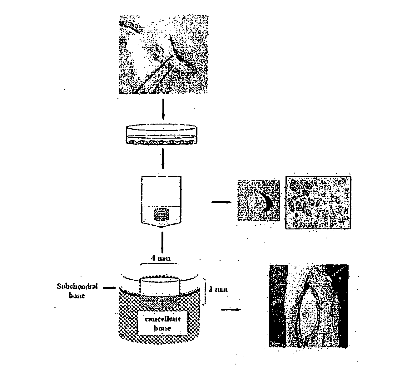
[0058]Articular and costal cartilages were obtained from 3 to 4 months old New Zealand White Rabbit, and then weighed. To harvest costal cartilage, animals were anesthetized by intravenous injection of a mixture of xylazine hydrochloride (2 mg / weight kg, Rompun, Bayer, Korea) and ketamine hydrochloride (6-10 mg / weight kg, ketalar, Yuhan Co., Korea). The skin in the region of the chest was shaved, washed with alcohol, and prepared with povidone-iodine solution. The back side of the right chest was opened, and 9th and 10th costal cartilages were exposed. The costal cartilages, after removing soft adhering tissues, were then weighed. These cartilages were minced into 1-2 mm3 pieces and rinsed 3 times in D-PBS (Dulbecco's phosphate buffered saline; Jeil Biotech services Inc., Taegu, Korea). After being rinsed, the minced cartilage was diges...

example 2

Confirmation on Whether Fully Dedifferentiated Costal Chondrocytes Show MSC's Properties

Materials and Methods

Isolation of Chondrocytes

[0075]Costal cartilages were obtained from 4 to 5 months old New Zealand White Rabbit. The skin in the region of the chest was shaved, washed with alcohol, and prepared with povidone-iodine solution. The back side of the right chest was opened, and 9th and 10th costal cartilages were exposed. The costal cartilages, after removing soft adhering tissues, were then weighed. These cartilages were minced into 1-2 mm3 pieces and rinsed 3 times in D-PBS (Dulbecco's phosphate buffered saline; Jeil Biotech services Inc., Taegu, Korea). After being rinsed, the minced cartilage was digested by the enzyme cocktail solution including collagenase D (2 mg / ml Roche Diagnostic GmbH, Germany), hyaluronidase (1 mg / ml, Roche), and DNase (0.75 mg / ml, Roche) under 37° C. and 5% CO2 for overnight. Then the solution was filtered through a 53 μm nylon mesh, isolated cells wer...

example 3

Evaluation on Expansion of Costal Chondrocytes and Their Differentiation into Cartilage According to Culture Condition (DMEM+10% FBSs, MSCGM or FGF-Containing MSCGM)

Materials and Methods

[0080]Isolation and Culture of Chondrocytes

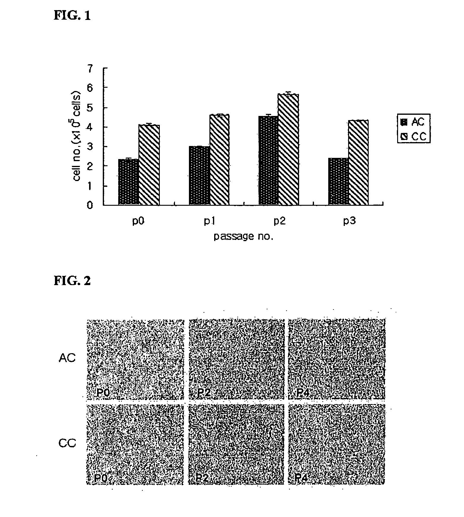
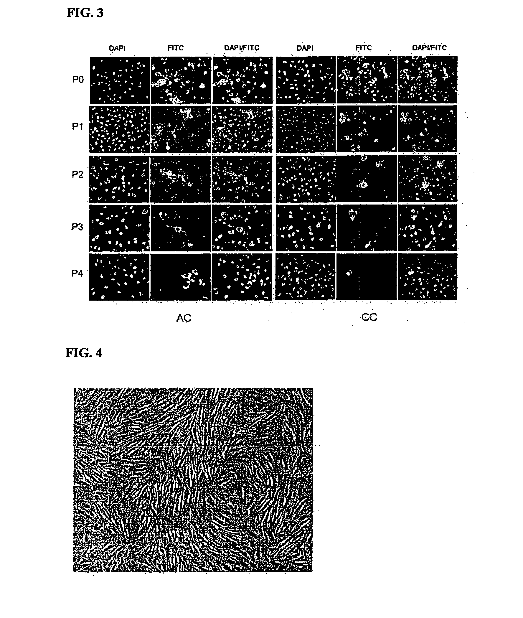
[0081]Chondrocytes were isolated by the method as described above, and viable cells were counted on Haemocytometer based on the trypan blue exclusion. The chondrocytes were plated at a cell density of 5×10⁵ cells / 100 mm diameter Petri dish, and cultured in DMEM (DMEM-FBS), MSCGM (Cambrex Bio Science Walkersville, Inc., MD, U.S.A.), or MSCGM added with 1 ng / ml of FGF (R&D System Inc., MN, U.S.A.).

[0082]Cell Expansion Rate According to the Medium

[0083]When the dish was filled with the cells, the cells were removed by Trypsin-EDTA (Gibco), and viable cells were counted on Haemocytometer based on the trypan blue exclusion to evaluate cell expansion rate. Then, the cells were plated at a cell density of 5×10⁵ cells / 100 mm diameter Petri dish, and cultured in the ...


PUM

PropertyMeasurementUnit
diameteraaaaaaaaaa
weightaaaaaaaaaa
weightaaaaaaaaaa

Abstract

The present invention relates to an artificial cartilage containing mesenchymal stem cell (MSC)-like dedifferentiated cells obtained by passage culturing costal chondrocytes, and a preparation process thereof.

Description

TECHNICAL FIELD[0001]The present invention relates to an artificial cartilage containing chondrocytes obtained from costal cartilage and a preparation process thereof.BACKGROUND ART[0002]Damage to articular cartilage is an exceedingly common problem affecting the joints of millions of people. The ability of adult cartilage to regenerate is limited due to avascularity and the absence of stem cell in this tissue. Although defects that extend to subchondral bone provoke the formation of a fibrous or fibrocartilagious tissue, it undergoes premature degeneration because the repair tissue is biochemically and biomechanically different from hyaline cartilage.[0003]A variety of clinical procedures have been developed to repair articular cartilage defects, but success has been limited. Bone marrow stimulation technique (fine joint plastic surgery and drill articular plastic surgery) interpenetrate subchondral bone, and stimulate multipotent stem cells within bone marrow to repair the defects...

Claims


Application Information

Patent Timeline
Patent Type & Authority Applications(United States)
IPC IPC(8): A61F2/08A61K35/12A61K35/28A61K35/32C12N5/077C12N5/0775
CPCA61K35/12A61K35/28C12N2501/115C12N5/0655C12N5/0668A61K35/32A61L27/20A61L27/3804A61L27/3852A61L2430/06C08L5/08C12N5/0652A61F2/28
Inventor SON, YOUNG SOOKLEE, JUNG SUNLEE, EUN KYUNGLEE, JIN YEON
Owner BIO SOLUTION CO LTD
Why Eureka
Try Eureka