


Quick Research
Generate reliable direction feasibility study reports for your R&D in just a few steps.

Technical Q&A
Discover and master advanced knowledge NOW. Basics, ideas, possibilities, all at once.

Find Solutions
As an expert in R&D theories, this can generate solutions to your technical problems instantly.

Evaluate Feasibility
Analyze your overall solution with one click, know your potential R&D risks in advance.

Monitor Landscape
Get weekly tech updates, stay abreast of the latest tech innovations and key insights.
Connective Tissue Growth Factor-2
a technology of connective tissue and growth factor, which is applied in the direction of peptides, drug compositions, fused cells, etc., can solve the problems of uncontrolled growth of cells that lose the ability to respond to tgf-, and become tumorigenic, so as to promote attachment, fixation and stabilization of tissue implants, and enhance wound healing
- Summary
- Abstract
- Description
- Claims
- Application Information
AI Technical Summary
Benefits of technology
Problems solved by technology
Method used
Image

Examples
example 1
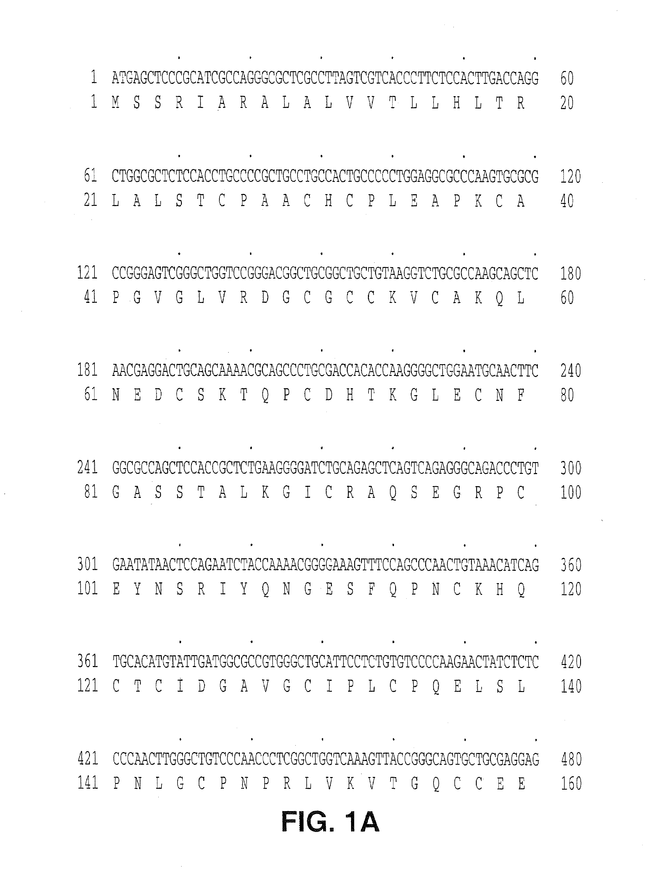
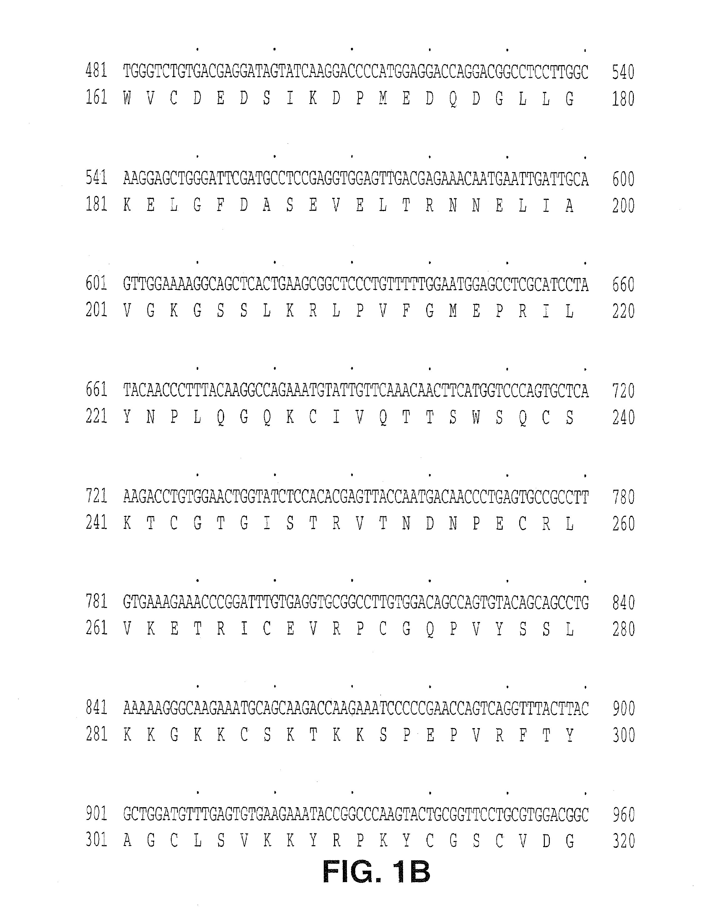
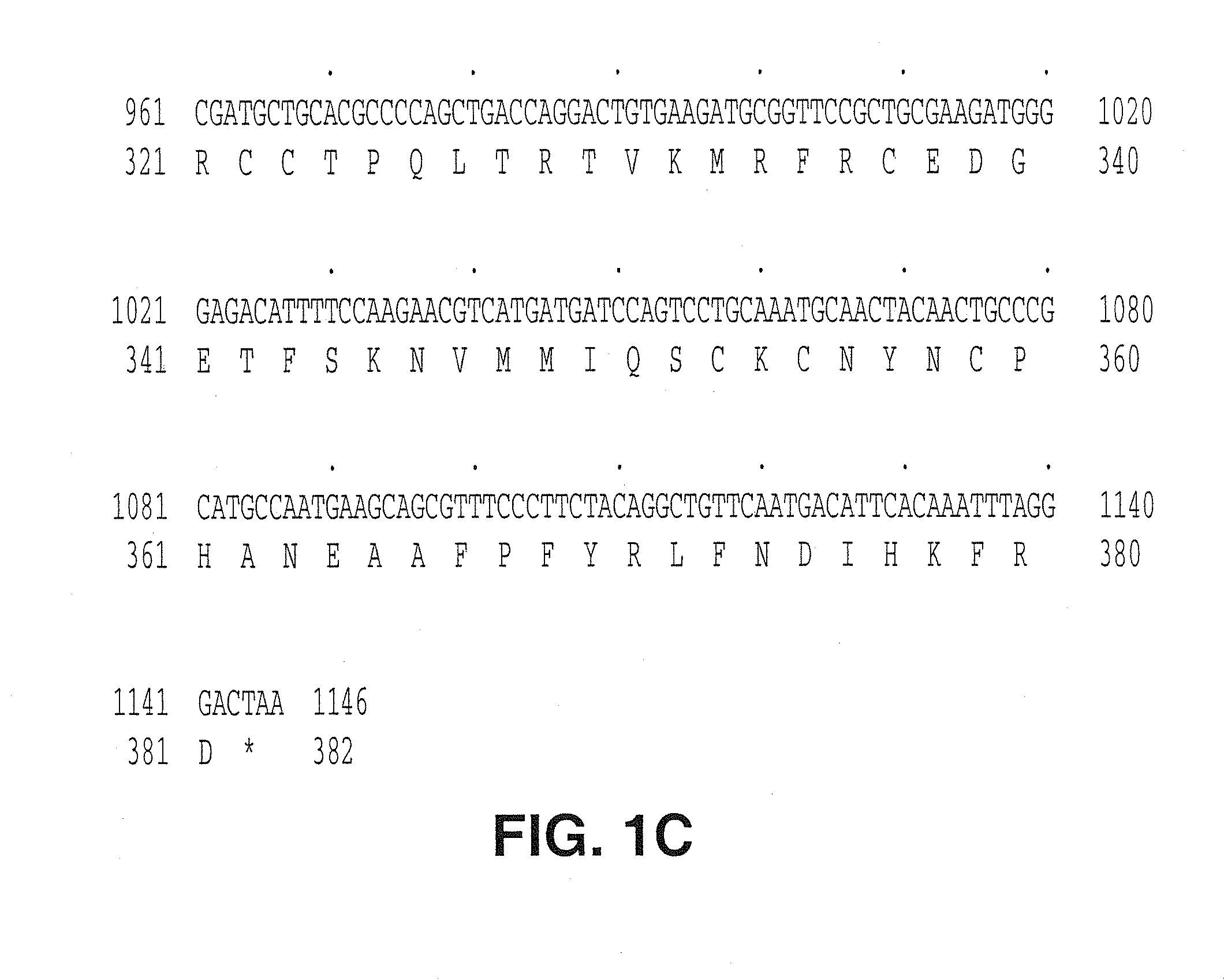
Cloning and Expression of CTGF-2 in a Baculovirus Expression System
[0117]The DNA sequence encoding the full length CTGF-2 protein, ATCC® # 75804, was amplified using PCR oligonucleotide primers corresponding to the 5′ and 3′ sequences of the gene:
[0118]The 5′ primer has the sequence CGCGGGATCCTGCGCGACACAATGAGCT (SEQ ID NO:3) and contains a BamHI restriction enzyme site (in bold) followed by 18 nucleotides resembling an efficient signal for the initiation of translation in eukaryotic cells (J. Mol. Biol. 1987, 196, 947-950, Kozak, M.). The initiation codon for translation “ATG” is underlined.
[0119]The 3′ primer has the sequence CGCGGGTACCAGGTAGCATTTAGTCCCTAA (SEQ ID NO:4) and contains the cleavage site for the restriction endonuclease Asp781 and 20 nucleotides complementary to the 3′ non-translated sequence of the CTGF-2 gene. The amplified sequences were isolated from a 1% agarose gel using a commercially available kit (GENECLEAN®, BIO 101 Inc., La Jolla, Calif.). The fragment was t...
example 2
Expression of Recombinant CTGF-2 in COS Cells
[0129]The expression of plasmid, CTGF-2 HA is derived from a vector pcDNAI / Amp (Invitrogen) containing: 1) SV40 origin of replication, 2) ampicillin resistance gene, 3) E. coli replication origin, 4) CMV promoter followed by a polylinker region, a SV40 intron and polyadenylation site. A DNA fragment encoding the entire CTGF-2 precursor and a HA tag fused in frame to its 3′ end was cloned into the polylinker region of the vector, therefore, the recombinant protein expression is directed under the CMV promoter. The HA tag correspond to an epitope derived from the influenza hemagglutinin protein as previously described (I. Wilson, H. Niman, R. Heighten, A Cherenson, M. Connolly, and R. Lerner, 1984, Cell 37, 767). The infusion of HA tag to our target protein allows easy detection of the recombinant protein with an antibody that recognizes the HA epitope.
[0130]The plasmid construction strategy is described as follows:
[0131]The DNA sequence en...
example 3
Expression via Gene Therapy
[0132]Fibroblasts are obtained from a subject by skin biopsy. The resulting tissue is placed in tissue-culture medium and separated into small pieces. Small chunks of the tissue are placed on a wet surface of a tissue culture flask, approximately ten pieces are placed in each flask. The flask is turned upside down, closed tight and left at room temperature over night. After 24 hours at room temperature, the flask is inverted and the chunks of tissue remain fixed to the bottom of the flask and fresh media (e.g., Ham's F12 media, with 10% FBS, penicillin and streptomycin, is added. This is then incubated at 37° C. for approximately one week. At this time, fresh media is added and subsequently changed every several days. After an additional two weeks in culture, a monolayer of fibroblasts emerge. The monolayer is trypsinized and scaled into larger flasks.
[0133]pMV-7 (Kirschmeier, P. T. et al, DNA, 7:219-25 (1988)) flanked by the long terminal repeats of the M...
PUM
 Abstract
Description
Claims
Application Information
 - R&D Engineer
- R&D Manager
- IP Professional
- Industry Leading Data Capabilities
- Powerful AI technology
- Patent DNA Extraction
Browse by: Latest US Patents, China's latest patents, Technical Efficacy Thesaurus, Application Domain, Technology Topic, Popular Technical Reports.
© 2024 PatSnap. All rights reserved.Legal|Privacy policy|Modern Slavery Act Transparency Statement|Sitemap|About US| Contact US: help@patsnap.com