Method for fixing antibody on the surface of medical instrument

Active Publication Date: 2010-05-06
LEPU MEDICAL TECH (BEIJING) CO LTD
View PDF5 Cites 7 Cited by
  • Summary
  • Abstract
  • Description
  • Claims
  • Application Information

AI Technical Summary

Benefits of technology

[0004]The object of the present invention is to provide a method for fixing antibody on the surface of medical instrument, which may improve the firm

Problems solved by technology

As the development of medical instruments, especially the emergence of drug-containing medical instrument, fixing biological products on the surface of the medical instrument has become an extensive and in-depth issue.
The method of physical adsorption is simple a

Method used


Image

  • Method for fixing antibody on the surface of medical instrument
  • Method for fixing antibody on the surface of medical instrument
  • Method for fixing antibody on the surface of medical instrument

Examples


example 1

[0055]The method of fixing antibody CD34 on the scaffold surface based on the antigen-antibody binding reaction and the fluorescence detection of the antibody activity have the steps as follows:

[0056](1) immerging the 316L stainless steel stent which has holes in surface into the phosphate buffer solution or the carbonate buffer solution, whose pH is 7.2˜7.4, containing 0.1˜100 μg / mL CD34 antibody such as type Ilmouse anti-human CD34 antibody produced by U.S. Stem Cell Technology Co., Ltd., to incubate for 30 min˜2 h at 25˜37° C. and then taking out the scaffold;

[0057](2) washing the scaffold 3 times for 5 min per time using the phosphate buffer solution, whose pH is 7.2˜7.4, and blowing the scaffold for 15 min at room temperature 25° C. and then preserving the scaffold at 4° C. after it becomes dry.

[0058](3) immerging the scaffold which has antibody on surface into the phosphate buffer solution, whose pH is 7.2˜7.4, containing 500 μL 5˜10% bovine serum albumin (BSA) to incubate for...

example 2

[0063]The method of fixing antibody CD34 on the scaffold surface based on the antigen-antibody binding reaction and the immunohistochemistry detection of the antibody activity, and the method of fixing antibody CD34 on the scaffold surface is the same as the steps 1 and 2 disclosed in example 1;

[0064](3) immerging the scaffold which has antibody on surface into the phosphate buffer solution containing 500 μL 5˜10% bovine serum is albumin (BSA) to incubate for 30 min˜2 h at 25˜37° C. and then taking out the scaffold to dry in the air;

[0065](4) immerging the scaffold into the phosphate buffer solution containing horseradish peroxidase (HRP) labeled goat anti-mouse IgG to incubate for 20˜30 min at 25˜37° C. and then taking out the scaffold;

[0066](5) washing the scaffold 3 times for 5 min per time by the phosphate buffer solution;

[0067](6) after using 3-amino-9-ethyl-carbazole (AEC) staining kit produced by China Huamei Bio-engineering Corporation for immunohistochemical staining, takin...

example 3

[0068]The steps of hemodynamics simulation experiment of scaffold with antibody in artificial body flow, the method of fixing the antibody on the scaffold surface and the firm degree detection are as follows:

[0069](1) putting the scaffold with the fixed mouse anti-human CD34 antibody on surface into fluid artificial simulated body flow, such as the phosphate buffer solution, whose pressure is 100 mmHg, flow rate is 91 cm / s and pH is 7.2˜7.4, to make a scouring experiment, when the scaffold is at a normal state or at a state after the balloon dilation;

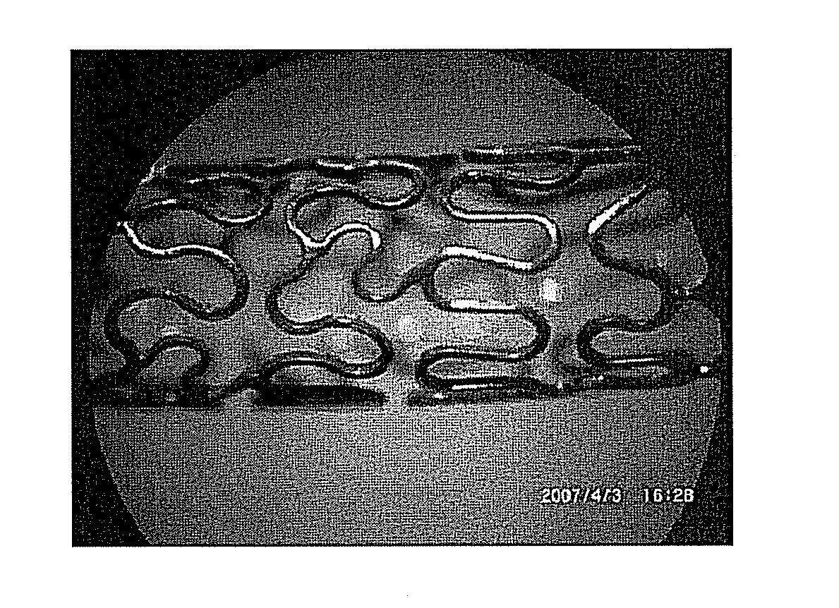
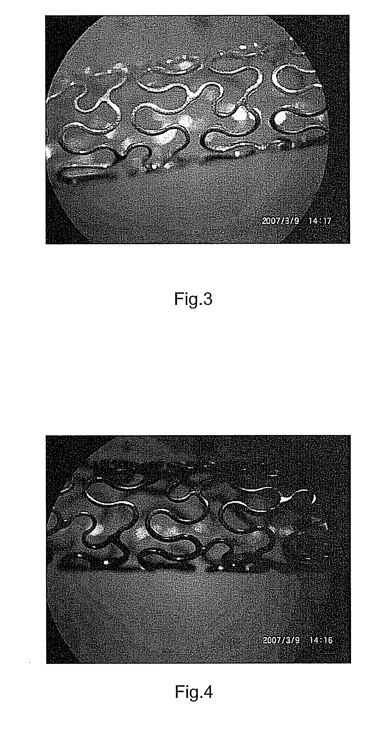
[0070](2) after the scaffold surface is scoured for 24 hours, detecting the activity of the antibody on the scaffold surface with the method disclosed in Example 2; the experimental results are shown in FIG. 5 and FIG. 6, which indicate that the method of absorbing the antibody using the holes in the surface of the scaffold has the advantage of high firm degree, and enables the antibody to be fixed on the scaffold surface even in the ar...


PUM

PropertyMeasurementUnit
Temperatureaaaaaaaaaa

Abstract

A method for fixing antibody on the surface of medical instrument, mainly includes: 1) pre-treating the instrument surface; 2) preparing holes: preparing multicrystal phase structure which has same size holes in the surface of the instrument by chemical corrosion, electrochemical corrosion, anodic oxidation, micro-arc oxidation, micro-arc nitridation; 3) post-treating the instrument surface; 4) fixing the antibody: immerging the bare metal scaffold which has holes in surface into a buffer solution containing antibody, adjusting the pH value of the antibody buffer solution, fixing the antibody on the surface of the instrument by the attraction between positive and negative charge and hole effect; and 5) confirming the effectiveness of the fixed antibody by artificial simulation hemodynamics and detecting method of antibody activity in scaffold surface. The method can promote the firm degree of the antibody which is fixed on the surface of the instrument, and keep high activity of the antibody on the surface of medical instrument.

Description

TECHNICAL FIELD[0001]The present invention belongs to the field of medical instrument, and relates to a method for fixing antibody on the surface of medical instrument.BACKGROUND [0002]It has been widely applied to biochip to fix biological products, such as protein, antibody, growth factor and gene plasmid, on the solid surfaces, such as the surface of metal, ceramics and polymeric material,. As the development of medical instruments, especially the emergence of drug-containing medical instrument, fixing biological products on the surface of the medical instrument has become an extensive and in-depth issue. Comparing with common protein, the antibody contained in biological product has advantages such as high stability, hard inactivation and specificity of antigen-antibody reaction and has been widely used as in vitro diagnostic reagent and has been applied to medical diagnosis. Thus, the method for firmly fixing the antibody on the surface of the medical instrument including the s...

Claims


Application Information

Patent Timeline
IPC IPC(8): B05D3/00
CPCA61L27/227A61L27/54A61L29/048A61L29/16A61L31/047A61L31/16A61L2300/256
Inventor YU, ZHANJIANG
Owner LEPU MEDICAL TECH (BEIJING) CO LTD
Why Eureka
Try Eureka