Luciferase gene optimized for use in imaging of intracellular luminescence

a luciferase and luminescence imaging technology, applied in the field of luciferase gene constructs, can solve the problems of inability to observe fluorescent proteins long-term, difficult quantification, impaired cells, etc., and achieve the effects of improving sensitivity, low gene introduction efficiency, and wide enhancement of intracellular amount of expressed luciferas

Active Publication Date: 2010-05-06
TOYOBO CO LTD +1
View PDF2 Cites 0 Cited by
  • Summary
  • Abstract
  • Description
  • Claims
  • Application Information

AI Technical Summary

Benefits of technology

[0059] According to the present invention, the intracellular amount of expressed luciferase can be widely enhanced, and it has become possible to perform bioluminescence imaging in an individual cell for a long term. In a system using luciferase, it is not necessary to consider cell damage due to the short term cell imaging exposed to the excitation light, long-term cell imaging becomes possible, and the system can be utilized for the treatment of various pathological conditions and drug discoveries.
[0060] According to the method of the present invention, particularly in the analysis of the promoter having weak transcription activity and the analysis of the promoter in the cell having low gene introduction efficiency, the sensitivity is improved and stable measurement becomes possible compared with those using conventional firefly luciferase. Furthermore, by enhancing the signal intensity according to the method of the present invention, it becomes possible to analyze living cells in the plate format, in the test in which it was difficult to treat multi-samples in the plate format due to the small amount of the samples. Thus, an application range to analyze living cells can be expanded in drug discovery screenings and cytotoxic evaluations of chemicals.

Problems solved by technology

However, quantification is difficult because of the requirement of an excitation light and uneven fluorescence efficiency, and the cells are impaired because of exposure to the excitation light.
Thus, fluorescent protein is not suitable for long-term observation.
GFP does not require a substrate and can be easily detected by irradiating the excitation light, but is not suitable for quantification.
Since the excitation light is irradiated, the cells are greatly damaged.
Thus, GFP is not suitable for long-term, continuous monitoring purposes.
However, there are few examples of firefly luciferase imaging.
This is because the stability of luciferase in mammalian cells is lower, the protein lifespan is shorter compared with the fluorescent protein, and the transcription efficiency is low; firefly luciferase is thus not suitable for practical use, as image analyzers of the cells correspond to the fluorescence, and no imaging system for efficiently measuring the luminescence is available.
This is particularly because the luminescence intensity of firefly luciferase in living cells is low, thus hindering the easy obtainment of luminescence signals.
However, one shortcoming of firefly luciferase is that the luminescent color is changed in conjunction with the intracellular pH value, and thus is not suitable for analyzing multiple gene expressions based on the diversity of luminescent colors (Yoshihiro Ohmiya, Yoshihiro Nakajima, Multiple Gene Transcription Activity Measurement System; Patent Document 1).
This method is complicated because the cell is lysed and luciferase further reacted with the luminescent reagent, compared with the method of detection adding luciferin to the cell culture medium.
Additionally, when multiple samples are measured simultaneously, the sample amount is reduced and measurement is difficult in a detection system with low sensitivity.
The fluorescent protein is suitable as an intracellular imaging probe for short-term measurement, but is not suitable for long-term analysis.
Luciferase is suitable for long-term measurement, but has not been established as an imaging tool.
In particular, stable measurements are difficult in luciferases derived from fireflies produced in North America and Japan because the luminescence spectra changes depending on environmental pH values.

Method used


Image

  • Luciferase gene optimized for use in imaging of intracellular luminescence
  • Luciferase gene optimized for use in imaging of intracellular luminescence
  • Luciferase gene optimized for use in imaging of intracellular luminescence

Examples


example 1

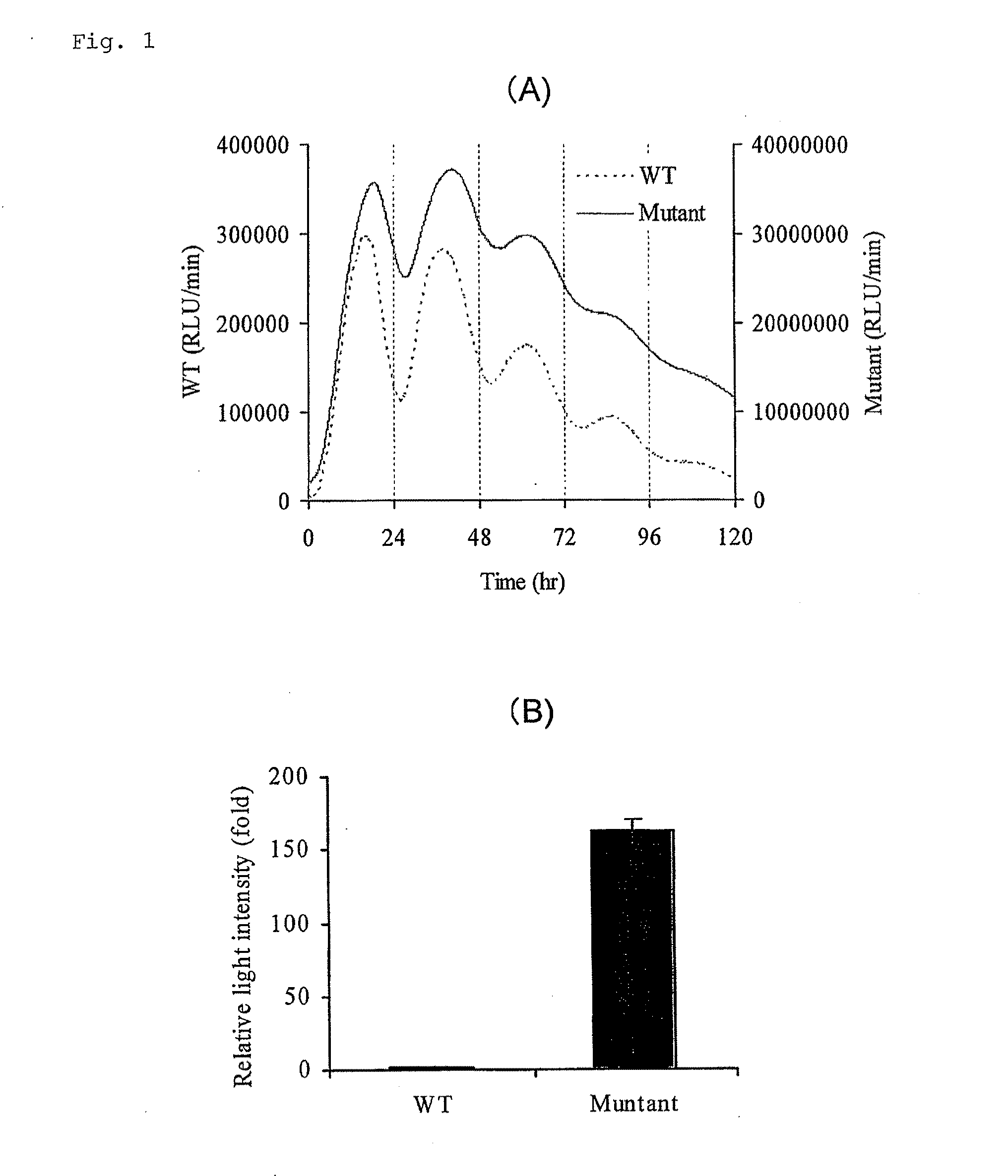
[0108] Short lifespan-type luciferase was made by ligating a PEST sequence (SEQ ID NO: 7) of murine ornithine decarboxylase to a wild-type (SEQ ID NO: 1) and improved-type (SEQ ID NO: 2) click beetle luciferase cDNA. Vectors in which these had been inserted downstream of a murine circadian clock gene Bmal1 promoter (GenBank Accession No. AB064982) were made. Subsequently, 1 μg of each vector was introduced into cultured fibroblast NIH3T3 cells seeded in a 35 mm culture dish by a lipofection method (LipofectAMINE PLUS, Invitrogen), and the cells were cultured at 37° C. for 24 hours and treated with a DMEM medium containing 100 nM dexamethasone for 2 hours. The medium was replaced with a DMEM medium containing 200 μM D-luciferin and 10% (w / v) bovine serum, and then one minute of luminescence was measured every 15 minutes for 5 days in real time using a real-time gene expression measurement apparatus (AB2500 supplied from ATTO Corporation) (FIG. 1A). The change in about a 24-hour cycle...

example 2

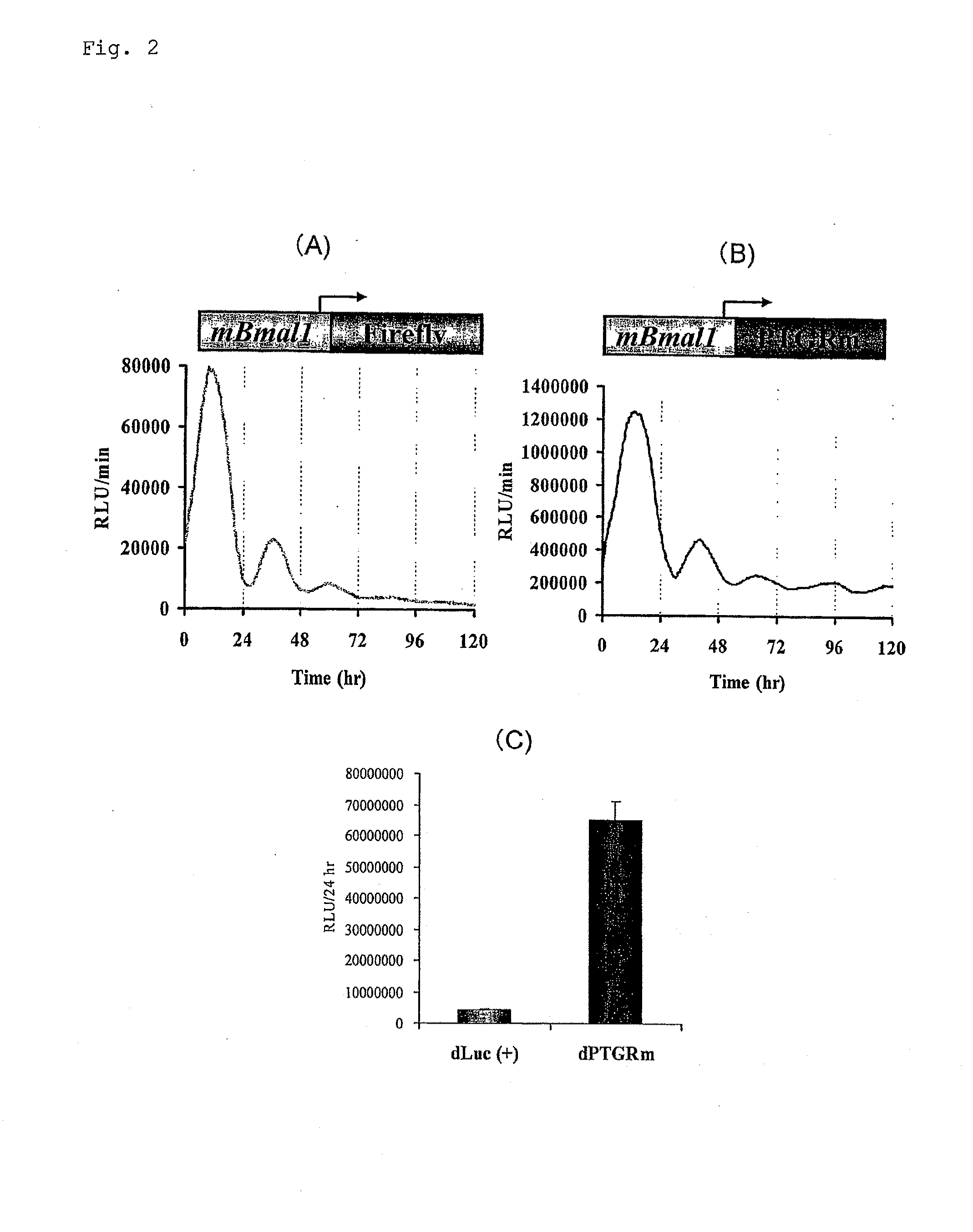
[0110] Short lifespan-type luciferase was made by ligating the PEST sequence (SEQ ID NO: 7) of murine ornithine decarboxylase to firefly luciferase cDNA (Luc(+), supplied from Promega) derived from Photinus pyralis produced in North America and improved-type click beetle luciferase cDNA (SEQ ID NO: 2). Vectors in which these had been inserted downstream of a murine circadian clock gene Bmal1 promoter (GenBank Accession No. AB064982) were made. Subsequently, 1 μg of each vector was introduced into cultured fibroblast rat1 cells seeded in a 35 mm culture dish by the lipofection method (LipofectAMINE PLUS), and the cells were cultured at 37° C. for 24 hours and treated with the DMEM medium containing 100 nM dexamethasone for 2 hours. The medium was replaced with a DMEM medium containing 200 μM D-luciferin and 10% (w / v) bovine serum, and then one minute of luminescence was measured every 15 minutes for 5 days using the real-time gene expression measurement apparatus (AB2500 supplied fro...

example 3

[0111] Expression vectors were made by inserting the aforementioned short lifespan-type firefly luciferase cDNA and short lifespan-type improved-type click beetle luciferase cDNA downstream of an SV40 promoter. The expression vector (200 ng) was introduced into the cultured fibroblast NIH3T3 cells seeded in a 24-well plate by the lipofection method, and the cells were cultured at 37° C. for 48 hours. Subsequently, the medium was replaced with the medium containing 100 μM protein synthesis inhibitor, cycloheximide, and after culturing for 30 minutes, the intracellular luminescence intensity was measured every one hour. The luminescence intensity was measured in the same way as in Example 1 (FIG. 3). In short lifespan-type firefly luciferase derived from Photinus pyralis produced in North America, the half life was one hour; however, in short lifespan-type improved click beetle luciferase, the half life was 4 hours, a fourfold extension.


PUM

PropertyMeasurementUnit
pHaaaaaaaaaa
luminescence wavelengthaaaaaaaaaa

Abstract

The present invention provides a gene construct encoding pH insensitive luciferase for visualizing intracellular information, wherein an intracellular expression activity is higher compared with a gene construct of luciferase derived from a firefly.

Description

INCORPORATION-BY-REFERENCE OF MATERIAL ELECTRONICALLY SUBMITTED [0001] Incorporated by reference in its entirety herein is a computer-readable nucleotide / amino acid sequence listing submitted herewith and identified as follows: 7,558 bytes ASCII (Text) file named “703019ReplacementSequenceListing.txt,” created Apr. 28, 2009. TECHNICAL FIELD [0002] The present invention relates to a gene construct of luciferase wherein a luminescence intensity in living cells is enhanced for the purpose of cell imaging, a combination thereof, a cell transformed with the gene construct, and a method for evaluating an interaction of two proteins in a cell. [0003] Furthermore, the present invention relates to a highly sensitive method for monitoring transcription activity in living cells using a gene construct of luciferase whose expression is enhanced, and more particularly relates to a method for a high-throughput analysis using a plate format. BACKGROUND ART [0004] In the field of life science, it is...

Claims


Application Information

Patent Timeline
Patent Type & Authority Applications(United States)
IPC IPC(8): C12Q1/68C07H21/04C12N5/08
CPCC12N9/0069
Inventor OHMIYA, YOSHIHIRONAKAJIMA, YOSHIHIROVIVIANI, VADIMNISHII, SHIGEAKIASAI, TOMOMI
Owner TOYOBO CO LTD
Why Eureka
Try Eureka