Multimodal imaging probes for in vivo targeted and non-targeted imaging and therapeutics

a multi-modal imaging and probe technology, applied in the field of multi-modal imaging probes, can solve the problems of insufficient contrast obtained in mri, small intra-tissue differences, and inability to provide distinguishable relaxation times, so as to facilitate the addition of additional moieties and increase the stability and half-life of such molecules

Inactive Publication Date: 2010-07-22
RGT UNIV OF CALIFORNIA
View PDF6 Cites 111 Cited by
  • Summary
  • Abstract
  • Description
  • Claims
  • Application Information

AI Technical Summary

Benefits of technology

[0046]The terms “nucleic acid” or “oligonucleotide” or grammatical equivalents herein refer to at least two nucleotides covalently linked together. A nucleic acid of the present invention is preferably single-stranded or double stranded and will generally contain phosphodiester bonds, although in some cases, as outlined below, nucleic acid analogs are included that may have alternate backbones, comprising, for example, phosphoramide (Beaucage et al. (1993) Tetrahedron 49(10):1925) and references therein; Letsinger (1970) J. Org. Chem. 35:3800; Sprinzl et al. (1977) Eur. J Biochem. 81: 579; Letsinger et al. (1986)Nucl. Acids Res. 14: 3487; Sawai et al. (1984) Chem. Lett. 805, Letsinger et al. (1988) J. Am. Chem. Soc. 110: 4470; and Pauwels et al. (1986) Chemica Scripta 26: 1419), phosphorothioate (Mag et al. (1991) Nucleic Acids Res. 19:1437; and U.S. Pat. No. 5,644,048), phosphorodithioate (Briu et al. (1989) J. Am. Chem. Soc. 111 :2321, O-methylphophoroamidite linkages (see Eckstein, Oligonucleotides and Analogues: A Practical Approach, Oxford University Press), and peptide nucleic acid backbones and linkages (see Egholm (1992) J. Am. Chem. Soc. 114:1895; Meier et al. (1992) Chem. Int Ed. Engl. 31: 1008; Nielsen (1993) Nature, 365: 566; Carlsson et al. (1996) Nature 380: 207). Other analog nucleic acids include those with positive backbones (Denpcy et al. (1995) Proc. Natl. Acad. Sci. USA 92: 6097; non-ionic backbones (U.S. Pat. Nos. 5,386,023, 5,637,684, 5,602,240, 5,216,141 and 4,469,863; Angew. (1991) Chem. Intl. Ed English 30: 423; Letsinger et al. (1988) J. Am. Chem. Soc. 110:4470; Letsinger et al. (1994) Nucleoside &Nucleotide 13:1597; Chapters 2 and 3, ASC Symposium Series 580, “Carbohydrate Modifications in Antisense Research”, Ed. Y. S. Sanghui and P. Dan Cook; Mesmaeker et al. (1994), Bioorganic &Medicinal Chem. Lett. 4: 395; Jeffs et al. (1994) J. Biomolecular NMR 34:17; Tetrahedron Lett. 37:743 (1996)) and non-ribose backbones, including those described in U.S. Pat. Nos. 5,235,033 and 5,034,506, and Chapters 6 and 7, ASC Symposium Series 580, Carbohydrate Modifications in Antisense Research, Ed. Y. S. Sanghui and P. Dan Cook. Nucleic acids containing one or more carbocyclic sugars are also included within the definition of nucleic acids (see Jenkins et al. (1995), Chem. Soc. Rev. pp169-176). Several nucleic acid analogs are described in Rawls, C & E News Jun. 2, 1997 page 35. These modifications of the ribose-phosphate backbone may be done to facilitate the addition of additional moieties such as labels, or to increase the stability and half-life of such molecules in physiological environments.

Problems solved by technology

Unfortunately, as naturally-occurring molecules in cells lack useful fluorescence properties for imaging, intrinsic differences between tissues are often too small to provide distinguishable relaxation times. This is why exogenous contrast agents are often used, most notably in the form of small amounts of paramagnetic impurities, such as chelated Ge.
However, even in such cases, the contrast obtained in MRI might not be strong enough, because the contrast agent by itself does not provide enough sensitivity.
Since increasing the local concentration of a contrast agent is not always a viable option in MRI, considerable efforts have focused in developing contrast agents with enhanced sensitivity.

Method used


Image

  • Multimodal imaging probes for in vivo targeted and non-targeted imaging and therapeutics
  • Multimodal imaging probes for in vivo targeted and non-targeted imaging and therapeutics
  • Multimodal imaging probes for in vivo targeted and non-targeted imaging and therapeutics

Examples


example 1

Paramagnetic Silica-Coated Nanocrystals as an Advanced MRI Contrast Agent

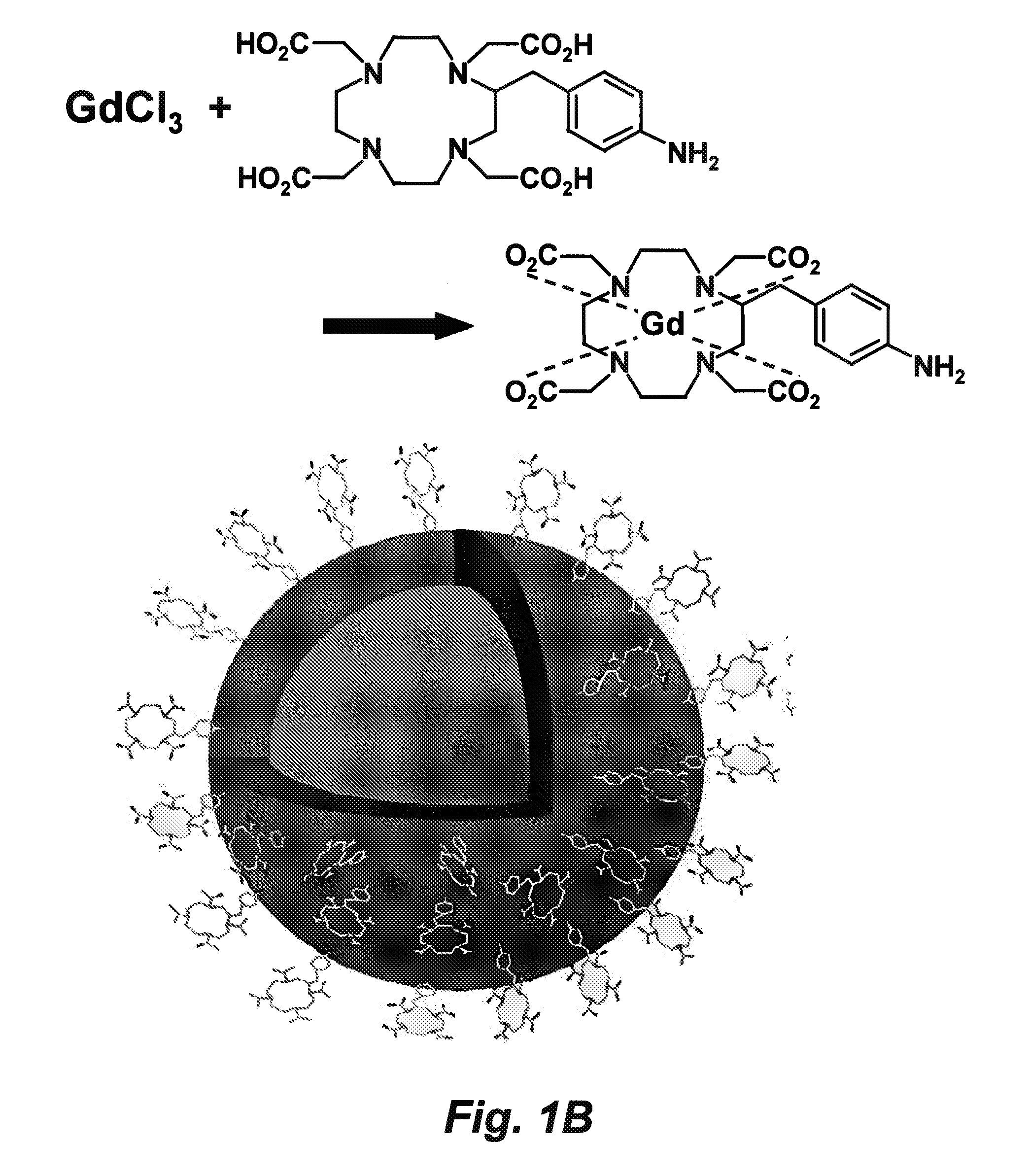
[0242]This example describes a robust and general method for embedding nanoparticles, such as quantum dots (QD) or colloidal gold (Au) nanocrystals, into a highly water-soluble thin silica shell doped with paramagnetic gadolinium (Gd3+) ions without negatively impacting the optical properties of the QD or Au nanoparticle cores. The ultrathin silica shell has been covalently linked to Gd3+ ions chelator, etraazacyclododecanetetraacetic acid (DOTA). The resulting complex has a diameter of 8 to 15 nm and is soluble in high ionic strength buffers at pH values ranging from approximately 4 to 11. For this system, nanoparticle concentrations exceed 50 μM, while most other nanoparticles might aggregate. In magnetic resonance imaging (MRI) experiments at clinical magnetic field strengths of 1.4 T (1H resonance frequency of 60 MHz), the gadolinium-DOTA (Gd-DOTA) attached to SiO2-coated QDs has a spin-lattice (T1) particl...

example 2

Multimodal Probes

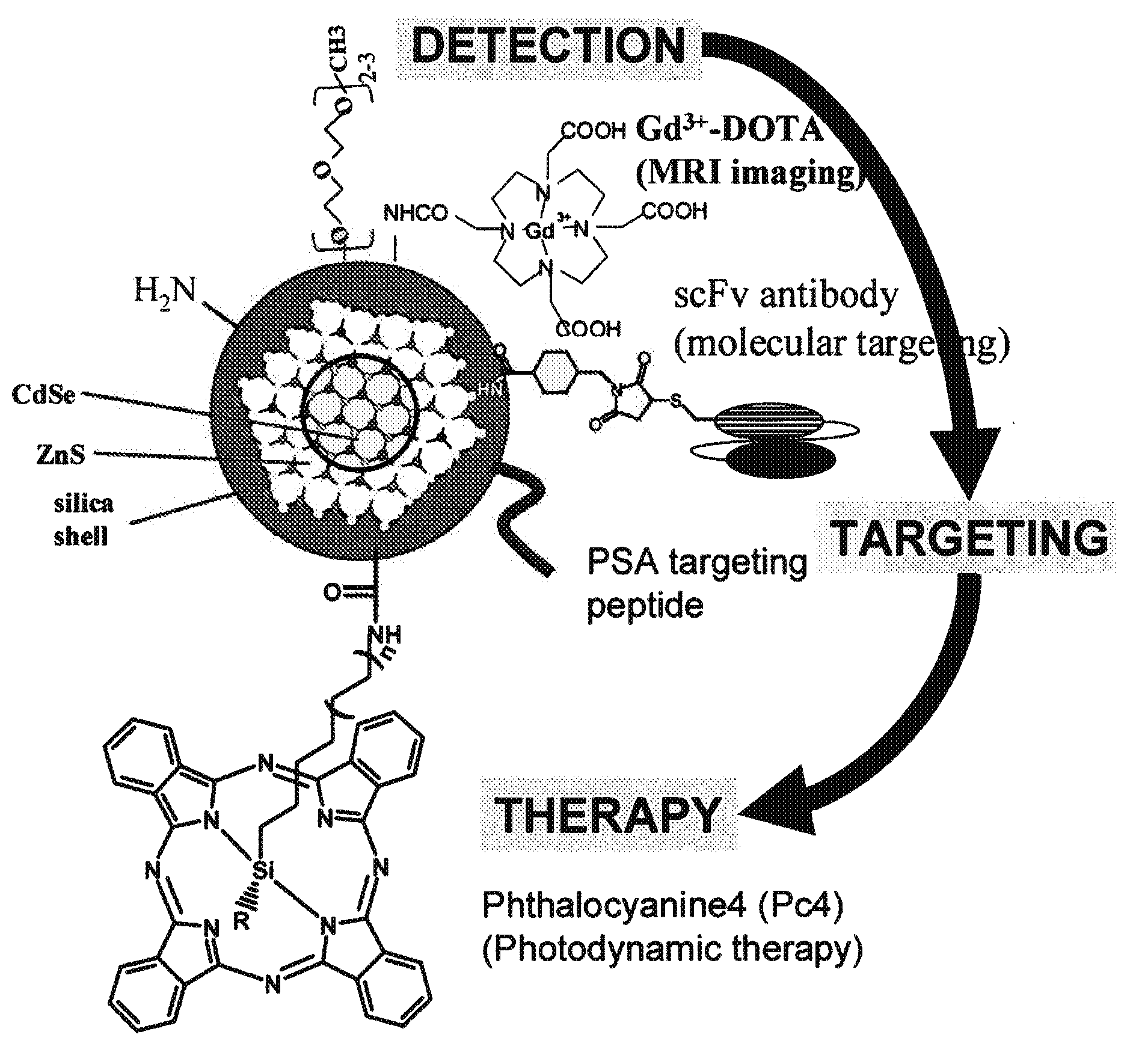
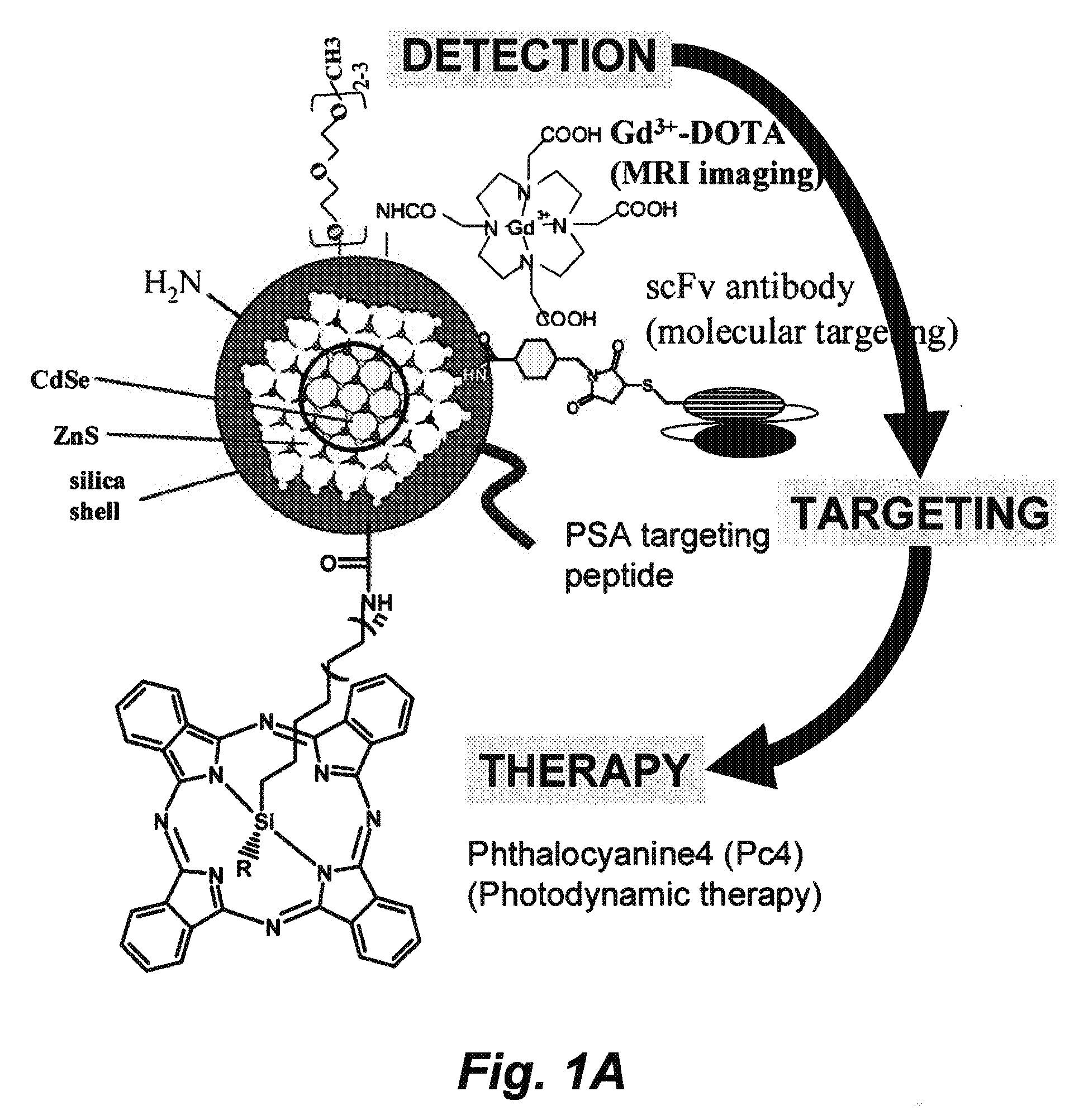
[0294]A nanoparticle is constructed as illustrated in FIG. 1A. DOTA, anti-PSA / PSMA antibody, and Pc4 are conjugated to the amine group, the thiol group, and the carboxyl group on the Qdot, respectively. The resulting nanocomposite has modalities of MRI, PET, NIR imaging, antibody-based targeting, and photodynamics therapeutics. This nanoconstruct offers sensitive and molecular targeted imaging and imaging-guided intervention. Other modifications include, but are not limited to the conjugation of an enhancer to the photodynamic chemicals, scFv antibodies targeting other prostate cancer antigens identified in the SPORE, and specific peptides / inhibitors against prostate cancer surface antigens.

[0295]Construction of the Gd and 64Cu-charged DOTA-Qdot (amine group). We have constructed silica-coated Qdots with different surface groups, such as amine, thiol, and carboxyll groups, and can tune the wavelength of Qdot to the absorption window of Pc4. Using EDC-NHS chemistry, ...

example 3

Targeting Breast And Prostate Cancer In Vivo With Multimodal Probes

[0298]The multimodal nanoparticle probe described in Example 2 is used to target cancer in vivo. To target prostate tumors, it is possible to utilize two scFvs. One scFv stains primary and metastatic tissue and binds to MEMD (CD166). MEMD has recently been found to be overexpressed in up to 84% of prostate cancer. The second scFv can e A33 which binds an unknown prostate tumor antigen. A33 has exquisite specificity for metastatic prostate tissue

[0299]Although there is still some controversy about prostate cancer screening in asymptomatic men, several organizations have formally made recommendations for serum Prostate Specific Antigen (PSA) testing. PSA screening appears to reduce the number of prostate cancers detected at late stage, and the incidence of metastatic prostate cancer. However, because PSA-screening does not distinguish between benign and malignant prostatic disease, a large fraction of biopsies that are...


PUM

PropertyMeasurementUnit
diameteraaaaaaaaaa
diameteraaaaaaaaaa
diameteraaaaaaaaaa

Abstract

In certain embodiments this invention provides a nanoparticle-based technology platform for multimodal in vivo imaging and therapy. The nanoparticle-based probes detects diseased cells by MRI, PET or deep tissue Near Infrared (NIR) imaging, and are capable of detecting diseased cells with greater sensitivity than is possible with existing technologies. The probes also target molecules that localize to normal or diseased cells, and initiates apoptosis of diseased cells.

Description

CROSS-REFERENCE TO RELATED APPLICATIONS[0001]This application claims benefit of and priority to U.S. Ser. No. 60 / 944,055, filed on Jun. 14, 2007, which is incorporated herein by reference in its entirety for all purposes.STATEMENT AS TO RIGHTS TO INVENTIONS MADE UNDER FEDERALLY SPONSORED RESEARCH AND DEVELOPMENT[0002]This invention was made during work supported by U.S. Department of Energy under Contract No. DE-ACO2-05CH11231 and Contract No. W-7405-Eng-48. The government has certain rights in this invention.FIELD OF THE INVENTION[0003]This invention relates to using multimodal imaging probes for use in in vivo targeted and non-targeted imaging, specifically as magnetic resonance imaging (MRI), positron emission tomography (PET), and Near Infrared imaging agents, and for in vivo targeted delivery of therapeutics.BACKGROUND OF THE INVENTION[0004]Recent years have witnessed a renaissance in fluorescence imaging driven by remarkable advances in molecular biology, and in detection tech...

Claims


Application Information

Patent Timeline
Patent Type & Authority Applications(United States)
IPC IPC(8): A61K51/12A61K49/18C12N5/071A61P35/00
CPCA61K47/48861A61K47/48884A61K49/0002A61K49/0065A61K49/0067A61K51/1244A61K49/106A61K49/1881B82Y5/00G01N21/6428A61K41/0071A61K49/085A61K47/6923A61K47/6929A61P35/00
Inventor CHEN, FANQING FRANK
Owner RGT UNIV OF CALIFORNIA
Why Eureka
Try Eureka