Methods of evaluating a test agent in a diseased cell model
a cell model and test agent technology, applied in the field of disease models, can solve the problems of inability to translate animal models into human immunology, inability to accurately predict human outcomes, and inability to use animal models at all, so as to improve the level of interferon gamma (ifn)-inducible protein 10, and protect against glanders
- Summary
- Abstract
- Description
- Claims
- Application Information
AI Technical Summary
Benefits of technology
Problems solved by technology
Method used
Image
Examples
example 1
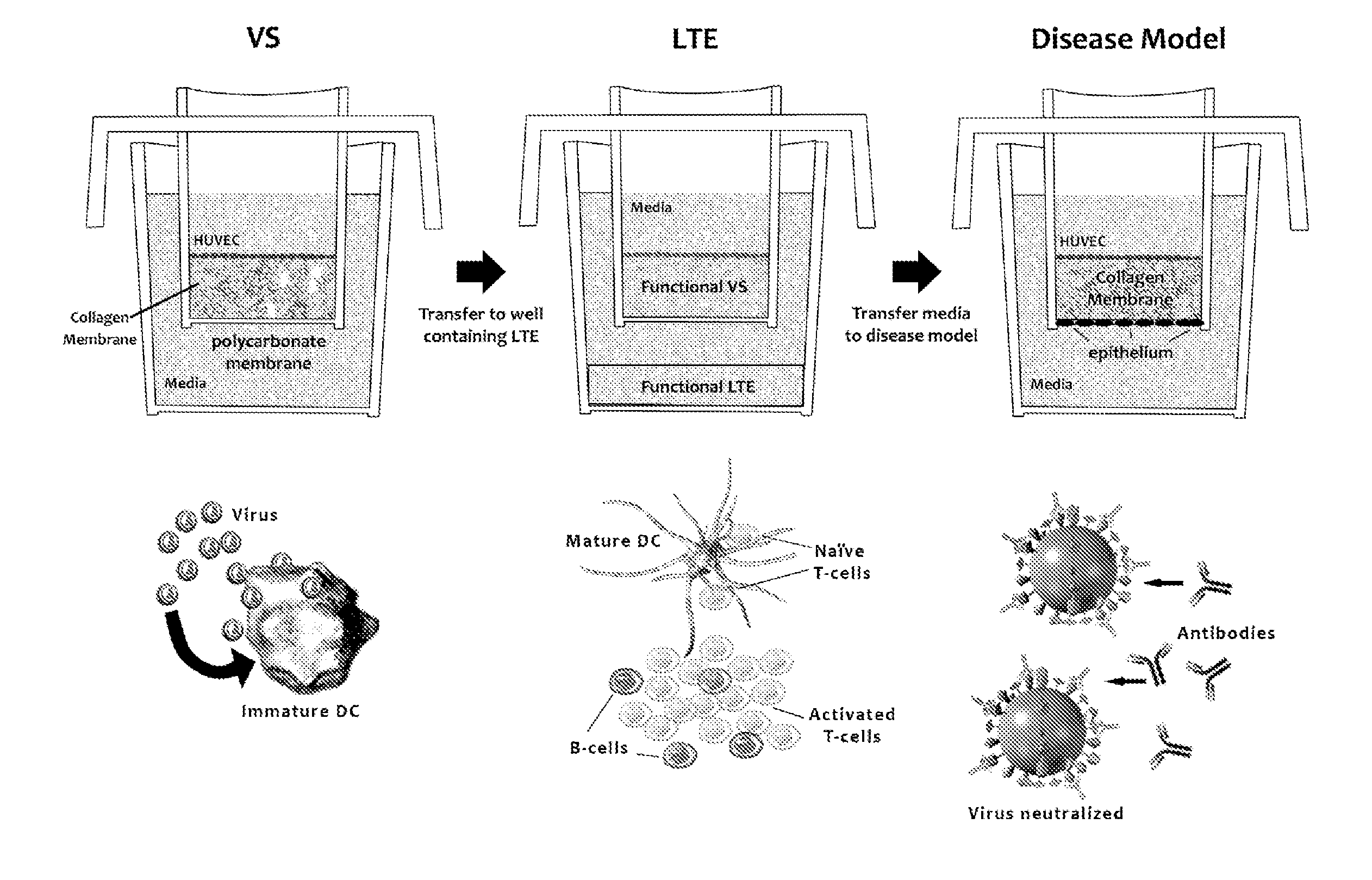
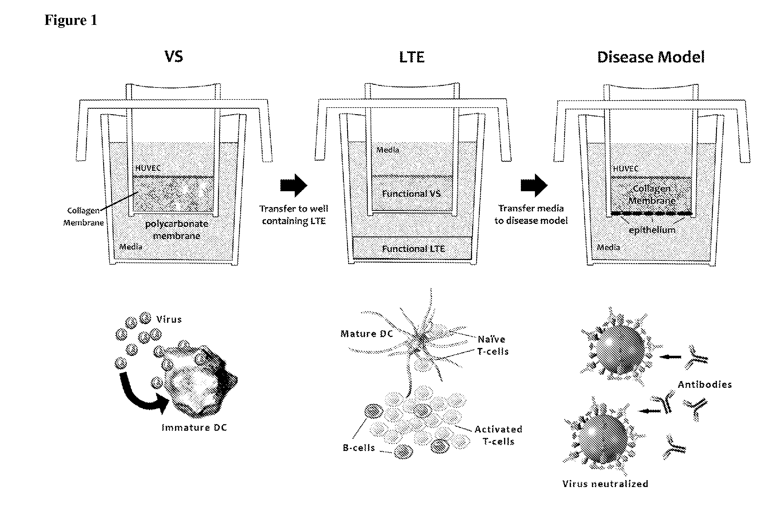
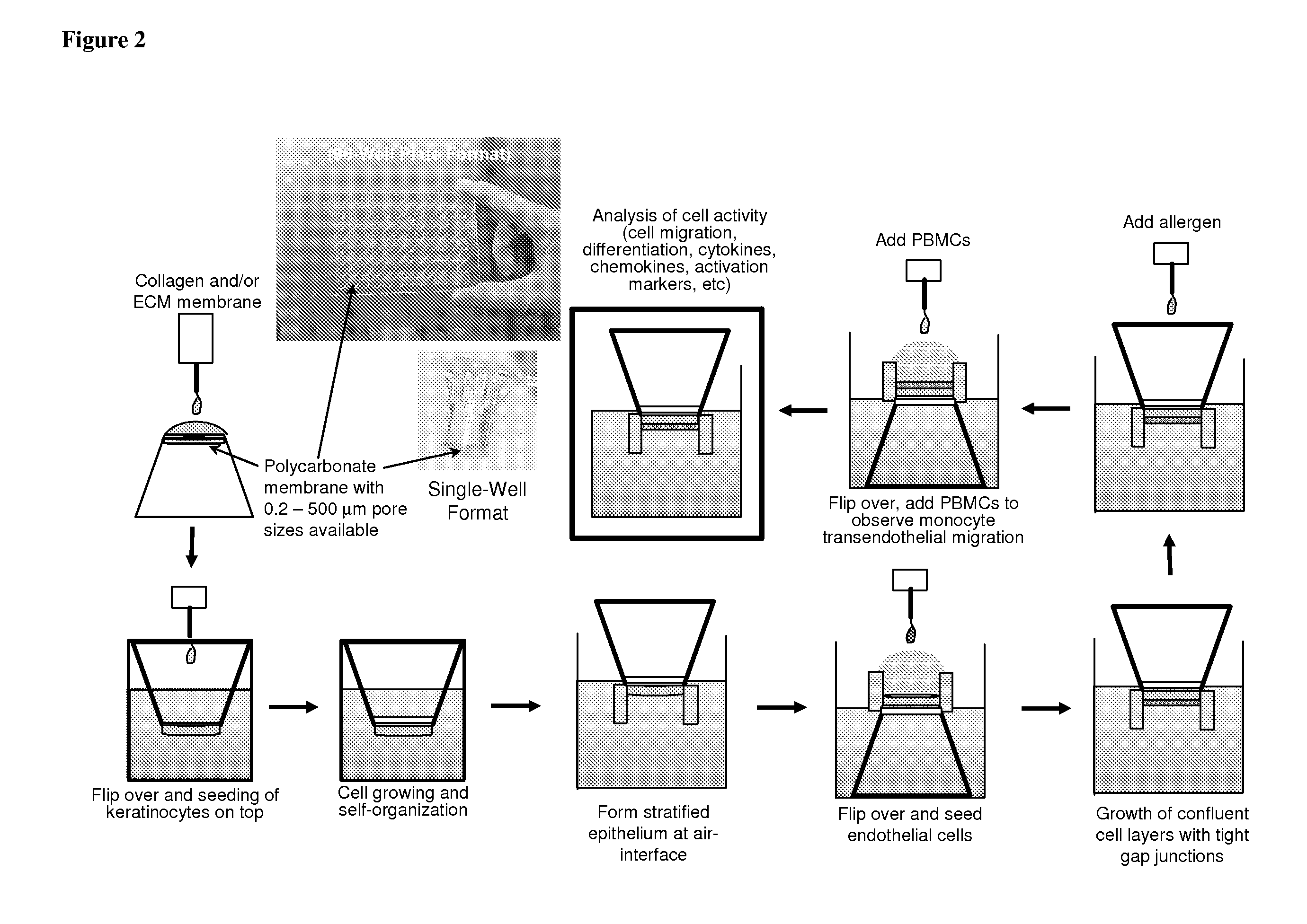
[0064]Generic tissue construct for a 3D in vitro disease model. FIG. 2 illustrates a 3D heterogeneous tissue construct, comprising the addition of cells on the top and bottom of the construct, to create endothelial and epithelial layers. This model is an improvement on our established 3D endothelial cell-only construct, which has been used for transendothelial migration and for monocyte to dendritic cell and macrophage differentiation (the vaccination site, VS).
[0065]The 3D model of this example can be used to study immunophysiological reactions when subjected to various diseases and vaccine formulations. This is a generic construct because most tissues involve a 3D extracellular matrix with associated endothelial and epithelial layers. The disease, whether viral, bacterial, or tumoral, is introduced into the generic tissue construct. The various immunocytes and biomolecules from the AIS (e.g., antibodies, T cells, cytokines, chemokines) can then be delivered to the disease model to...
example 2
[0066]Tumor modeling in the AIS using melanoma cells. Many in vitro model systems have been used for examining the effects of anti-cancer therapeutics and tumor growth in adult and childhood cancers, using both primary cells and various cell lines (see, e.g., Houghton et al. (2002) Clin Cancer Res 8, 3646-57). Such models have proven useful for assessing tumor metabolic states, inhibition of proliferation, and decreases in overall biomass (see, e.g., Monks et al., (1991) J Natl Cancer Inst 83, 757-66; Scherf et al., (2000) Nat Genet. 24, 236-44).
[0067]Animal models of human cancers have not been good predictors of human therapeutic outcome because of species differences (see, e.g., Houghton et al., (2002) Clin Cancer Res 8, 3646-57; Bridgeman et al., (2002) Cancer Res 60, 6573-6; Batova et al., (1999) Cancer Res 59, 1492-7).
[0068]As with any tumor model, the primary end goal is to increase patient survival and overall well being and to decrease tumor burden. The most predictive mode...
example 3
[0071]Heterogeneous tissue constructs with the addition of cells on the top and bottom of the tissue construct to form endothelial and epithelial layers. A schematic representation of the development of the generic disease model and how it can be tested with a particular disease is shown in FIG. 3. As an example, we used a polycarbonate membrane support structure to prepare a 3D ECM matrix, comprising either collagen, synthetic or natural materials (e.g., hydrogels, PLA, PLGA, gelatin, hyaluronic acid), or combinations thereof. We have established an ECM that is capable of supporting two cell layers. We first grow a layer of epithelial cells (e.g., human keratinocytes) on one side of the matrix. An advantage of this model is that other epithelial cells can be used, such as respiratory epithelial cells, skin epithelial cells, or intestinal / oral epithelial cells (as schematically illustrated in FIG. 3). The basement membrane zone between the epithelium and the matrix is important to t...
PUM
 Abstract
Description
Claims
Application Information
 - R&D
- Intellectual Property
- Life Sciences
- Materials
- Tech Scout
- Unparalleled Data Quality
- Higher Quality Content
- 60% Fewer Hallucinations
Browse by: Latest US Patents, China's latest patents, Technical Efficacy Thesaurus, Application Domain, Technology Topic, Popular Technical Reports.
© 2025 PatSnap. All rights reserved.Legal|Privacy policy|Modern Slavery Act Transparency Statement|Sitemap|About US| Contact US: help@patsnap.com


