

Use of particles comprising an alcohol
a technology of alcohol and particles, applied in the field of use of particles comprising alcohol, can solve the problems of limiting medical therapy, reducing the therapeutic-to-toxicity ratio, unstable particles, and less suitable for drug delivery
- Summary
- Abstract
- Description
- Claims
- Application Information
AI Technical Summary
Benefits of technology
Problems solved by technology
Method used
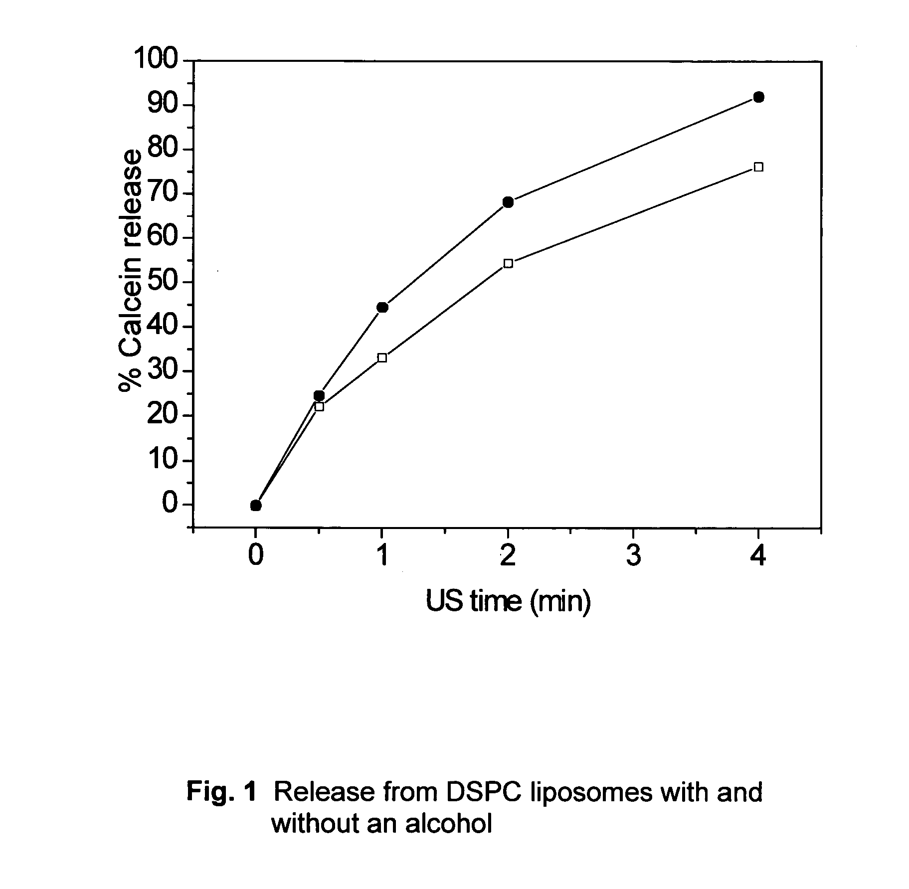
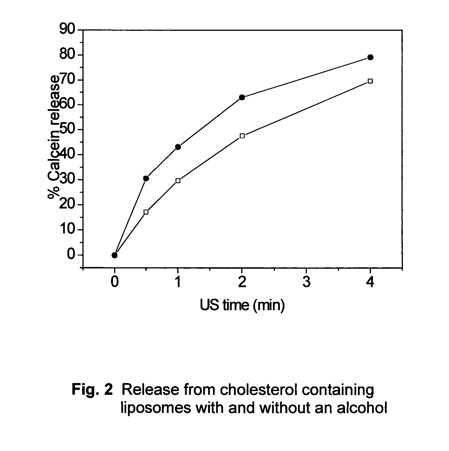
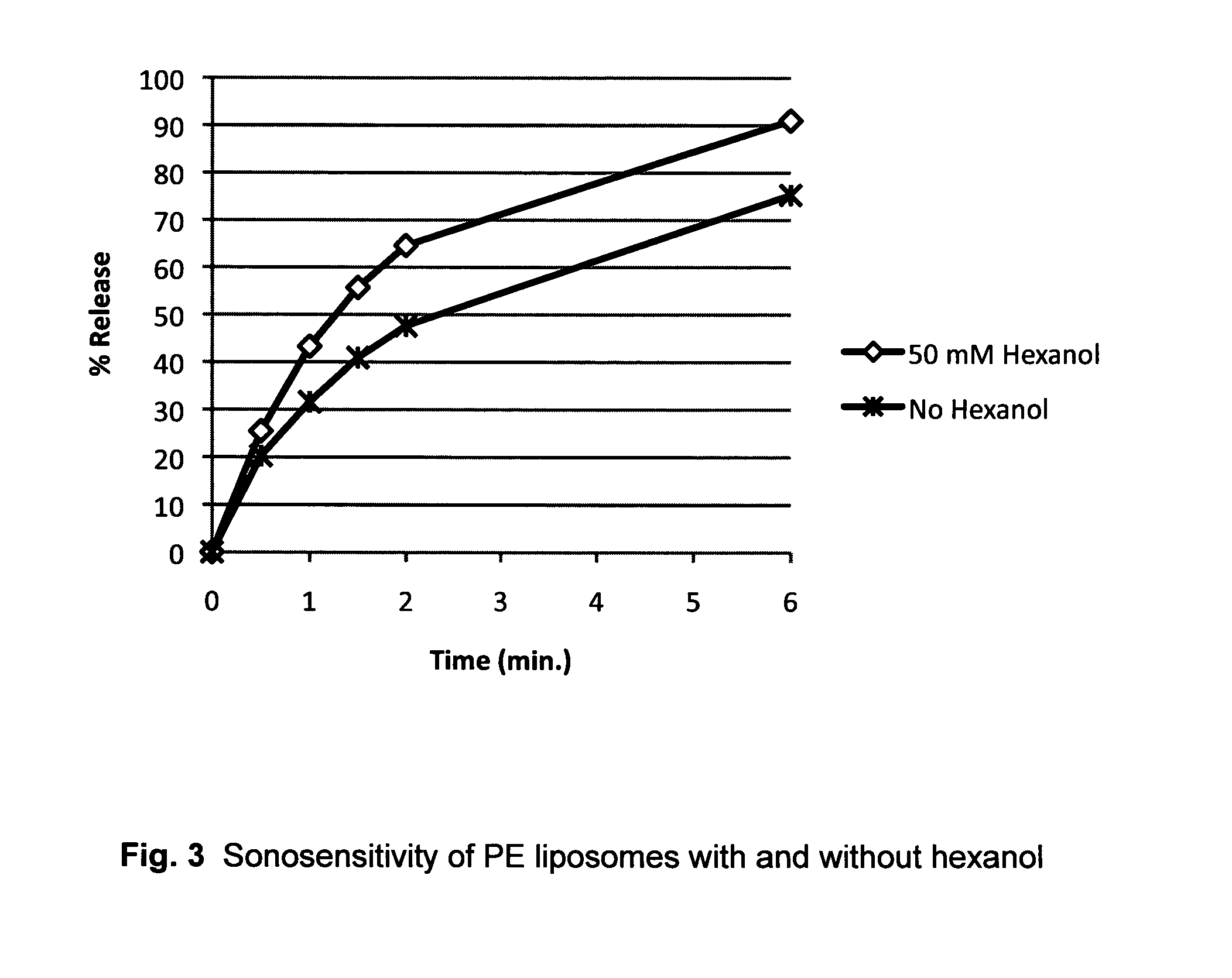
Image

Examples
example 1
Preparation of Liposomes
[0056]DSPC and DSPE-PEG 2000 were purchased from Genzyme Pharmaceuticals (Liestal, Switzerland). Cholesterol, calcein, HEPES, sodium azide and sucrose were obtained from Sigma Aldrich. Hexanol was supplied by BDH Chemicals Ltd. (Poole, England).
[0057]Calcein carrying liposomes (liposomal calcein) of different membrane composition were prepared using the thin film hydration method (Lasic 1993). The nominal lipid concentration was 16 mg / ml. Liposomes were loaded with calcein via passive loading, the method being well known within the art. The hydration liquid consisted of 10 mM HEPES (pH 7.4) and 50 mM calcein. In liposomes containing hexanol, the hydration liquid was supplemented with a given amount of hexanol 2 days prior to usage in the lipid film hydration step.
[0058]The size of the liposomes were made 80-90 nm by extrusion (Lipex, Biomembrane Inc. Canada) at 65° C. (PC liposomes) through polycarbonate (Nuclepore) filters of consecutive smaller size.
[0059]E...
example 2
Characterisation of Liposomes
[0060]Liposomes were characterised with respect to key physicochemical properties like particle size, pH and osmolality by use of well-established methodology.
[0061]The average particle size (intensity weighted) and size distribution were determined by photon correlation spectroscopy (PCS) at a scattering angle of 173° and 25 deg C. (Nanosizer, Malvern Instruments, Malvern, UK). The width of the size distribution is defined by the polydispersity index. Prior to sample measurements the instruments was tested by running a latex standard (60 nm). For the PCS measurements, 10 μL of liposome dispersion was diluted with 2 mL sterile filtered isosmotic sucrose solution containing 10 mM HEPES (pH 7.4) and 0.02% (w / v) sodium azide. Duplicates were analysed.
[0062]Osmolality was determined on non-diluted liposome dispersions by freezing point depression analysis (Fiske 210 Osmometer, Advanced Instruments, MA, US). Prior to sample measurements, a reference sample wi...
example 3
US Mediated Release Methodology
[0064]Liposome samples were exposed to 20 kHz ultrasound up to 4 min in a custom built sample chamber as disclosed in Huang and MacDonald (Huang and Macdonald 2004). The US power supply and converter system was a ‘Vibra-Cell’ ultrasonic processor, VC 750, 20 kHz unit with a 6.35 cm diameter transducer, purchased from Sonics and Materials, Inc. (USA). Pressure measurements were conducted with a Bruel and Kjaer hydrophone type 8103.
[0065]The system was run at the lowest possible amplitude at 20% of maximum amplitude. This translates to a transducer input power of 0.9-1.2 W / cm2 and a peak-to-peak transducer pressure of about 460 kPa.
[0066]For the US measurements, liposome dispersions were diluted in a 1:500 volume ratio, with isosmotic sucrose solution containing 10 mM HEPES (pH 7.4) and 0.02% (w / v) sodium azide. Duplicates were analysed.
[0067]The release assessment of calcein is based on the following well-established methodology: Intact liposomes contai...
PUM
| Property | Measurement | Unit |
|---|---|---|
| size | aaaaa | aaaaa |
| size | aaaaa | aaaaa |
| molecular weight | aaaaa | aaaaa |
Abstract
Description
Claims
Application Information