Biologically active sutures for regenerative medicine

Active Publication Date: 2011-01-27
WISCONSIN ALUMNI RES FOUND
View PDF19 Cites 23 Cited by
  • Summary
  • Abstract
  • Description
  • Claims
  • Application Information

AI Technical Summary

Benefits of technology

[0013]In yet another embodiment, the present disclosure is directed to a method for preparing a biologically active material for use in tissue regeneration. The method comprises: functionalizing at least one surface of a bioresorbable material to expose carboxylate anions on the surface; contacting the functionalized s

Problems solved by technology

Bone-tendon healing presents a particularly challenging problem that must be addressed in myriad orthopedic applications, including cruciate ligament reconstruction, rotator cuff repair, patellar tendon repair, and avulsion injury repair.
Although widely successful in enhancing knee stability, the process of cruciate ligament reconstruction is plagued by significant limitations.
This tunnel widening is indicative of bone resorption instead of the desired tendon-bone healing, and it creates significant reconstructive challenges in the 5-10% of cases that require revision surgery.
A second limitation in ACL reconstruction is the extensive amount of time required for full patient recovery, which is typically a 6 month timeframe.
Taken together, these limitations cause a significant increase in patient morbidity and loss of physical activity, and these issues are typical in other clinical scenarios that require bone-tendon healing.
However, although these approaches have been useful in a wide variety of biomedical applications, their application to functional bone and tendon healing is pragmatically limited.
Plastic microspheres do not represent a stand alone device for tissue ingrowth and are difficult to process into structural orthopedic devices while retaining protein biological activity.
Hydrogels are also non-ideal carriers, as growth factors typically transport out of the hy

Method used


Image

  • Biologically active sutures for regenerative medicine
  • Biologically active sutures for regenerative medicine
  • Biologically active sutures for regenerative medicine

Examples


example 1

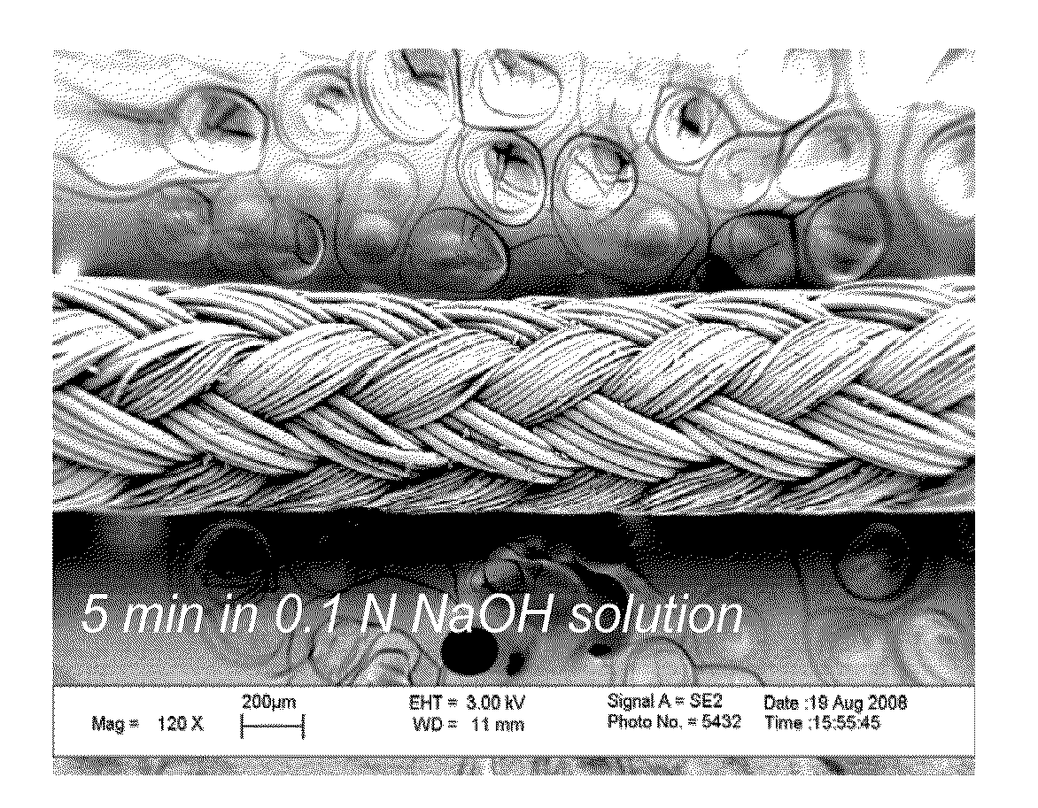
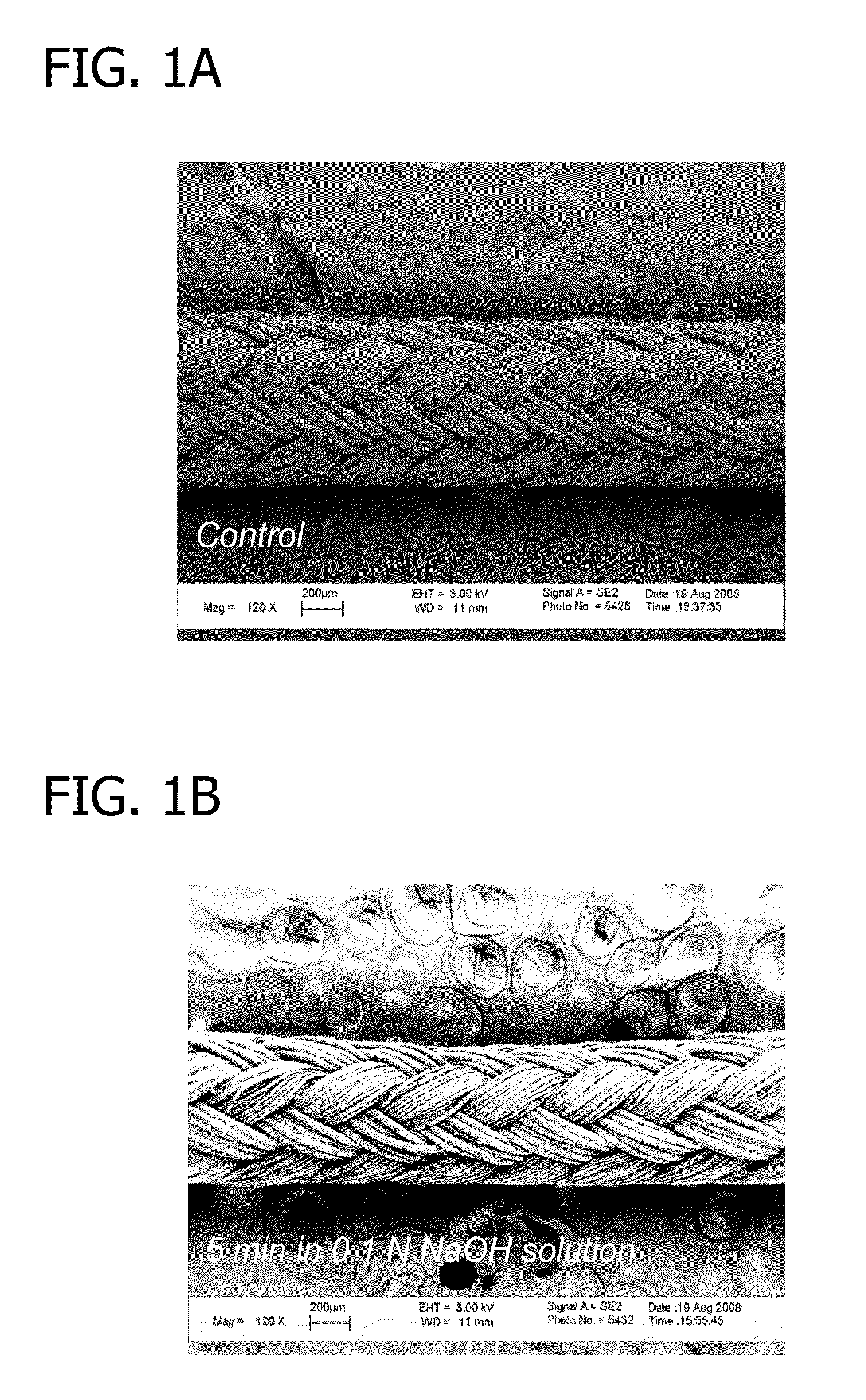
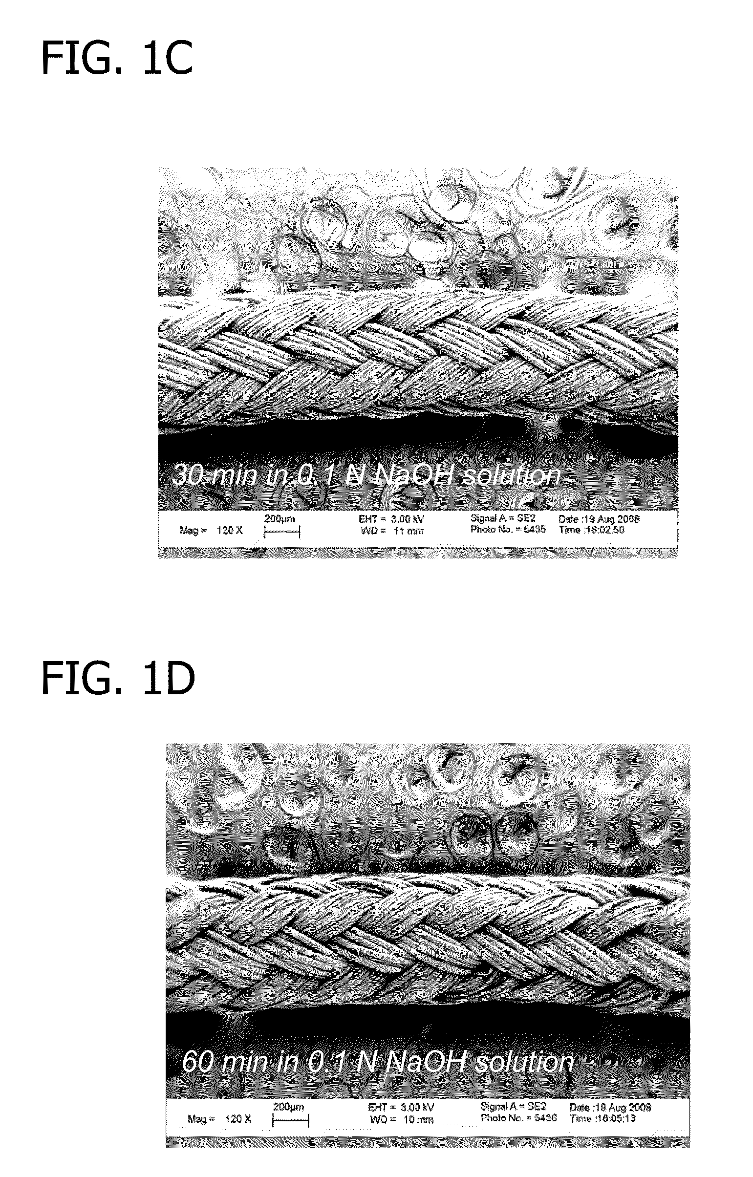
[0092]In this example, a hydroxyapatite mineral layer was applied to the surface of sutures.

[0093]To begin, ORTHOCORD™ (USP No. 2; DePuy Mitek, Raynham, Mass.) sutures were treated with 0.1 N NaOH solution for 5 minutes, 30 minutes, or 1 hour. The resulting sutures were hydrolyzed and contained carboxylic acid groups on the suture surface. FIG. 1 depicts a 120× magnification (using LEO 1530 FE-SEM, Zeiss, Germany) of an untreated ORTHOCORD™ suture (FIG. 1A), an ORTHOCORD™ suture treated with 0.1 N NaOH solution for 5 minutes (FIG. 1B), an ORTHOCORD™ suture treated with 0.1 N NaOH solution for 30 minutes (FIG. 1C), and an ORTHOCORD™ suture treated with 0.1 N NaOH solution for 60 minutes (FIG. 1D). As can be seen from FIGS. 1A-1D, the hydrolysis procedure caused no appreciable morphological or mass change in the suture.

[0094]The hydrolyzed, carboxylic acid-rich sutures were then incubated in modified simulated body fluid (mSBF; 141 mM NaCl, 4 mM KCl, 0.5 mM MgSO4, 1.0 mM MgCL2, 4.2 mM...

example 2

[0096]In this example, the hydroxyapatite mineral layer applied to the sutures in Example 1 was evaluated.

[0097]The morphology and composition of the mineralized surface of sutures, prepared as described in Example 1 (60 minute hydrolysis), were characterized using LEO 1530 field-emission scanning electron microscopy (FE-SEM; Zeiss, Germany) equipped with energy dispersive spectroscopy (EDS), respectively.

[0098]Scanning electron microscopy results are shown in FIGS. 3A-3C. FIG. 3A shows a SEM micrograph of uncoated ORTHOCORD™ suture (scale bar: 500 μm), and FIGS. 3B and 3C show SEM micrographs of a mineralized ORTHOCORD™ suture, prepared using the 7-day mineralization procedure described in Example 1 (FIG. 3B scale bar: 500 μm; FIG. 3C scale bar: 5 μm). As can be seen from FIGS. 3B and 3C, the mineral phase uniformly covers the entire surface of the suture (FIG. 3B) and has plate-like microstructure with nanoscale porosity (FIG. 3C) similar to human bone material.

[0099]Energy disper...

example 3

[0104]In this example, the ability of a hydroxyapatite-coated suture to incorporate protein was evaluated.

[0105]To begin, a mineralized ORTHOCORD™ suture, prepared as described in Example 1 (−7 mm, 60 minute hydrolysis), was incubated for 4 hours at 37° C. in a 150 μL solution of either lysozyme (14,300 Da, isoelectric point (pI) 10.7, hydrodynamic radius (RH) 1.9 nm, charge 8.5) or cytochrome c (11700 Da, pI 10.2, RH 1.8 nm, charge 9.5) in PBS (pH 7.4) to allow for incorporation of the protein molecules into the mineralized layer. The concentration of lysozyme or cytochrome c in the protein solutions varied from 100-1000 μg / mL. Specifically, protein concentrations of 100 μg / mL, 200 μg / mL, 400 μg / mL, 600 μg / mL, 800 μg / mL, and 1000 μg / mL were used.

[0106]Fourier transform infrared (FT-IR) spectrometry (Equinox 55 spectrometer, Bruker AXS, Germany) was carried out on the mineralized suture incubated in the lysozyme solution. The results are shown in FIG. 4A. Specifically, FIG. 4A plots...


PUM

PropertyMeasurementUnit
Ratioaaaaaaaaaa
Biological propertiesaaaaaaaaaa
Biodegradabilityaaaaaaaaaa

Abstract

The present disclosure generally relates to biodegradable and bioresorbable materials having a mineral layer on the surface of the material. More particularly, the disclosure relates to biodegradable and bioresorbable orthopedic devices having a degradable mineral layer on the surface thereof that can be used as a delivery vehicle for biological substances. Also provided are various methods of using the mineralized devices in tissue regeneration, including bone tissue engineering, and methods for producing the mineralized devices.

Description

BACKGROUND OF THE DISCLOSURE[0001]The present disclosure generally relates to biodegradable and bioresorbable materials having a mineral layer on the surface of the material. More particularly, the disclosure relates to biodegradable and bioresorbable orthopedic devices having a degradable mineral layer on the surface thereof that can be used as a delivery vehicle for biological substances. Also provided are various methods of using the mineralized devices in tissue regeneration, including bone tissue engineering, and methods for producing the mineralized devices.[0002]The field of orthopedic tissue engineering has developed rapidly in response to the expanding need for skeletal tissue replacements to treat injury, disease, and birth defects. Costs of musculoskeletal conditions represent an average of 3% of the gross domestic product of developed countries, an estimated $254 billion annually in the United States, and bone and joint diseases account for half of all chronic conditions...

Claims


Application Information

Patent Timeline
IPC IPC(8): A61B17/04
CPCA61B17/06166A61L17/145A61B2017/00893A61L27/32A61L27/54A61L27/58A61L29/106A61L29/148A61L29/16A61L31/086A61L31/148A61L31/16A61L17/005A61L17/06A61B2017/00889
Inventor MURPHY, WILLIAM L.LEE, JAE SUNGMARKEL, MARK D.GRAF, BEN K.BAER, GEOFFREYLU, YAN
Owner WISCONSIN ALUMNI RES FOUND
Why Eureka
Try Eureka