Bone Xenograft

a bone xenograft and bone technology, applied in the field of bone xenograft, can solve the problems of inability to transplantable autologous bone tissue for large defects, poor bone id filling effect, and high postoperative morbidity

Inactive Publication Date: 2011-05-05
APERION BIOLOGICS
View PDF11 Cites 2 Cited by
  • Summary
  • Abstract
  • Description
  • Claims
  • Application Information

AI Technical Summary

Problems solved by technology

Id Transplantable autologous bone tissue for large defects, particularly in children, is often unavailable, however.
In addition, autologous bone transplantation may result in postoperative morbidity such as pain, hemorrhage, wound problems, cosmetic disability, infection or nerve damage at the donor site.
Further, difficulties in fabricating the desired functional shape from the transplanted autologous bone tissue may result in less than optimal filling of the bone defect Id.
In particular, bone xenografts may result in increased rates of fracture, resorption and nonunion secondary to immunologic rejection.

Method used


Image

  • Bone Xenograft
  • Bone Xenograft
  • Bone Xenograft

Examples


example 1

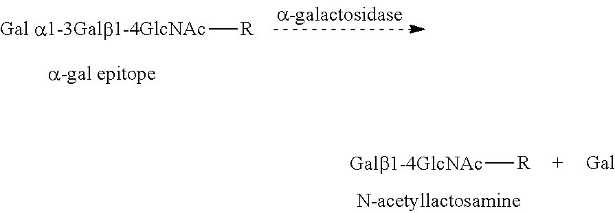
Assay for α-Gal Epitopes' Elimination from Bone by α-Galactosidase

[0063]In this example, an ELISA assay for assessing the elimination of α-gal epitopes from bone is conducted.

[0064]A monoclonal anti-Gal antibody (designated M86) which is highly specific for α-gal epitopes on glycoproteins is produced by fusion of splenocytes from anti-Gal producing knock-out mice for α 1,3 galactosyltransferase, and a mouse hybridoma fusion partner.

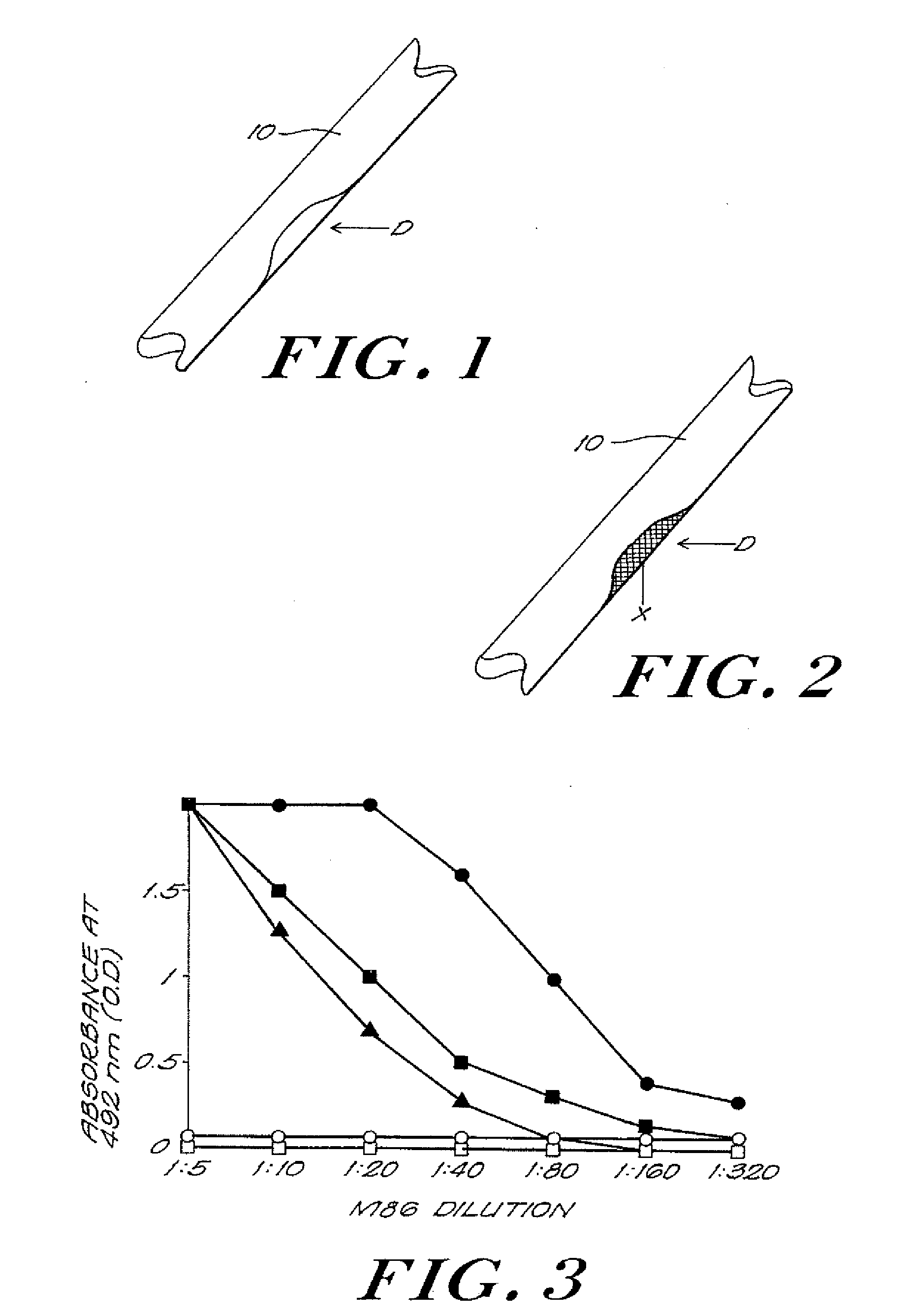
[0065]The specificity of M86 for α-gal epitopes on glycoproteins is illustrated in FIG. 3. M86 binds to synthetic α-gal epitopes linked to -bovine serum albumin (BSA), to ▴-bovine thyroglobulin which has 11 α-gal epitopes, R. G. Spiro et al., Occurrence of α-D-galactosyl residues in the thyroglobulin from several species. Localization in the saccharide chains of complex carbohydrates, 259 J. Biol. Chem. 9858 (1984); or to ▪-mouse laminin which has 50 α-gal epitopes, R. G. Arumugham et al., Structure of the asparagine-linked sugar chains of laminin. 883 B...

example 2

Assessment of Primate Response to Implanted Bone Treated with α-Galactosidase

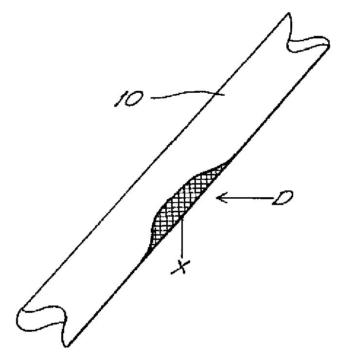
[0067]In this example, porcine bone implants are treated with α-galactosidase to eliminate α-galactosyl epitopes, the implants are transplanted into cynomolgus monkeys, and the primate response to the implants is assessed. An exemplary bone portion 10 with a defect D is shown in FIG. 1.

[0068]Porcine bone specimens are sterilely prepared and the surrounding attached soft tissues surgically removed. The bone specimens are washed for at least five minutes with an alcohol, such as ethanol or isopropanol, to remove synovial fluid and lipid soluble contaminants.

[0069]The bone specimens are frozen at a temperature ranging from about −35° C. to about −90° C., and preferably at a temperature up to about −70° C., to disrupt, that, is to kill, the specimens' bone cells.

[0070]Each bone specimen is cut into two portions. The first bone portion is immersed in a buffer, such as citrate buffer solution, with a pH ranging f...

example 3

Assessment of Primate Response to Implanted Bone Treated with α-Galactosidase, Sialic Acid-Cytosine Monophosphate and Sialyltransferase

[0080]In this example, porcine bone implants are treated with α-galactosidase to eliminate α-gal epitopes, as described in Example 2. The implants are further treated with sialic acid-cytosine monophosphate (SA-CMP) and sialyltransferase to cap carbohydrate chains with sialic acid. Sialytransferase facilitates the transfer of the sialic acid from the SA-CMP compound to the xenograft. The sialic acid links to and thus caps the carbohydrate chains. The cytosine monophosphate provides the necessary energetic level to the sialic acid for such linking and capping. Capping with sialic acid interferes with the ability of the subject's immune system to recognize the xenograft as foreign. The negative charge of the sialic acid further interferes with the ability of the bone antigens to bind with non-anti-Gal anti-bone antibodies (i.e., antibodies binding to b...


PUM

PropertyMeasurementUnit
temperatureaaaaaaaaaa
sizeaaaaaaaaaa
concentrationsaaaaaaaaaa

Abstract

The invention provides an article of manufacture comprising a substantially non-immunogenic bone xenograft (X) for the implantation into a defect (D) located in a bone portion (10) of a human. The invention further provides methods for preparing a bone xenograft (X) by removing at least a portion of bone from a non-human animal to provide a xenograft (X); washing the xenograft (X) in saline and alcohol; and subjecting the xenograft (X) to at least one of the treatments including exposure to ultraviolet radiation, immersion in alcohol, ozonic, and freeze / thaw cycling. In addition to or in lieu of the above treatments, the methods include a cellular disruption treatment, and digestion of the carbohydrate moieties of the xenograft (X) with a glycosidase followed by treatment for sialylation.

Description

FIELD OF THE INVENTION[0001]The present invention relates to the field of bone transplantation, and in particular, to replacement and repair of damaged or defective human bone using a substantially immunologically compatible bone from a non-human animal.BACKGROUND OF THE INVENTION[0002]Human bone, a hard connective tissue consisting of cells embedded in an extracellular matrix of mineralized ground substance and collagen fibers, (Stedman's Medical Dictionary, Williams & Wilkins, Baltimore, Md. (1995)), is the most frequently transplanted tissue in humans. J. M. Lane et al., Current Approaches to Experimental Bone Grafting, 18 Orthopedic Clinics of North America (2) 213 (1987). In the United States alone more than 100,000 bone graft or implant procedures are performed every year to repair or to replace osseous defects resulting from trauma, infection, congenital malformation, or malignancy. Id.[0003]Bone grafts and implants are often formed of autologous bone. Id Transplantable autol...

Claims


Application Information

Patent Timeline
Patent Type & Authority Applications(United States)
IPC IPC(8): A61K9/00C12N5/077C12N13/00A61K35/12A61P19/00A61F2/28A61F2/30A61L27/36
CPCA61F2/28A61F2/3094A61F2002/2835A61L27/3608A61L27/3847A61L27/365A61L27/3687A61L27/3691A61L27/3633A61P19/00
Inventor STONE, KEVIN R.GALILI, URI
Owner APERION BIOLOGICS
Why Eureka
Try Eureka