Ruthenium purple biosensor

Active Publication Date: 2011-05-05
UNIVERSITY OF WARWICK
View PDF0 Cites 10 Cited by
  • Summary
  • Abstract
  • Description
  • Claims
  • Application Information

AI Technical Summary

Problems solved by technology

Although these are important signaling agents in the nervous system, the polymer films on these sensors were insufficiently robust to survive implantation into neural tissue (Poitry et al., 1997) and the sensors had mixed success at detecting release from biological tissue (Poitry et al., 1997; Cosnier et al., 1997).
Adenosine, in particular, does not conform to the conventional paradigm of chemical neurotransmission in the nervous system.
HPLC analysis of collected superfusate has been used to study adenosine release, however this method has very limited time and spatial resolution (Pedata et al., 1993).
However it is too large to implant into nervous tissue without causing considerable damage and subsequent tissue reaction that may confound and invalidate physiological measurements.
Furthermore the large size of the sensing assembly (500 μm when used in its finally constructed format) introduces diffusional delays which slows sensor responsiveness.
A baby with more than 5 μM of hypoxanthine per litre of blood is at a severe risk of foetal hypoxia.
At present, blood samples are taken to a laboratory for analysis, which causes a delay in obtaining the results.
However, the use of PB as an electron acceptor provides problems, most notably due to its intolerance to the presence of Na+ ions in physiological fluids.
Na+ ions are prominent in physiological fluids such as blood, which clearly renders PB less suitable as a biosensor material.
Other smaller cations in a buffer solution, such as Na+

Method used


Image

  • Ruthenium purple biosensor
  • Ruthenium purple biosensor
  • Ruthenium purple biosensor

Examples


Embodiment Construction

[0168]Apparatus

[0169]A CH1 660B potentiostat was used to electrochemically deposit the different polymers and test the biosensor. The biosensor was used in vivo with a potentiostat interfaced to a PC by an A to D converter board (Data Translation). In all cases an Ag / AgCl was used as reference electrode and a Pt counter electrode. The electrochemical cell for deposition consisted of a capillary of 1.5 mm diameter and 2 cm length.

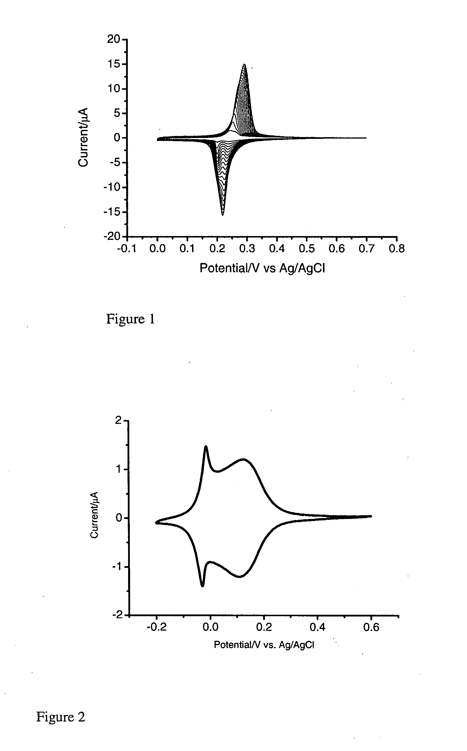
[0170]Electrodeposition of RP on the Surface of a Gold Electrode:

[0171]In a three electrode system, a pre-treated working gold electrode is dipped into a mixture (FeCl3 (1 mM)+KCl (40 mM, pH 2) and K4Ru(CN)6 (1 mM)+KCl (40 mM, pH 2), followed by electrochemical cycling from −0.2 to +0.7 V (vs. Ag / AgCl) for forty cycles at 50 mV / s (FIG. 1). The resulting RP modified electrode is heated at 80° C. overnight. In order to stabilise the RP membrane, electrochemical cycling is performed in a solution of RuCl3.

[0172]Polyaniline, where used, was formed using 10 μm an...


PUM

PropertyMeasurementUnit
Temperatureaaaaaaaaaa
Electrical conductivityaaaaaaaaaa

Abstract

The invention relates to a biosensor comprising an electrically conductive substrate, with a first layer comprising Ruthenium Purple formed on the substrate, a second layer comprising polyaniline or a derivative thereof comprising one or more non-polar substituents formed on the first layer, and a third layer comprising one or more enzymes trapped within a matrix formed on the second layer. The biosensor is for use in the detection of analytes such as purines and derivatives thereof, particularly in the detection of hypoxanthine.

Description

[0001]The present invention relates to a biosensor, which can be used for the detection of chemicals, such as purines and derivatives thereof, for example hypoxanthine, adenosine, ATP and / or inosine. This application claims priority from GB 0700157.1, filed 4 Jan. 2007, which is incorporated herein in its entirety.BACKGROUND OF THE INVENTION[0002]Biosensors contain an electrode with a biorecognition molecule such as enzymes, antibodies, organelles and whole cells to allow the specific identification of analytes such as metabolytes, ions, gases and organic vapours. Such biorecognition elements are typically attached to a transducer, such as an electrochemical, piezoelectric, optoelectronic, fibreoptic, thermistor, diode or surface acoustic device, to enable the binding of the analyte to the biorecognition element to be detected. Amperometric biosensors, for example, are reviewed in the article by Palmisano F., et al. (Fresenius J. Anal. Chem. (2000), Vol. 366, pages 586-601). A wide ...

Claims


Application Information

Patent Timeline
IPC IPC(8): A61B5/00G01N27/327C23C28/00
CPCC12Q1/004
Inventor DALE, NICHOLASTIAN, FAMING
Owner UNIVERSITY OF WARWICK
Why Eureka
Try Eureka