
Chondrocyte-like cell, and method for producing same

a technology of chondrocytes and cells, applied in the field of chondrocytelike cells, can solve the problems of reducing presenting a definitive therapy, and a big challenge in the aging society of osteoarthritis, so as to reduce the burden on patients, and improve the quality of li

Inactive Publication Date: 2011-10-13
IPS ACAD JAPAN
View PDF2 Cites 3 Cited by
  • Summary
  • Abstract
  • Description
  • Claims
  • Application Information

AI Technical Summary

Benefits of technology

[0067]The present invention provides chondrocyte-like cells that have a proliferative ability and the properties of the chondrocytes, and thus can provide a medical means effective for the treatment of cartilage disease that involve cartilage damage such as in osteochondrosis deformans. Further, the present invention enables production of chondrocyte-like cells from the somatic cells of patients suffering from osteoarthritis and a wide range of other cartilage diseases including a growth cartilage disease such as chondrodystrophy, and can thus contribute to elucidating the pathology of the disease by performing various analyses. The chondrocyte-like cells produced from humans are particularly suitable as material for the discovery and development of drugs.
[0068]Further, because the present invention can be used to obtain chondrocyte-like cells from skin tissue-derived somatic cells such as skin fibroblasts and subcutaneous adipose tissue-derived stromal cells, the invention is highly useful in the clinic from the standpoint of reducing burdens on patients and cell donors.

Problems solved by technology

The symptoms of osteoarthritis include joint pain during joint movement (movement pain) and a restricted range of motion (restricted motion), which lower the quality of daily life.
Osteoarthritis thus poses a big challenge in the aging society.
These methods, however, are only supportive, and do not represent a definitive therapy, because the chondrocytes have only weak repairing capabilities (see Non-Patent Literature 1), and cannot regenerate cartilage tissue.
However, artificial joints have a number of drawbacks, including a heavy burden put on patients during the procedure, deterioration due to wear, a tendency to dislocate, and possible revision surgery necessitated by a loosened artificial joint.
However, because chondrocytes are limited in number and cause dedifferentiation through monolayer expansion (see Non-Patent Literature 4), recent studies focus more on the development of a technique that induces formation of cartilage tissue with the use of bone marrow-derived mesenchymal stem (MS) cells or embryonic stem (ES) cells (see Non-Patent Literatures 5 to 7).
However, MS cells have only limited proliferative capabilities, and recent studies suggest that the cartilage produced from MS cells is unstable, and lacks sufficient cartilage properties (see Non-Patent Literatures 8 and 9).
However, because of the pluripotency of iPS cells, use of iPS cells for cartilage tissue regeneration requires the establishment of a technique that enables the cells to differentiate into a homogeneous chondrocyte population, and there are still technical problems that need to be solved for practical applications in cartilage tissue regeneration.

Method used


Image

  • Chondrocyte-like cell, and method for producing same
  • Chondrocyte-like cell, and method for producing same
  • Chondrocyte-like cell, and method for producing same

Examples


example 1

Production of Chondrocyte-Like Cells from Dermal Fibroblasts and Embryonic Fibroblasts

[0118]1. Production of Col11a2-β Geo Transgenic Mice

Methods

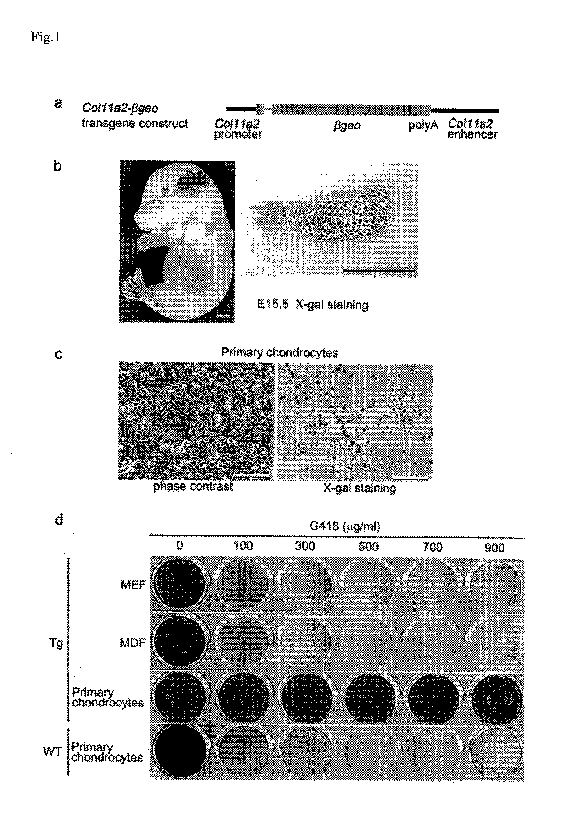
[0119]First, transgenic mice were produced that express β-geo (fused gene of β-galactosidase gene and neomycin resistant gene) under the control of Col11a2 promoter / enhancer sequences shown in FIG. 1, a, using the following procedure.

[0120]742LacZInt, an α2 (XI) collagen gene-based expression vector, includes a mouse Col11a2 promoter (−742 to +380), SV40 RNA splicing sites, a β-galactosidase reporter gene, an SV40 polyadenylation signal, and a 2.34-kb first intron sequence of Col11a2 as a enhancer (Reference Literature 1). In order to produce a β-geo introducing gene, a 0.8-kb neomycin resistant gene fragment was ligated to the 3′-end of a 3.1-kb cDNA fragment that codes for LacZ. The β-geo fragment was incorporated in a 742LacZInt expression vector at the Not I site by replacing the LacZ gene, and a Col11a2-β geo plasmid was produced.

[0121...

example 2

Formation of Cartilage Tissue from Chondrocyte-Like Cells

Methods

[0186]Eleven chondrocyte-like cells (MK-5 to MK-15) were obtained by transfecting the MDFs with the c-Myc, Klf4, and Sox9 genes using the method of Example 1. Two of these chondrocyte-like cell lines (MK-7 and MK-10) were digested with trypsin / EDTA, and suspended in a DMEM medium containing 10 volume % FBS to prepare a cell suspension (1×10⁷ cells / ml). 0.1 ml of the cell suspension was subcutaneously injected to the back of a nude mouse (female, 6 weeks of age, BALE / cA Jc1-nu / nu). The injection site was removed in week 16 post-injection in the MK-7 cell-injected mouse, and in week 8 post-injection in the MK-10 cell-injected mouse. The removed sites were fixed with 4% paraformaldehyde, and embedded in paraffin. Then, tissue slices were produced, and stained with safranine O, fast green, and iron haematoxylin.

Results

[0187]The results are presented in FIG. 9. The subcutaneous adipose tissue of the MK-7 cell- and MK-10 cell...

example 3

Analysis of Genomic DNA of Chondrocyte-Like Cells

Results

[0189]Experiments were conducted to evaluate the identity of the chondrocyte-like cells (MK-1, MK-3, and MK-4) obtained in Example 1, and of the chondrocyte-like cells (MK-5, MK-7, MK-10, and MK-15) obtained in Example 2, as follows.

[0190]First, genomic DNA was obtained from the chondrocyte-like cells using an ordinary method, and fragmented by digestion with EcoRI and BamHI. The genomic DNA fragments were developed by electrophoresis on agarose gel, transferred to a nylon membrane, and subjected to southern hybridization using Klf4 cDNA probes.

[0191]The results are presented in FIG. 10. As can be seen in FIG. 10, the chondrocyte-like cells obtained in Examples 1 and 2 showed different band patterns for different cell lines, each independently representing an established cell line.


PUM

PropertyMeasurementUnit
Therapeuticaaaaaaaaaa

Abstract

Disclosed is a cell which enables the reproduction of a cartilage tissue and has a proliferative ability. Also disclosed is a technique for providing a cell supply source which can be used in a definitive treatment of osteochondrosis deformans. A chondrocyte-like cell which has the same properties as those of a chondrocyte and can proliferate can be produced by selecting a combination of an Myc family gene and / or a Klf family gene and a SOX9 gene and introducing the combination into a somatic cell. The chondrocyte-like cell can be used for a medical purpose of cartilage regeneration.

Description

TECHNICAL FIELD[0001]The present invention relates to chondrocyte-like cells that are induced from somatic cells and have proliferative capabilities and the properties of the chondrocytes, and to processes for producing the chondrocyte-like cells. The invention also concerns cell preparations for cartilage tissue regeneration, implants, implant producing processes, cartilage disease therapeutic methods, and drug efficacy determining methods for determining the efficacy of a tested substance for cartilage disease, all using the chondrocyte-like cells. The invention also relates to chondrocyte-like cell preparation compositions used to induce somatic cells to the chondrocyte-like cells.BACKGROUND ART[0002]Articular cartilage has a role as a joint lubricant for absorbing impact at the diarthrodial joints during articular movement. The mechanical functions of the cartilage are imparted by the cartilage extracellular matrix constructed from type II and type XI collagens, and collagenous ...

Claims


Application Information

Patent Timeline
IPC IPC(8): A61K35/32C12N5/10A61K49/00A01K67/00A61P19/04C12N15/85A61K35/12
CPCA61K31/713A61K35/12A61L27/3817A61L27/3852C12N15/85C12N2501/60C12N2510/00A61K35/32A61K49/0004C12N5/0655A61P19/02A61P19/04
Inventor TSUMAKI, NORIYUKI
Owner IPS ACAD JAPAN
Learn More