Neuregulin induced proliferation of cardiomyocytes

a technology of cardiomyocytes and neutrophils, applied in the direction of skeletal/connective tissue cells, peptide/protein ingredients, drug compositions, etc., can solve the problems of inadequate human myocardial regeneration, affecting the survival rate of patients with myocardial infarction, so as to reduce slow down the onset of cardiac damage, and increase the cell cycle activity

Inactive Publication Date: 2012-05-17
CHILDRENS MEDICAL CENT CORP
View PDF15 Cites 5 Cited by
  • Summary
  • Abstract
  • Description
  • Claims
  • Application Information

AI Technical Summary

Benefits of technology

[0007]The present invention provides methods and compositions for increasing proliferation, increasing cell cycle activity, and / or inducing division of post-mitotic mammalian differentiated cardiomyocytes. The invention can be used to slow, reduce, prevent or treat the onset of cardiac damage caused by, for example, myocardial ischemia, hypoxia, stroke, or myocardial infarction in vivo. The invention can also be used in a subject with chronic ischemic heart disease. In addition, the methods of the invention can be used in pharmaceutical compositions to enhance proliferation of differentiated cardiomyocytes in vitro and / or in vivo, or can be used ex vivo in tissue grafting.
[0009]Neuregulin stimulates mononuclear cardiomyocytes, present in the adult mammalian heart, to undergo the full mitotic cell cycle division. Without being limited to any particular mechanism of action, neuregulin is understood to activate ErbB4 located in the cardiomyocyte cell membrane. Neuregulin-induced cardiomyocyte proliferation results from activation of ErbB4 tyrosine kinase signaling pathways. After myocardial infarction, recombinant neuregulin induces cardiomyocyte cell cycle re-entry, improves cardiac remodeling and function, reduces fibrosis and infarct size, and increases angiogenesis. These results demonstrate that neuregulin and the pathways it regulates are new targets for innovative strategies to treat injured heart tissue.
[0011]The neuregulin composition can also be formulated into a pharmaceutical composition with a pharmaceutically acceptable carrier, diluent or medium for treating damaged heart tissue. The neuregulin composition of the invention can further comprise at least a fragment of the neuregulin composition of SEQ ID NO:1 or a sequence that is at least 80%, 85%, 90%, 91%, 92%, 93%, 94%, 95%, 96%, 97%, 98%, 99% identical to that of the SEQ ID NO:1 fragment. In another embodiment, the neuregulin composition comprises a polypeptide comprising the neuregulin fragment of SEQ ID NO:2 or a functional variant or a sequence that is at least 80%, 85%, 90%, 91%, 92%, 93%, 94%, 95%, 96%, 97%, 98%, 99% identical to that of SEQ ID NO:2. In other embodiments, the neuregulin composition can comprise a polypeptide comprising the neuregulin fragment of SEQ ID NO:3, SEQ ID NO:4, SEQ ID NO:5, or SEQ ID NO:6 or a functional variants thereof or a sequence that is at least 80%, 85%, 90%, 91%, 92%, 93%, 94%, 95%, 96%, 97%, 98%, 99% identical to that of SEQ ID NO:3, SEQ ID NO:4, SEQ ID NO:5, or SEQ ID NO:6. In yet another embodiment, the neuregulin composition can comprise at least an epidermal growth factor-like (EGF-like) domain of neuregulin and the neuregulin can activate ErbB4 or a fragment that is at least 80%, 85%, 90%, 91%, 92%, 93%, 94%, 95%, 96%, 97%, 98%, 99% identical to an epidermal growth factor-like domain of neuregulin and the fragment can activate ErbB4. The neuregulin composition can also induce or facilitate heterodimerization of ErbB4 and ErbB2 receptors or homodimerization of ErbB4 receptors.
[0012]In another embodiment, the neuregulin composition can be administered in an amount and regime effective to stimulate mitotic division. The administration regime can be a duration sufficient to induce cell cycle re-entry of the heart muscle cells. Data has shown that administration for at least 12 weeks can stimulate division by inducing the heart muscle cells to re-enter the cell cycle, increase DNA synthesis and induce cytokinesis.
[0016]In some embodiments, the subject in need of heart tissue repair has undergone myocardial ischemia, hypoxia, stroke, or myocardial infarction. The method of repairing heart tissue can also comprise replacing damaged heart tissue with proliferating cardiomyocytes, improving myocardial function in the subject and reducing myocardial hypertropy to repair the heart tissue.

Problems solved by technology

Cardiovascular diseases are a leading cause of death, resulting in almost 40% of deaths annually in the United States.
Inadequate human myocardial regeneration poses a significant public health problem.
Because the human heart is incapable of adequate muscle regeneration, survivors of a myocardial infarction typically develop heart failure, arrhythmias, thrombosis, and other complications.
It has been a significant challenge to develop effective treatments for cardiac repair because adult mammalian cardiomyocytes are highly differentiated cells and presumed to be essentially unable to proliferate.
Thus the loss of cardiomyocytes after damage caused by events such as myocardial infarction generally results in compensatory responses that are inadequate to restore function.
Unreplaced loss of cardiomyocytes leads to heart failure, a significant health problem worldwide.
Current therapies are also limited in their effectiveness.

Method used


Image

  • Neuregulin induced proliferation of cardiomyocytes
  • Neuregulin induced proliferation of cardiomyocytes
  • Neuregulin induced proliferation of cardiomyocytes

Examples


example 1

Materials and Methods

Cell Cycle Activity In Vitro

[0083]Experiments were approved by the Animal Care and Use Committee. Ventricular cardiomyocytes were isolated from male Wistar rats (12 week old, 300 g, Charles River Laboratories). We added NRG1 (EGF-like domain, amino acids 176-246 (SEQ ID NO:5); 100 ng / mL, R&D Systems), the peptide consisting of the four fasciclin 1 domains of human periostin (500 ng / mL; BioVendor) (SEQ ID NO:7), FGF1 (100 ng / mL; R&D Systems), HB-EGF (10 ng / mL; R&D Systems), or PDGF-BB (10 ng / mL; Peprotech). For detection of DNA synthesis, we added BrdU (30 μM) for the last three days. The c-ErbB2 / Neu blocking antibody was from Calbiochem.

Mouse Strains

[0084]ErbB4F / F mice were obtained from the NIH-sponsored Mutant Mouse Repository at University of California Davis and were originally produced by Dr. Kent Lloyd (Golub et al., 2004). The α-MHC-MerCreMer mice were obtained from the Jackson Laboratories and originally generated by Dr. Jeffrey Molkentin (Sohal et al., ...

example 2

NRG1 Stimulates Mononucleated Cardiomyocytes to Proliferate

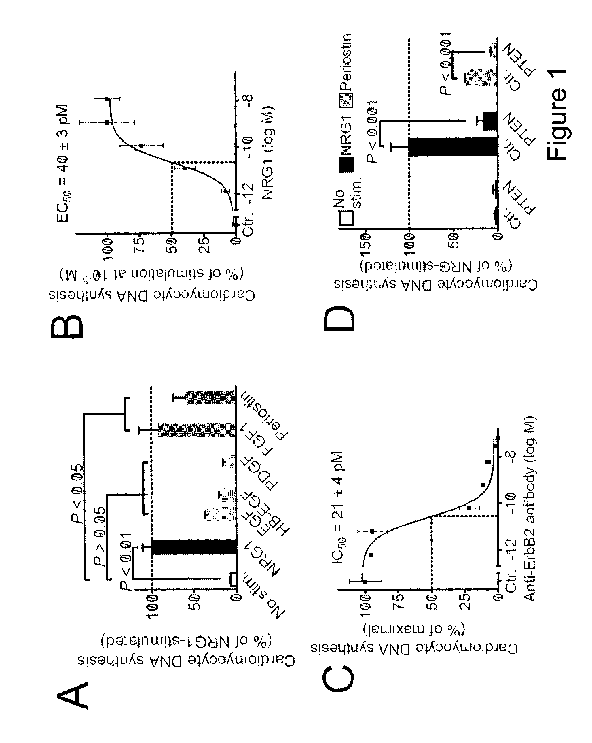
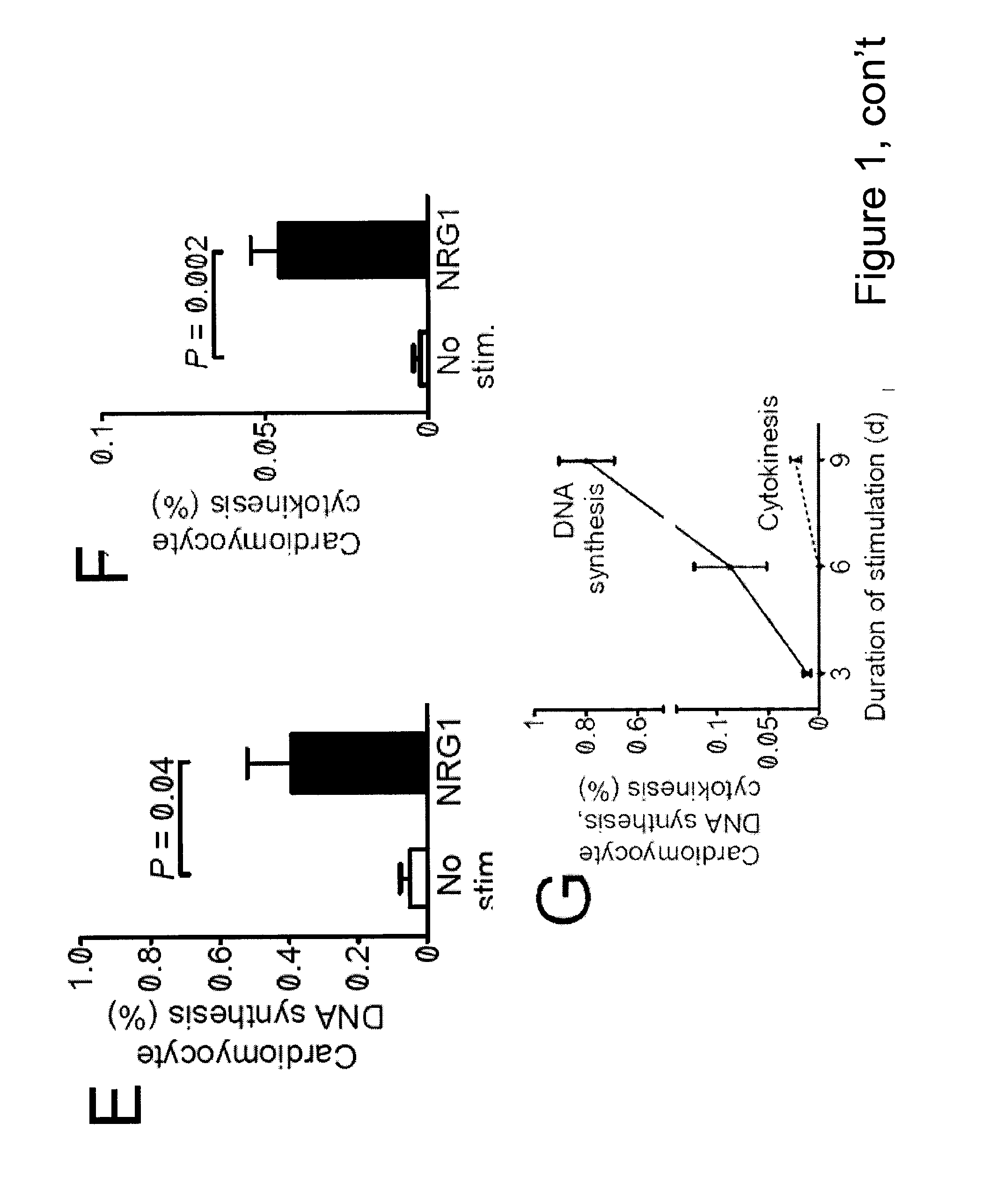
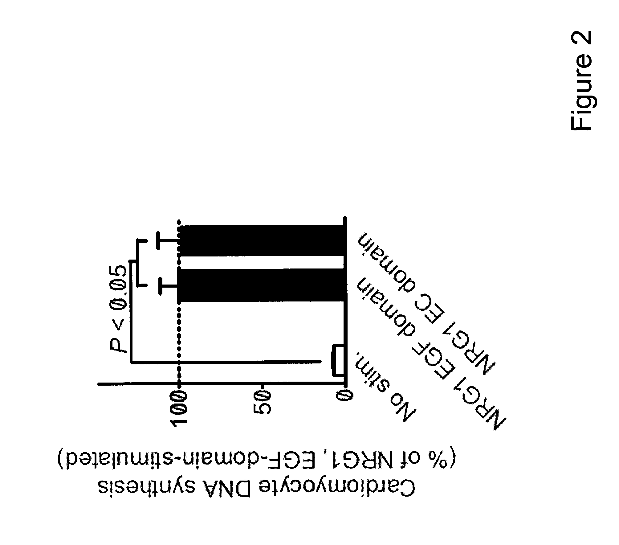
[0094]To identify factors that promote myocardial regeneration, we screened extracellular factors for their ability to induce DNA synthesis in primary adult rat ventricular cardiomyocytes. Three extracellular factors induced cardiomyocyte cell cycle reentry. Two have been previously identified: fibroblast growth factor-1 and periostin. The novel factor was the epidermal growth factor-like domain of NRG1β (FIG. 1A, FIG. 2). NRG1 induced concentration-dependent DNA synthesis, which best fit a sigmoidal function, suggesting a receptor-mediated process (FIG. 1B). The half-maximal stimulation EC50 was at 40±3 pM (n=3, FIG. 1B), indicating a high-affinity interaction with the receptor. In cardiomyocytes, NRG1 binds to ErbB4, which leads to formation and activation of ErbB2 / ErbB4 hetero- or ErbB4 / ErbB4 homodimers. To determine whether ErbB2 is required for cardiomyocyte cell cycle reentry, we added a fixed concentration of NRG1 (12...

example 3

ErbB4 Controls Postnatal Cardiomyocyte Proliferation In Vivo

[0099]To determine whether the NRG1 / ErbB2 / ErbB4 complex controls cardiomyocyte proliferation in postnatal hearts in vivo, we disrupted the complex by genetically inactivating the ErbB4 gene. We treated α-MHC-MerCreMer+ / +; ErbB4F / F mice (test) and α-MHC-MerCreMer+ / +; ErbB4Wt / F (control littermates) with tamoxifen. Following ErbB4 inactivation on postnatal days 2-4, we analyzed the effect on postnatal day 19. Cardiomyocyte differentiation was not affected, as demonstrated by two observations: the formation of bi- and multinucleated cardiomyocytes was not altered (FIG. 4A), and cardiomyocytes from test and control mice had indistinguishable morphology (FIG. 4B). To determine whether ErbB4 is required for postnatal cardiomyocyte cell cycling, we quantified cardiomyocytes that incorporated BrdU (FIG. 4B). After 5 injections of BrdU on postnatal days 16-18, test mice had no detectable cardiomyocyte BrdU uptake, while control mice...


PUM

PropertyMeasurementUnit
step sizeaaaaaaaaaa
thicknessaaaaaaaaaa
areaaaaaaaaaaa

Abstract

The present invention provides methods for inducing division of postmitotic mammalian differentiated cardiomyocytes. The invention can be used to repair heart tissue damaged by, for example, myocardial ischemia, hypoxia, stroke, myocardial infarction or chronic ischemic heart disease in vivo. In addition, the methods of the invention can be used to induce heart muscle cells to divide in vitro, in vivo and / or ex vivo, which can then be used in heart tissue repair.

Description

FIELD OF THE INVENTION[0001]The human heart is incapable of adequate regeneration or repair after injury. Thus the invention discloses methods for inducing division of post-mitotic cells for repairing heart tissue.BACKGROUND OF THE INVENTION[0002]Cardiovascular diseases are a leading cause of death, resulting in almost 40% of deaths annually in the United States. Inadequate human myocardial regeneration poses a significant public health problem. It is estimated that 13 million Americans have coronary artery disease, and more than half a million experience a myocardial infarction every year. Human cardiac tissue responds to injury, e.g. myocardial infarction, with scar formation. Because the human heart is incapable of adequate muscle regeneration, survivors of a myocardial infarction typically develop heart failure, arrhythmias, thrombosis, and other complications.[0003]Adult human hearts do not regenerate after injury; instead, the defect is replaced by fibrotic tissue. Most eviden...

Claims


Application Information

Patent Timeline
Patent Type & Authority Applications(United States)
IPC IPC(8): A61K35/34A61K38/22A61P9/00C12N5/077
CPCA61K38/1808A61K35/34A61P9/00
Inventor KUHN, BERNHARD
Owner CHILDRENS MEDICAL CENT CORP
Why Eureka
Try Eureka