Age related macular degeneration treatment

a macular degeneration and age-related technology, applied in the field of age-related macular degeneration (amd) diseases of the retina, can solve the problems of difficulty in adapting to low light, reduced color intensity, trouble recognizing, etc., and achieves the effects of reducing blood cholesterol, reducing edema, and reducing drusen formation

Inactive Publication Date: 2012-06-21
SHANTHA TOTADA R +2
View PDF7 Cites 13 Cited by
  • Summary
  • Abstract
  • Description
  • Claims
  • Application Information

AI Technical Summary

Benefits of technology

[0050]A method for treating age related macular degeneration (AMD) using an insulin preparation applied topically to the conjunctival sac of the affected eye. Another aspect of this invention is using antiangiogenic adjuvant therapeutic agents such as bevacizumab, ranibizumab, pegaptanib, etanercept, instilled in to the afflicted eye conjunctival sac with insulin to prevent further formation of new blood vessels, and shrink the existing pat

Problems solved by technology

This is the most common cause of blindness.
Troubles reading street signs, doing things at home or work because the lights seem dimmer, Trouble recognizing the faces of friends and family, trouble with close work such as reading, sewing or picking out matching clothes, Diminished color intensity, Difficulty adapting to low light, especially for sensitive vision tasks like reading.
Patients often first notice this when looking at mini-blinds in their home and trouble discerning colors; specifically dark ones from dark ones and light ones from light ones.
Slow recovery of visual function after exposure to bright light.
People with age related macular degeneration might find difficulty in doing simple everyday activities requiring sharp vision.
Above the age of 65, individuals lose at least 10% of their central vision resulting in the visual impairment related to the development of macular degeneration.
It is not possible, for example, to read without central vision.
Pictures that attempt to depict the central visual loss of macular degeneration with a black spot do not really do justice to the devastating nature of the visual loss.
If you have other genes and you smoke, you could be up to seven times more likely than non-smokers to get the disease.” The reason being, cataract removal creates a higher risk for AMD with the removal of the lens allows previously filtered light to pass unobstructed to the retina.
These supplements have not been shown to prevent AMD; however, these supplements slow the progression of the established disease.
This fluid can increase, build up pressure, and press on the RPE and retina, resulting in their detachment leading to defective vision and blindness (FIG. 7).
Ultimately, the fluid may be absorbed and drying which leads to scarring.
With no appropriate treatment, many of them become legally blind in both types of AMD.
This condition is the leading cause of loss vision in US above the age sixty years or older.
Treatment with additional supplemental vitamins and minerals may slow the pr

Method used


Image

  • Age related macular degeneration treatment
  • Age related macular degeneration treatment
  • Age related macular degeneration treatment

Examples


example 1

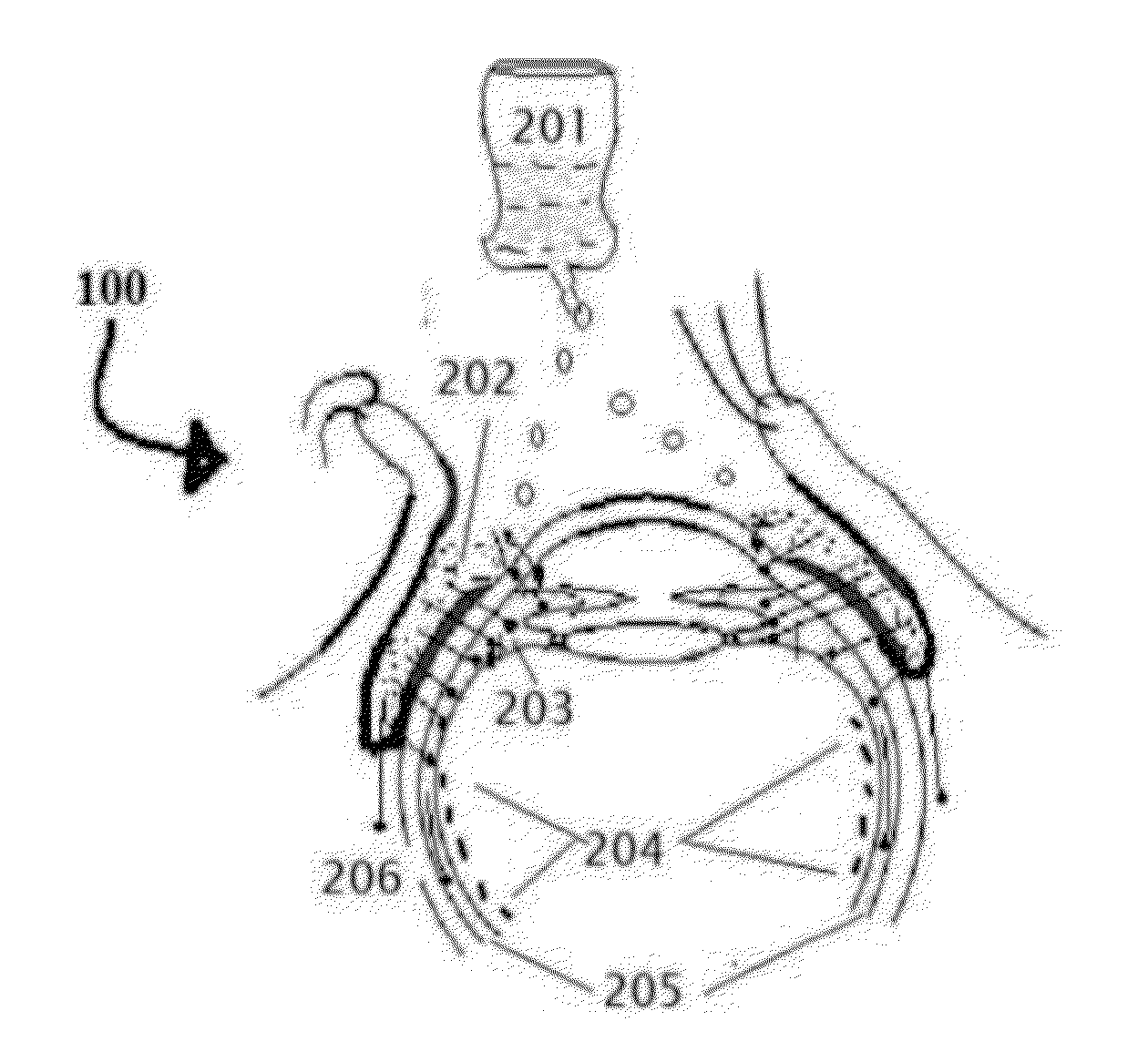
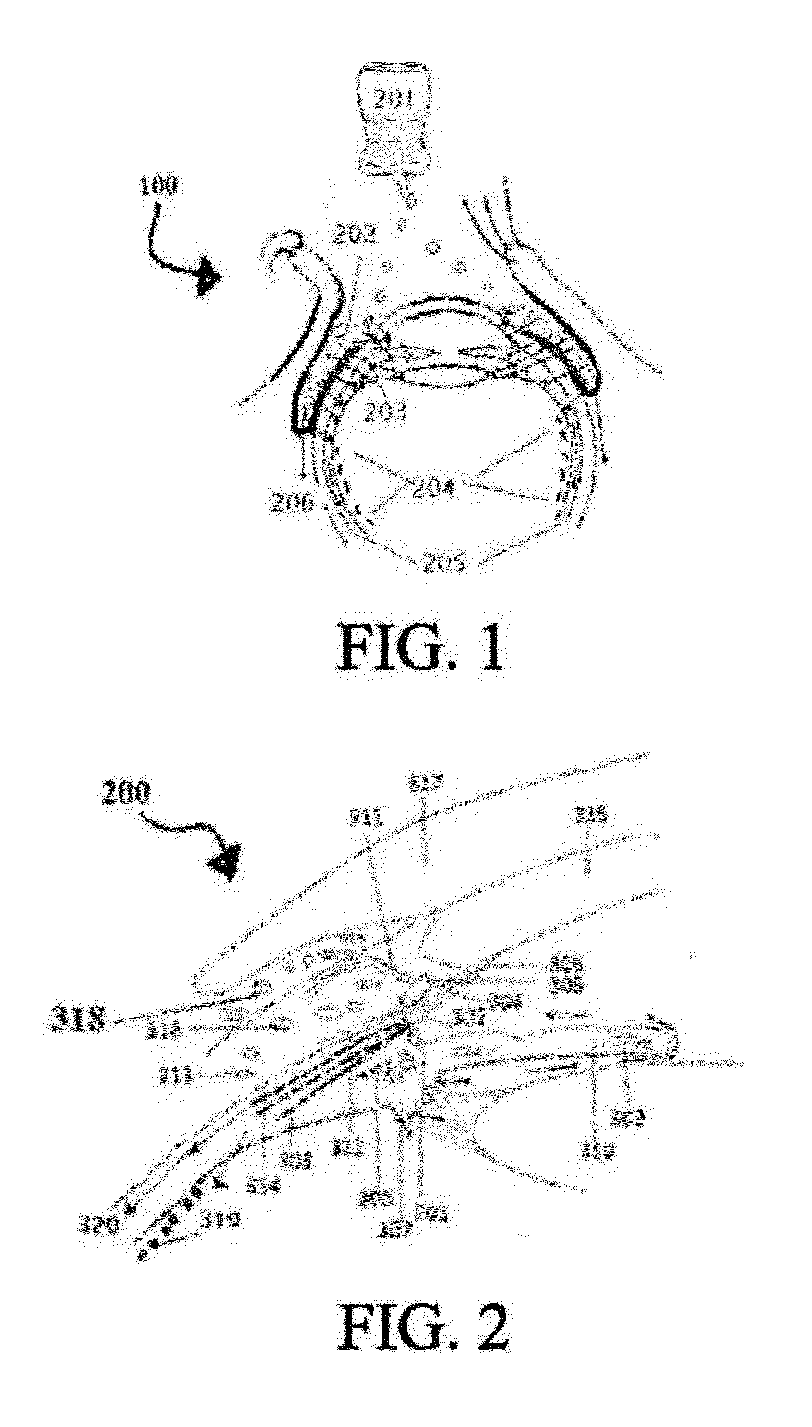
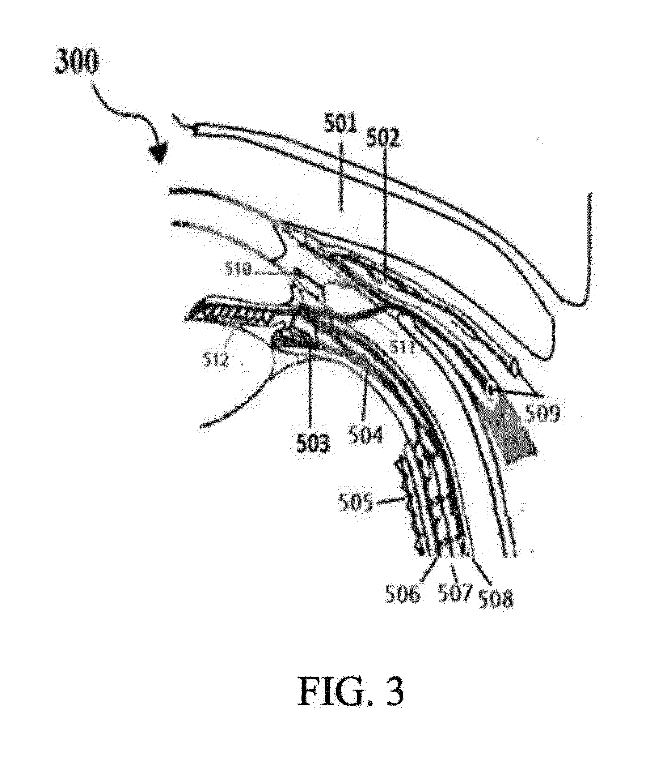
[0263]Select the patient; establish the type of Age related macular degeneration and its etiology, if possible, which the person is suffering from. The complete and thorough examination of the eye as described above is imperative. Record the preliminary examination results on the patient chart. The patient examined for any corneal, conjunctival, and retinal BV afflictions by using marker dyes and other ophthalmological examinations.[0264]I. Position the patient in a supine posture or sitting with the head hyper extended with a support.[0265]II. Prepare 0.05% povidone iodine in normal saline. Instill one or two drops to the conjunctional sac, wait 5 minutes for it to act on conjunctival lining and oxidize reduced glutathione to prevent it breaking the disulfide bonds of insulin.[0266]III. Using a dropper or dropper bottle containing the insulin formulations. Insulin is prepared in 5 ml normal saline insulin dropper or plastic squeeze instiller. Instill two or three drops of insulin p...

example 2

[0272]This is a 70-year-old patient diagnosed with wet AMD in right eye with CNV, associated with slight edema. The left eye had early symptoms of dry AMD, with still had good vision. It has had Drusen deposits in the macula, but no angiogenesis. The patient refused to undergo once every six-week intravitreal injection of anti-angiogenesis monoclonal antibody, Bevacizumab (trade name AVASTIN™, Genentech / Roche). AVASTIN (bevacizumab) is a recombinant humanized monoclonal IgG1 antibody that binds to and inhibits the biologic activity of human vascular endothelial growth factor (VEGF) in vitro and in vivo. It blocks angiogenesis, the growth of new blood vessels. This therapeutic agent used in doses of 8.3 to 10 mg in 0.3 ml solution injected in to the vitreous. It is not FDA approved for treating wet AMD, but many ophthalmologists use it off label. One of the advantages of these monoclonal antibodies is that it many times less expensive compared to another FDA approved monoclonal antib...

example 3

[0284]I. Follow the instruction as described in the above EXAMPLE 1.[0285]II. Instead of using Bevacizumab, use Ranibizumab monoclonal antibodies ophthalmic drops in similar way as described in example 2.


PUM

PropertyMeasurementUnit
Fractionaaaaaaaaaa
Fractionaaaaaaaaaa

Abstract

A method for treating age related macular degeneration (AMD) using an insulin preparation applied topically to the conjunctival sac of the affected eye. Another aspect of this invention is using antiangiogenic adjuvant therapeutic agents such as bevacizumab, ranibizumab, pegaptanib, etanercept, instilled in to the afflicted eye conjunctival sac with insulin to prevent further formation of new blood vessels, and shrink the existing pathologically formed blood vessels and reduce the edema in wet AMD. This method incorporates putting the patients on low fat diet, aerobic exercise, ketamine-a NMDA blocker, reducing the blood cholesterol using adjuvant therapeutic agents selected from Statins, that are inhibitors of 3-hydroxy-3-methylglutaryl coenzyme A, (i.e. HMG-Co A) reductase which in turn reduce drusen formation that leads to AMD, combined with insulin ophthalmic drops.

Description

CROSS REFERENCE TO RELATED APPLICATIONS[0001]This is a continuation in part of U.S. patent application Ser. No. 12 / 940,247, filed Nov. 5, 2010 the complete disclosure is hereby incorporated by reference.FIELD OF THE INVENTION[0002]This invention relates to the treatment of age related macular degenerative (AMD) diseases of the retina affecting the vision in humans or the animals.BACKGROUND OF THE INVENTION[0003]Age related macular degeneration (AMD) is a retinal eye disease that affects the macula lutea with fovea centralis involved in central vision. This is the most common cause of blindness. The fovea centralis of the macula is a small spot in the central area of the retina located at the back of the eye. The macula is responsible for sight in the centre of the field of vision.[0004]Structure of the Fovea: To understand the AMD, it is important know the histological structure of the Fovea. The center of the fovea is known as the foveal pit (Polyak S L. The retina. Chicago: Univer...

Claims


Application Information

Patent Timeline
IPC IPC(8): A61K38/28A61P27/02A61P9/00A61K39/395
CPCA61K9/0048A61K2039/54A61K31/52A61K31/56A61K31/5685A61K31/728A61K33/18A61K38/063A61K38/13A61K38/28A61K38/30A61K39/395A61K45/06A61K31/4706C07K16/241C07K16/22A61K39/3955A61K2300/00A61P27/02A61P9/00
Inventor SHANTHA, TOTADA R.SHANTHA, JESSICASHANTHA, ERICA MAYA
Owner SHANTHA TOTADA R
Why Eureka
Try Eureka