Multiplex Amplification for the Detection of Nucleic Acid Variations

a nucleic acid variation and multi-amplification technology, applied in the field of digital amplification methods, can solve the problems of serious pathologies, serious birth defects, and errors in the resulting cell, and achieve the effects of high background noise, high cross-reactivity, and high levels of non-specific amplification

Inactive Publication Date: 2013-01-24
THE UNIV OF BRITISH COLUMBIA +1
View PDF0 Cites 47 Cited by
  • Summary
  • Abstract
  • Description
  • Claims
  • Application Information

AI Technical Summary

Benefits of technology

[0016]The present application is based, in part, on the discovery that the amplification from multiple genetic loci in a single digital amplification reaction is possible and may be accomplished by the use of sequence specific primers that result in the amplification of a common internal probe target sequence, thus allowing the detection of the amplified nucleic acids using a single probe type. In certain embodiments, the common internal probe target sequence may be a naturally occurring repetitive sequence on the original nucleic

Problems solved by technology

Occasionally, during the processes of DNA replication, DNA repair, or recombination, errors occur in which the resulting cell comprises too many (or too few chromosomes), chromosomes with large deletions or duplications, etc.
When such errors occur during meiosis, chromosomal abnormalities may cause serious birth defects.
The occurrence after birth may also result in serious pathologies, including cancer.
The presence of a third chromosome results in over-expression of genes implicated in development, giving rise to phenotypical and cognitive abnormalities.
In addition, women can be stratified according to their risk of carrying a fetus with T21 by several screening methods (such as ultrasonography and maternal serum biochemistry) but these techniques have limited sensitivity and high false positive rates.
The amniocentesis procedure consists of inserting a needle into the uterus to collect a sample of amniotic fluid for karyotyping of fetal cells and carries significant risk of complications including infection, amniotic fluid leakage and, in 0.1% of the cases, miscarriage (Spencer, 2007).
This high false positive rate results in unnecessary anxiety, increased chance of complications and miscarriage (from unnecessary follow up testing), and increased cost of health care.
The use of cell-free fetal DNA in maternal plasma in noninvasive methods of prenatal diagnosis has been readily applied to sex-linked and certain single-gene disorders, but its use for fetal chromosomal aneuploidies has been a challenge (Costa et al.
First, fetal nucleic acids coexist in maternal plasma with a high background of maternal nucleic acids that can often interfere with analysis (Lo, Tein et al.
Second, fetal nucleic acids circulate in maternal plasma in a cell-free form, making it difficult to derive chromosome dosage information.
Unfortunately, this approach is limited by its reliance on the heterozygosity of SNPs that lie on the chromosome of interest and are solely expressed by the placenta.
The method is not applicable to a fetus homozygous for a single SNP allele.
Moreover, the number of suitable mRNA SNPs that are sufficiently high expressed and informative is limited (Zimmerman et al.
Regardless, an insufficiently low fraction of fetal DNA in maternal samples remains the current barrier

Method used


Image

  • Multiplex Amplification for the Detection of Nucleic Acid Variations
  • Multiplex Amplification for the Detection of Nucleic Acid Variations
  • Multiplex Amplification for the Detection of Nucleic Acid Variations

Examples


example 1

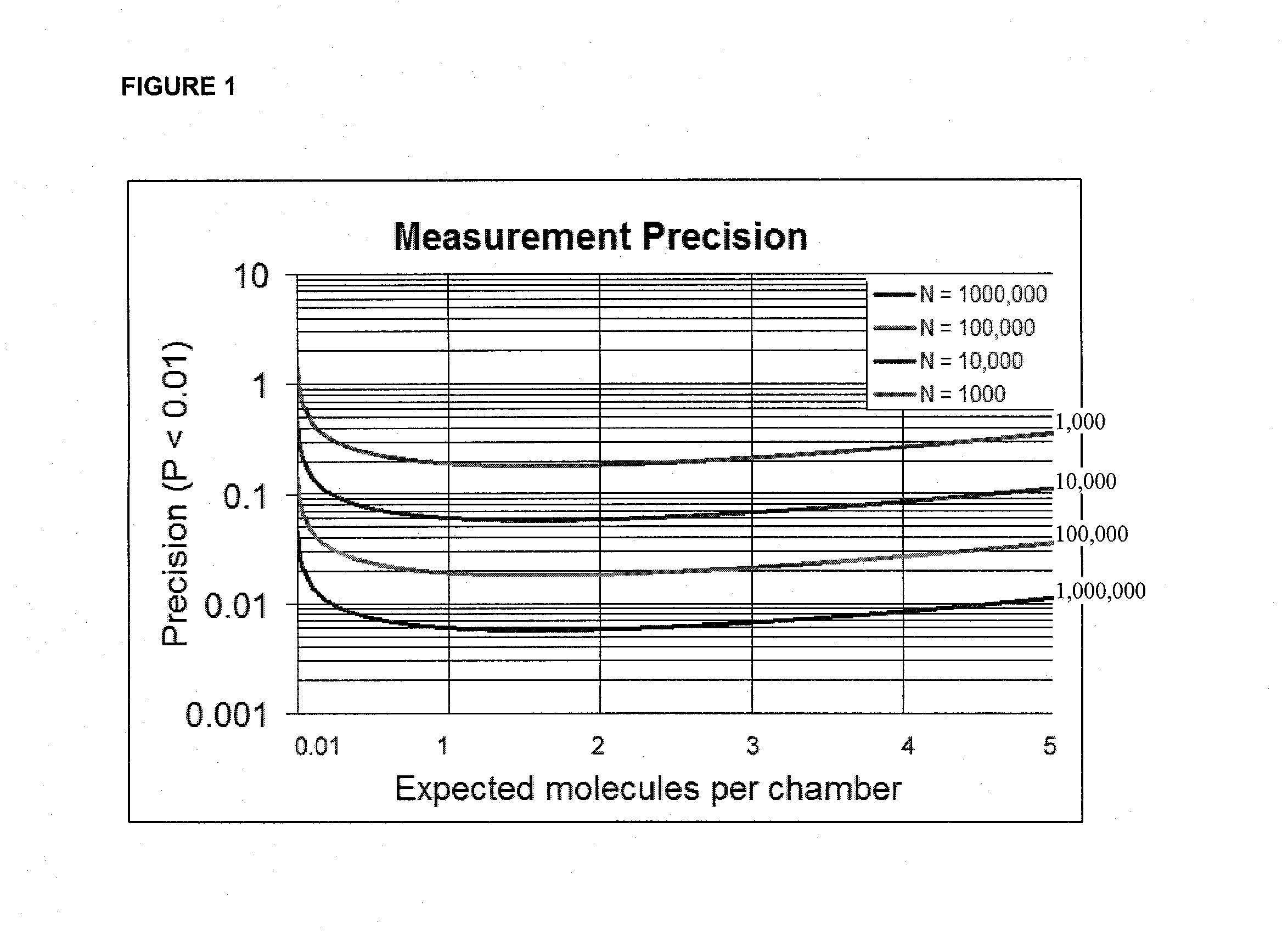
Calculation of Digital PCR Precision

[0133]The theoretical precision of digital PCR analysis depends on the total number of chambers (N) and the expected number of molecules per chamber (A). In the case of NA >>1 the number of molecules present in each chamber is a random variable k that is well described as an independent Poisson process. The probability of detecting at least one molecule is P(k>0)=1−e−λ. Let x be a random variable describing the number of chambers having at least one molecule in an array of N chambers. P(x) is therefore given by a binomial distribution with mean N(1−e−λ) and variance σ2=N(e−λ−e−2λ). Under the condition of large N this is well approximated as a Gaussian distribution:

P(x)=12πN(-λ--2λ)exp(-(x-N(1--λ))22N(-λ--2λ)

[0134]We define the precision of a digital PCR measurement as the minimum difference in concentration Δλ that can be reliably detected with less than 1% false positive and less than 1% false negative. This corresponds to a 4.6 σ separation in t...

example 2

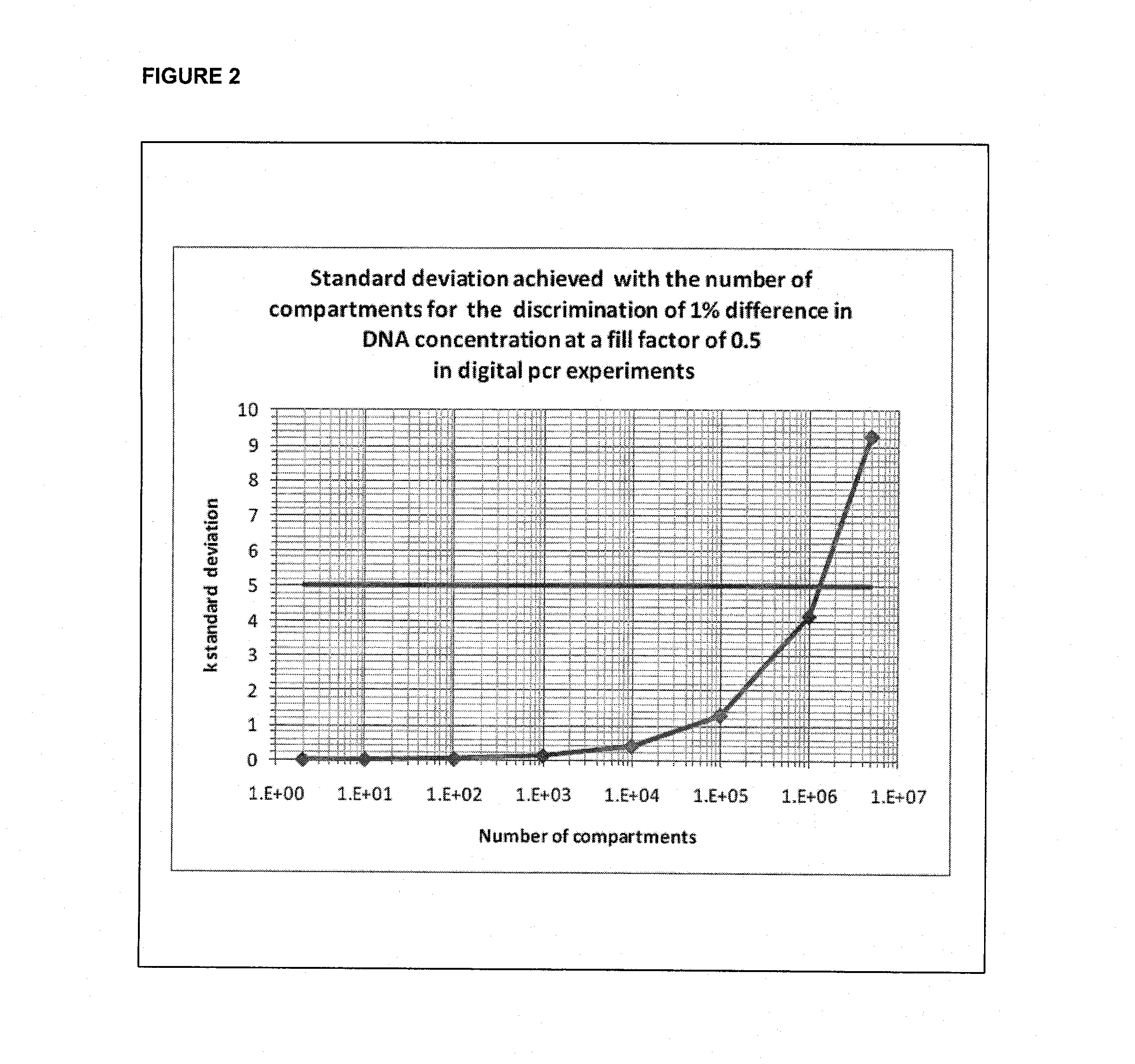
Calculation of the Separation in the Measured Mean of Two Alleles Varying by 1% Using Digital PCR as a Function of the Number of Discrete Subsamples (for Example, Chambers)

[0138]FIG. 2 shows a numerical calculation of the separation in the measured mean of two alleles varying by 1% using digital PCR as a function of the number of chambers. Difference is normalized by the expected standard deviation (sigma) as determined by the combined effect of 5 stochastic Poisson variation (curved line). The calculation was performed for template concentration corresponding to positive amplification in 50% of wells. 5 sigma separation (horizontal line) is achieved at approximately 1,000,000 chambers. Standard deviation achieved with the number of compartments for the discrimination of 1% difference in DNA concentration at a fill factor of 0.5 in digital PCR experiments.

example 3

Theoretical Calculations of Sample Molecule Content and Blood Sample Volume

[0139]Fetal DNA is reported to occur in maternal blood and represents approximately 2 to 6 percent of the total DNA present in the cell-free serum during the first trimester (Lo et al. 1997; Lo et al. 1998; Wachtel et al. 2001; Lee et al. 2002). However, later publications have suggested that the fetal contribution may be 9.7%, 9.0%, and 20.4% for the first, second, and third trimesters, respectively (Lun et al. 2008). For example, each genome copy of the T21 fraction contributes an extra copy of chromosome 21, which allows for a direct non-invasive maternal blood test by measuring the ratio of chromosome copy numbers present in maternal serum. If we assume a 2% fraction of fetal DNA in a maternal blood sample, the expected enrichment of chromosome 21 with respect to the other chromosomes in the pool is (0.02×1) / (1×2)=1%. Such a small difference in relative concentrations is undetectable by qRT-PCR which can ...


PUM

PropertyMeasurementUnit
Digital informationaaaaaaaaaa
Volumeaaaaaaaaaa
Volumeaaaaaaaaaa

Abstract

Kits, primers, and methods are provided herein for detecting relative target source to reference source ratios in a biological sample, by distributing the biological sample into discrete subsamples, wherein the biological sample includes, a plurality of target molecules on a target source; and a plurality of reference molecules on a reference source; providing target primers directed to one or more of the plurality of target molecules and reference primers directed to one or more of the plurality of reference molecules; performing digital amplification with the target primers and the reference primers; and detecting the presence or absence of amplified target products with target probes and detecting the presence or absence of amplified reference products with reference probes, wherein the ratio of amplified target products to amplified reference products is indicative of a relative amount of target source to reference source in a biological sample.

Description

CROSS REFERENCE TO RELATED APPLICATIONS[0001]This application claims the benefit of U.S. Provisional Patent Application Ser. No. 61 / 282,298 entitled “MULTIPLEX AMPLIFICATION USING A COMMON PRIMER-DERIVED INTERNAL PROBE SEQUENCE” and Ser. No. 61 / 282,299 entitled “MULTIPLEX AMPLIFICATION USING A COMMON TEMPLATE-DERIVED INTERNAL PROBE SEQUENCE”, both filed on 15 Jan. 2010.FIELD OF THE INVENTION[0002]The present invention relates to digital amplification methods. In particular, the invention relates to methods for detecting relative target source to reference source ratios in a biological sample.BACKGROUND[0003]Chromosomal abnormalities and imbalances are responsible for a significant portion of genetic disorders in humans throughout their lives. Occasionally, during the processes of DNA replication, DNA repair, or recombination, errors occur in which the resulting cell comprises too many (or too few chromosomes), chromosomes with large deletions or duplications, etc. When such errors o...

Claims


Application Information

Patent Timeline
IPC IPC(8): C12Q1/68G01N21/64
CPCC12Q1/6851C12Q1/6883C12Q2600/156C12Q2600/16C12Q2537/143C12Q2537/165C12Q2563/159
Inventor HANSEN, CARL L. G.PETRIV, OLEHHEYRIES, KEVINLIVAK, KENNETH J.
Owner THE UNIV OF BRITISH COLUMBIA
Why Eureka
Try Eureka