
Anti-s1p antibody treatment of patients with ocular disease

a technology for ocular disease and anti-s1p antibody, which is applied in the field of ocular disease treatment methods, can solve the problems of insufficient treatment options, complex search for effective treatments, and insufficient treatment options for scar formation and inflammation, so as to reduce the size of choroidal neovascularization lesion, reduce or resolve retinal pigment epithelial detachment, and reduce the thickness of the retina

Active Publication Date: 2013-03-07
APOLLO ENDOSURGERY INC
View PDF3 Cites 2 Cited by
  • Summary
  • Abstract
  • Description
  • Claims
  • Application Information

AI Technical Summary

Benefits of technology

The invention provides methods of using an anti-S1P antibody or fragment to treat diseases such as age-related macular degeneration. The methods may involve reducing the size of a choroidal neovascularization lesion, decreasing retinal pigment epithelial detachment, reducing retinal thickness, and improving visual acuity in patients with choroidal neovascularization. The technical effect is to provide a new treatment option for patients with choroidal neovascularization and age-related macular degeneration.

Problems solved by technology

Multiple hypotheses exist but the exact etiology and pathogenesis of AMD are still not well understood.
The multiple cellular and cytokine interactions which are associated with exudative AMD greatly complicate the search for effective treatments.
While CNV and edema are manageable in part by anti-VEGF therapeutics, potential treatments to mitigate scar formation and inflammation have not been adequately addressed [Bylsma and Guymer (2005), Clin Exp Optom, vol 88: 322-34 and Pauleikhoff (2005), Retina, vol 25: 1065-84].
The results in a larger patient population may be less robust.
Tissue damage can result from either component.
These other aspects of the pathologic process are not addressed by current anti-VEGF treatments.
There is no effective treatment for the atrophic (dry) form of AMD.
Secondary to RPE dysfunction, macular rods and cones degenerate leading to the irreversible loss of vision.
However, new vessels when uncontrolled, are commonly defective and promote leakage, hemorrhaging and inflammation.
The formation of subretinal fibrosis leads to irreversible damage to the photoreceptors and permanent vision loss.

Method used


Image

  • Anti-s1p antibody treatment of patients with ocular disease
  • Anti-s1p antibody treatment of patients with ocular disease
  • Anti-s1p antibody treatment of patients with ocular disease

Examples


example 1

Phase I Clinical Trial of Anti-S1P mAb (iSONEP) in Patients with Exudative AMD

[0066]Subjects with all sub-types of CNV secondary to AMD were enrolled in a Prospective Open-Label Dose-Escalating Multi-Center Phase I clinical trial to evaluate the safety, maximum tolerated dose (MTD) and preliminary activity of iSONEP in subjects with exudative AMD.

[0067]The primary objectives of the trial were safety, tolerability and establishment of MTD and dose-limiting toxicity (DLT). The secondary objectives were to characterize pharmacokinetics (PK), evaluate immunogenicity, investigate signs of biologic activity [CNV lesion area by fluorescein angiography (FA) and retinal thickness by optical coherence tomography (OCT) using the STRATUS imaging device and software (Zeiss). OCT is a non-invasive, non-contact method giving a cross sectional image of the living retina and its substructures in real time]. Visual acuity was also evaluated, measured as best corrected visual acuity using the Early Tr...

example 2

Safety Results of Phase I Trial

[0089]ISONEP was found to be well tolerated at all doses. No dose-limiting toxicity (DLT) was observed at any dose and the MTD was not reached. No significant adverse effects related to ISONEP were observed. Five adverse effects were potentially related to ISONEP: one occurrence of conjunctival hemorrhage, two instances of eye pain, and a 3 ms increase in QTcB interval and bundle branch block (observations on ECG) in the same subject, who had been treated with ISONEP at 0.2 mg and had a prior history of arrhythmia. All adverse effects resolved without sequelae. Thus iSONEP met its primary endpoint of being well tolerated in all 15 patients at dose-levels ranging from 0.2 mg. to 1.8 mg. per intravitreal injection (three patients per dose level). No drug-related serious adverse events were reported in any of the patients.

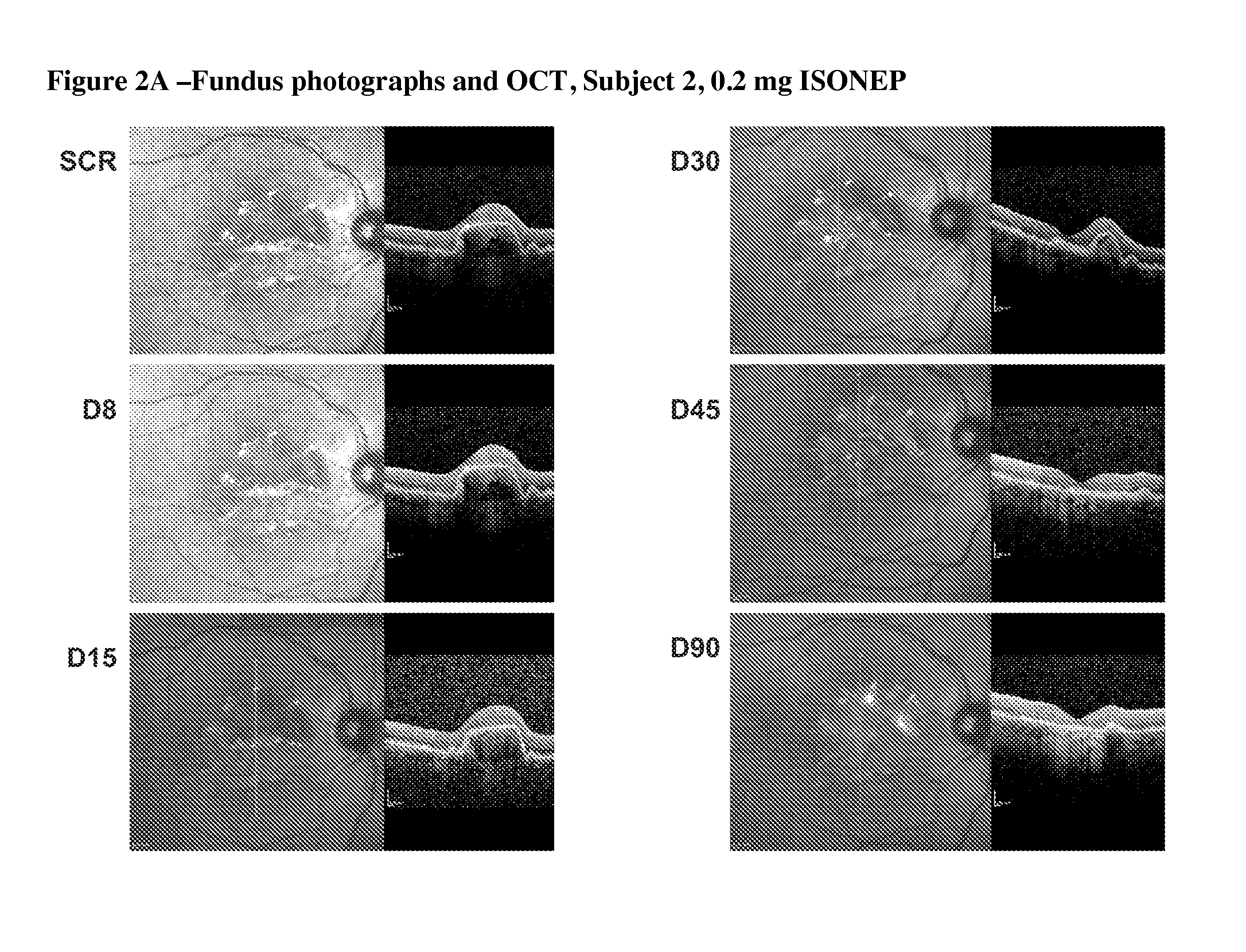
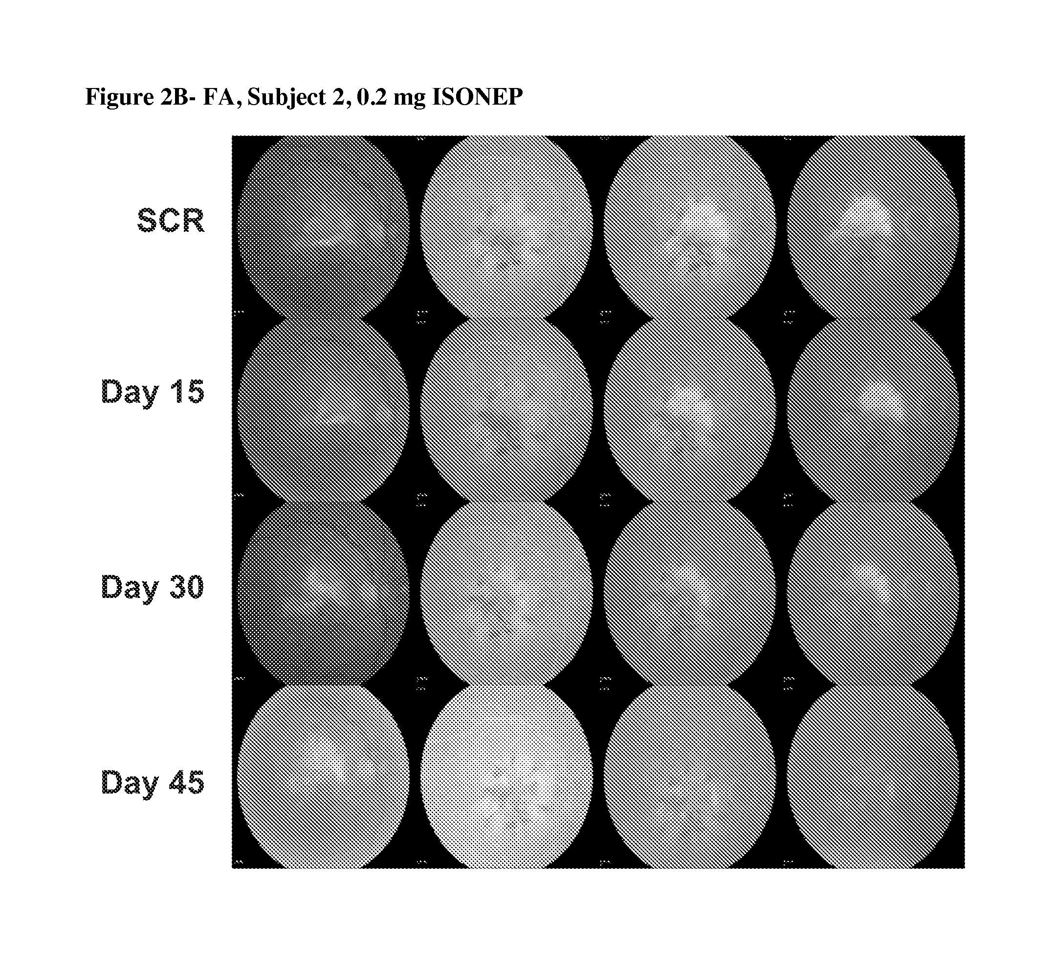
[0090]Interestingly, preliminary indications of biologic activity were seen in this Phase I trial. Five of the fifteen subjects on stud...

example 3

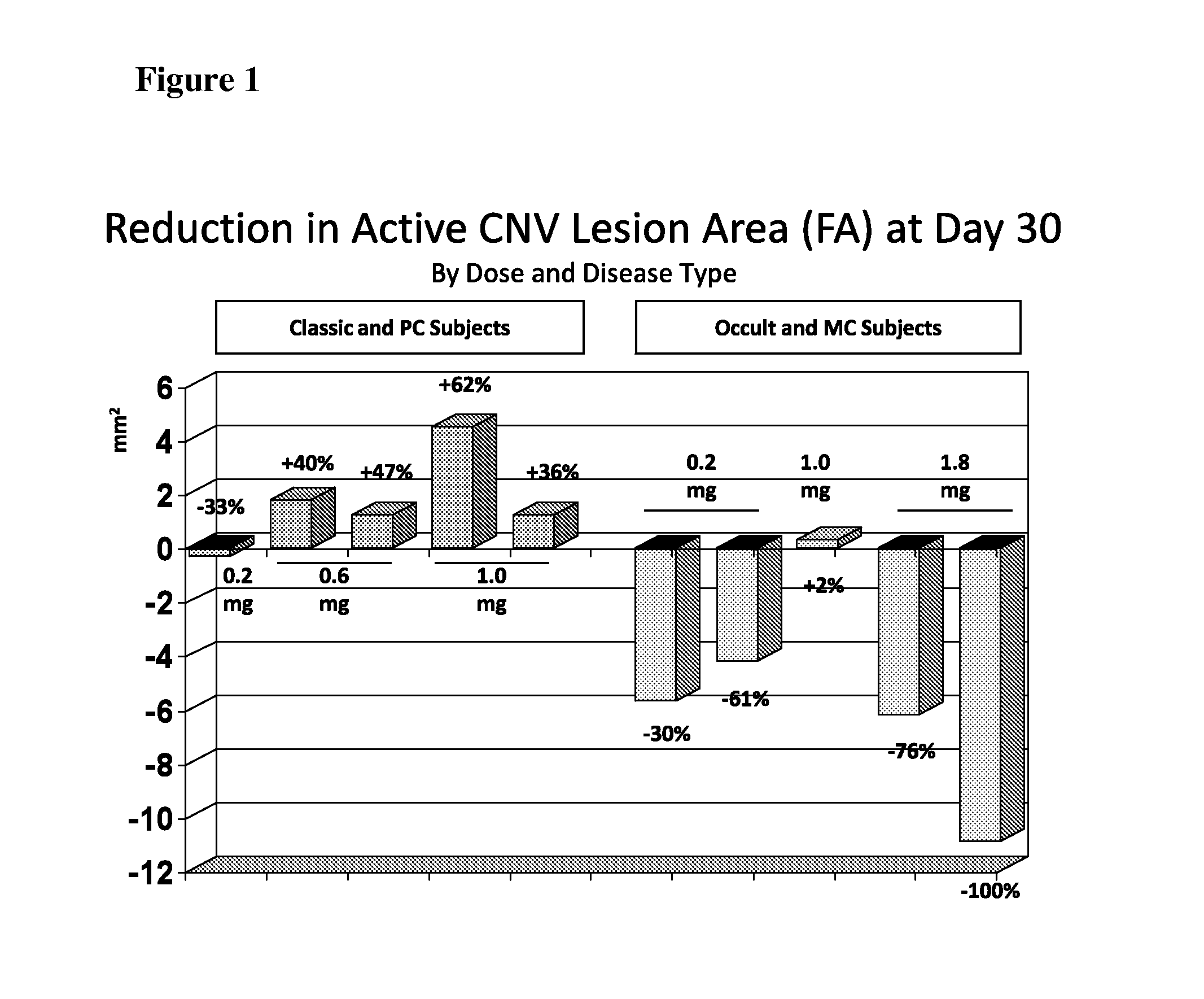
Reduction in Active CNV Lesion Area After Single Treatment

[0091]CNV lesion area was determined by fluorescence angiography (FA), with reading of images performed at the Digital Angiography Reading Center (DARC), New York, N.Y., a fully digital reading center for FA images. Each image was evaluated by two readers.

[0092]A regression in CNV was observed after treatment with ISONEP. CNV is the underlying cause of the disease that eventually leads to degeneration of the macula, the area of the retina responsible for central vision. Of ten patients, three experienced a reduction in lesion area exceeding 5 mm2 and two experienced a reduction of greater than 75%—all after a single dose of ISONEP. This type of clinical benefit is not typical, as the published data [Heier J S et al. (2006) Ophthalmology 113:642e1-642e4] suggest that, even with repeated Lucentis dosing, the total physical size of CNV lesion(s) does not show much reduction. Lucentis and Avastin target the protein VEGF, a valida...


PUM

PropertyMeasurementUnit
Fractionaaaaaaaaaa
Fractionaaaaaaaaaa
Molar densityaaaaaaaaaa

Abstract

The present invention relates to methods that involve administration of an anti-S1 P antibody or antibody fragment or derivative to a subject having or suspected of having an ocular disease or condition, including one involving choroidal neovascularization, in order to achieve a desired effect. Such effects include reducing the size of a choroidal neovascularization lesion in the eye, decreasing or resolving retinal pigment epithelial detachment, decreasing central retinal lesion thickness, and preserving or improving visual acuity. Pharmaceutical compositions comprising an anti-S1 P antibody for ocular administration are also provided. The compositions and methods are particularly useful for treating subjects having age-related macular degeneration, particularly exudative or wet age-related macular degeneration.

Description

RELATED APPLICATION[0001]This application claims the benefit of and priority to U.S. provisional patent application Ser. No. 61 / 330,276, filed 30 Apr. 2010, the contents of which are hereby incorporated by reference for any and all purposes.TECHNICAL FIELD[0002]The present invention relates to methods of treatment for ocular disorders using antibodies, including humanized antibodies, to sphingosine-1-phosphate (S1P), as well as to anti-S1P antibody fragments and derivatives. Such antibodies can be successfully used in the treatment of ocular diseases or conditions, including age-related macular degeneration (AMD), choroidal neovascularization (CNV), and retinal pigment epithelium (RPE) detachment (also called retinal pigment epithelial detachment or PED).SEQUENCE LISTING[0003]The instant application contains a Sequence Listing submitted via the Electronic Filing Sytem on Apr. 29, 2011 and, is hereby incorporated by reference in its entirety. Said ASCII copy, created on Apr. 20, 2011...

Claims


Application Information

Patent Timeline
IPC IPC(8): A61K39/395A61P27/02
CPCA61K2039/505A61K2039/507C07K2317/76C07K16/22C07K2317/73C07K16/18A61P27/02
Inventor STOLLER, GLENN L.SAFONOV, MARINAPANCOAST, SCOTT R.HSU, JAMES LEIGH
Owner APOLLO ENDOSURGERY INC
Learn More