Preparation of microfluidic device on metal nanoparticle coated surface, and use thereof for nucleic acid detection
a technology of microfluidic devices and coating surfaces, which is applied in the direction of fluorescence/phosphorescence, instruments, material analysis, etc., can solve the problems of increasing reaction kinetics, rapid analysis, and fluorescence quenching, so as to avoid or minimize the need for spacer molecules, improve detection efficiency, and enhance the effect of dramatic fluorescen
- Summary
- Abstract
- Description
- Claims
- Application Information
AI Technical Summary
Benefits of technology
Problems solved by technology
Method used
Image
Examples
example 1
Nanostructured Ag Substrate Fabrication
[0110]Nanostructured Ag substrates were fabricated by covalent attachment of Ag nanoparticles to thiolated glass substrates. Microscope glass slides (VWR, Cat. No. 48300-025) were first diced into individual chips with dimensions of 1.7 cm×2.5 cm using a diamond scribe. These glass chips were cleaned by soaking them in piranha solution (sulfuric acid: hydrogen peroxide 3:1; Caution: piranha solution is caustic and reacts vigorously with organics) for 15 min. The glass chips were then rinsed with distilled, deionized (DDI) water, soaked in a 10 M NaOH solution for 5 min, rinsed again with DDI water, and finally dried under nitrogen gas.
[0111]Surfaces of the clean glass chips were next silanized by immersion in a 1% MPTS (3-mercaptopropyl trimethoxysilane), 95% methanol, and 4% 1 mM acetic acid solution at room temperature for 30 min. The chips were then sonicated (300-W Vibracell probe sonicator, Sonic & Material Inc.) in a 95% ethanol: 5% water...
example 2
Microfluidic Device Fabrication
[0114]Once the probe spots were immobilized on the substrate surfaces as described in Example 1, the substrates were covalently bound to the channel-embedded PDMS replicates.
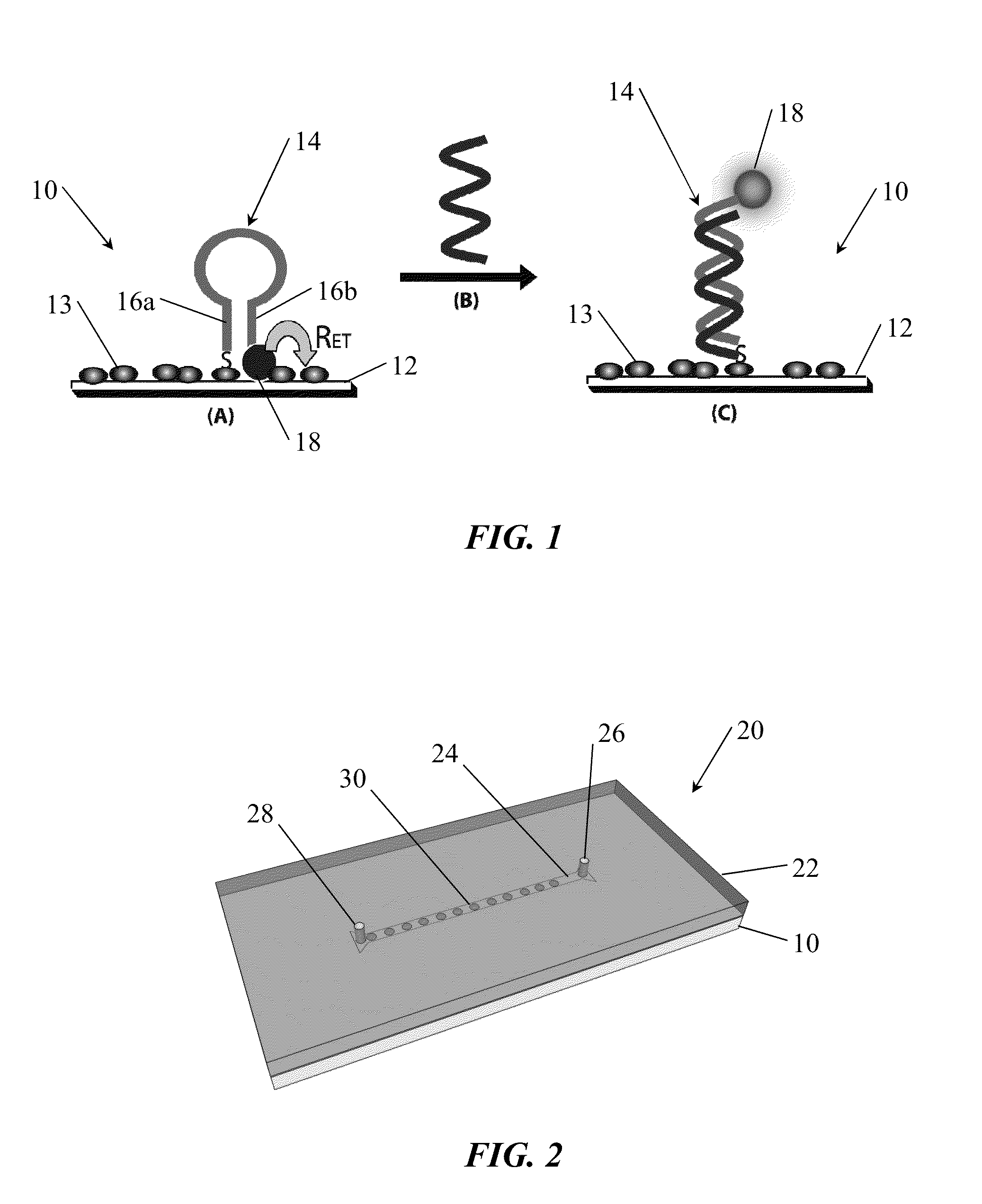
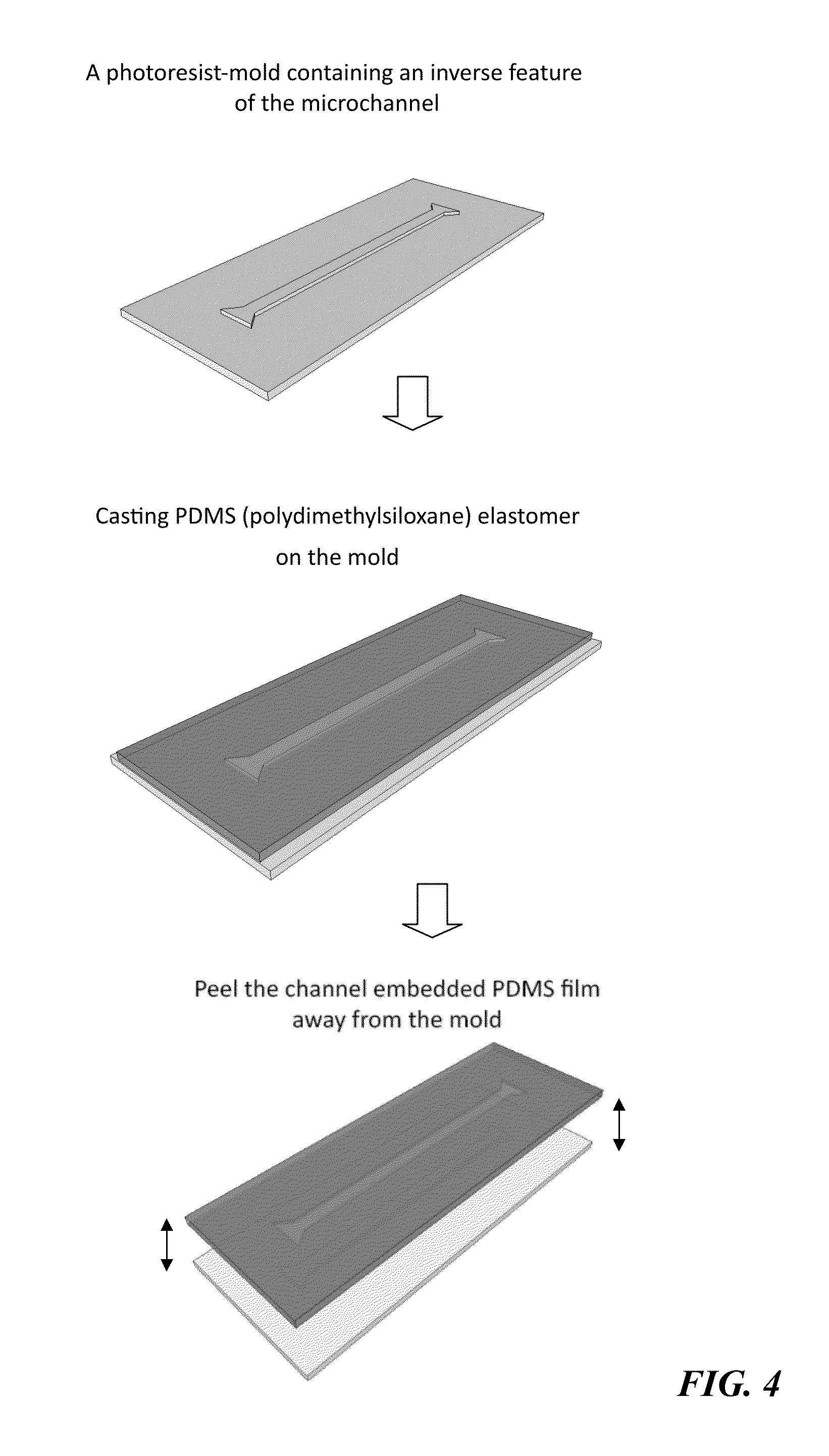
[0115]The microfluidic device was prepared with a single microchannel 2 cm in length, 1 mm in width, and 50 μm in height (FIG. 2), connected to two isosceles trapezoid fluidic reservoirs (top base: 1 mm, bottom base: 3.75 mm, and height: 1.25 mm) at both ends. Total fluid volume of the channel was 1 μl. The channel-embedded PDMS replicates were fabricated by casting 20 grams of the PDMS elastomer mixture (at a prepolymer: curing agent ratio of 10:1 (w / w), Sylgard 184 kit, Dow Corning, Midland, Mich.) over a SU-8 photoresist mask (see FIG. 4). The photoresist mask has an inverse feature of the microchannels, and was patterned using standard soft lithography at Stanford Microfluidics Foundry (Stanford, Calif.).
[0116]After initial casting, the overlaying PDMS mixture was cured at 110°...
example 3
DNA Detection in a Microfluidic Device
[0120]After successfully addressing the challenge of assembling a leakage-free device, the utility of this microfluidic system for arrayed DNA detection was examined. In this example, different probe sequences were spotted on the substrate surface using a microarrayer. This approach not only provides the potential for multiplex detection, but also dramatically reduces reagent consumption.
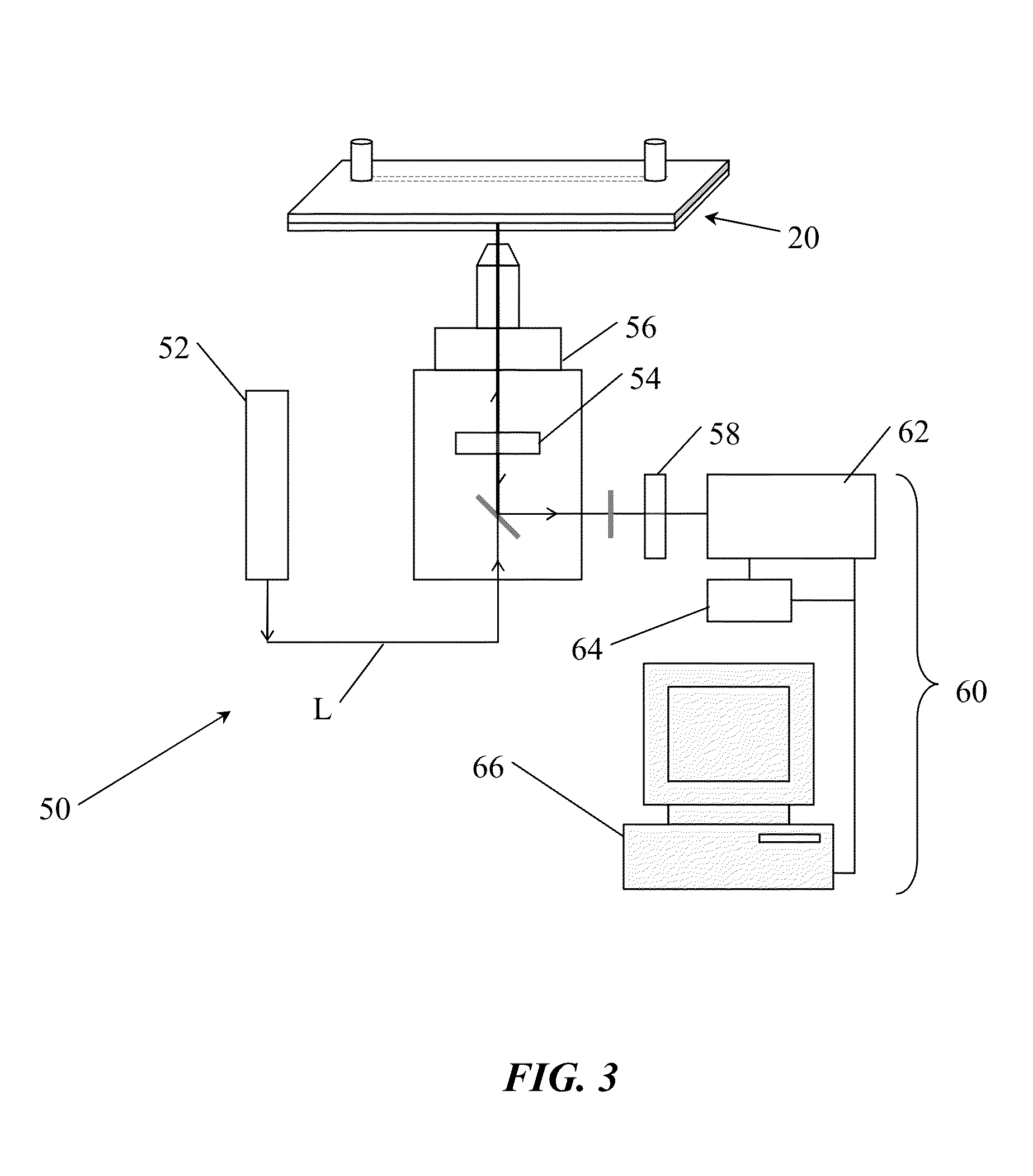
[0121]In this example, arrays of the Bacillus anthracis probe (2 columns, each column consisted of 10 probe spots) were first printed on the substrate surface and the substrate was next integrated with a channel-embedded PDMS replicate using the assembly protocols described above (FIG. 6). As a final step prior to detection, hairpin reformation was promoted by adding 1 μl of buffered saline (500 mM NaCl, 20 mM cacodylic acid, and 0.5 mM EDTA, pH=7) into the channel for 45 min (room temperature, in the dark).
[0122]Using a standard fluorescence microscope for imag...
PUM
| Property | Measurement | Unit |
|---|---|---|
| surface roughness | aaaaa | aaaaa |
| surface roughness | aaaaa | aaaaa |
| thick | aaaaa | aaaaa |
Abstract
Description
Claims
Application Information
 - R&D
- Intellectual Property
- Life Sciences
- Materials
- Tech Scout
- Unparalleled Data Quality
- Higher Quality Content
- 60% Fewer Hallucinations
Browse by: Latest US Patents, China's latest patents, Technical Efficacy Thesaurus, Application Domain, Technology Topic, Popular Technical Reports.
© 2025 PatSnap. All rights reserved.Legal|Privacy policy|Modern Slavery Act Transparency Statement|Sitemap|About US| Contact US: help@patsnap.com


