

Continuous Flow Bioreactor for Magnetically Stabilized Three-Dimensional Tissue Culture
a bioreactor and three-dimensional technology, applied in the field of continuous flow bioreactor for magnetically stabilized three-dimensional tissue culture, can solve the problem of not using traditional solid scaffolding for cell cultur
- Summary
- Abstract
- Description
- Claims
- Application Information
AI Technical Summary
Benefits of technology
Problems solved by technology
Method used
Image

Examples
example 1
Preparation of Magnetic Fluid
[0117]Dodecanoic acid (2 g) was added to a 200 mL aqueous solution of 0.12 M ferrous chloride and 0.24 M ferric chloride in a 600 mL beaker. After adding slowly 40 mL of 25% ammonia solution, the mixture was placed in a water bath at 50° C. and stirred at 1300 rpm with an overhead mixer from G. K. Heller Corporation (Floral Park, N.Y.). The process was allowed to run for 30 min, while removing the lather that was continuously formed. The precipitate was collected over a magnet and then rinsed with 0.5% ammonia aqueous solution several times. A 100 mL volume of 1 g / L dodecanoic acid suspension in DI water was transferred to the precipitate and the mixture was stirred at 1300 rpm in the water bath at 80° C. for 30 min. The magnetic fluid that was formed was stored at room temperature in a sealed container shielded from light until further use.
Preparation of Agarose Magnetic Beads
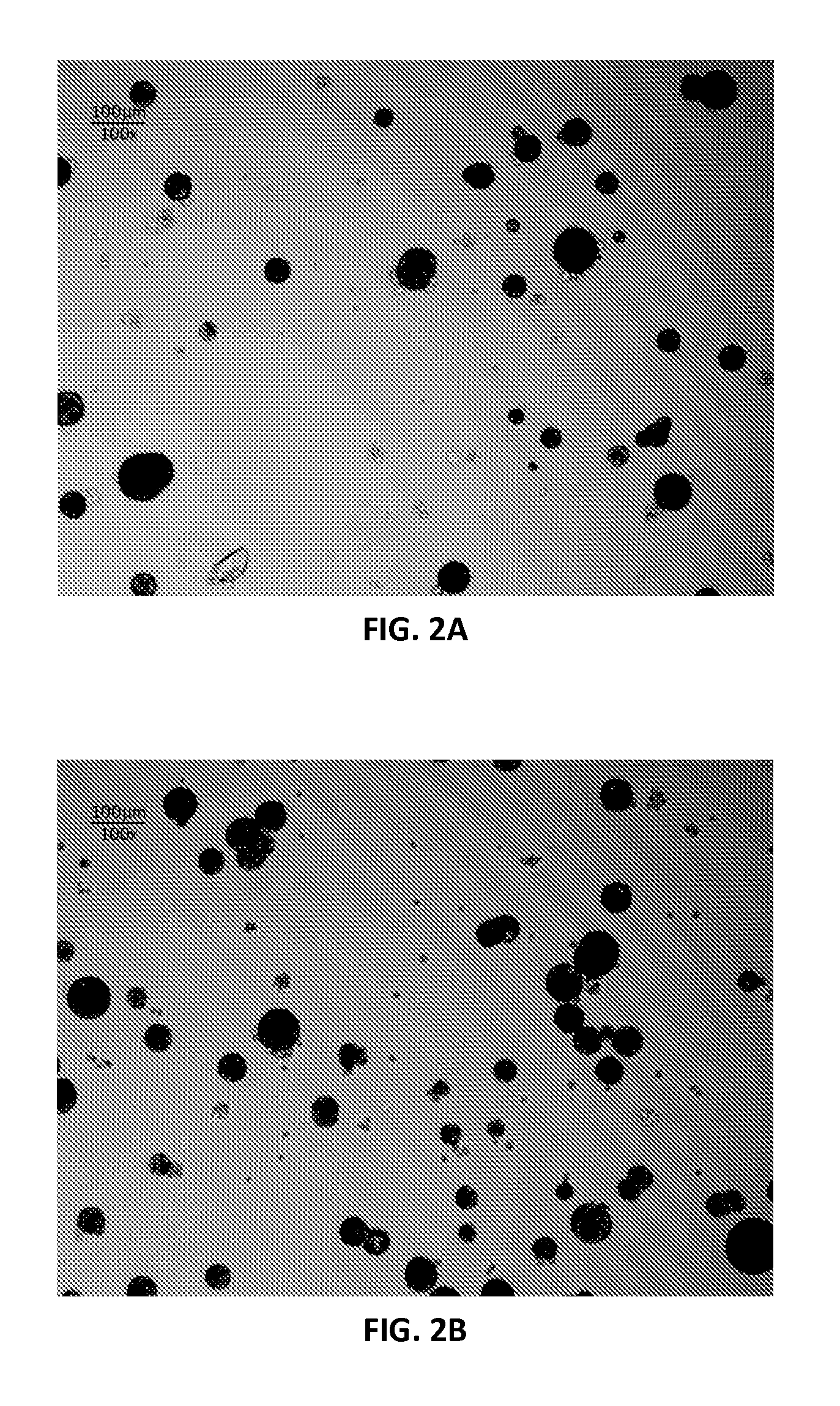
[0118]The beads were prepared by emulsification. A 160 mL soybean oil solution...
example 2
Growing Fibroblasts with Magnetic Beads in a Glass Tube
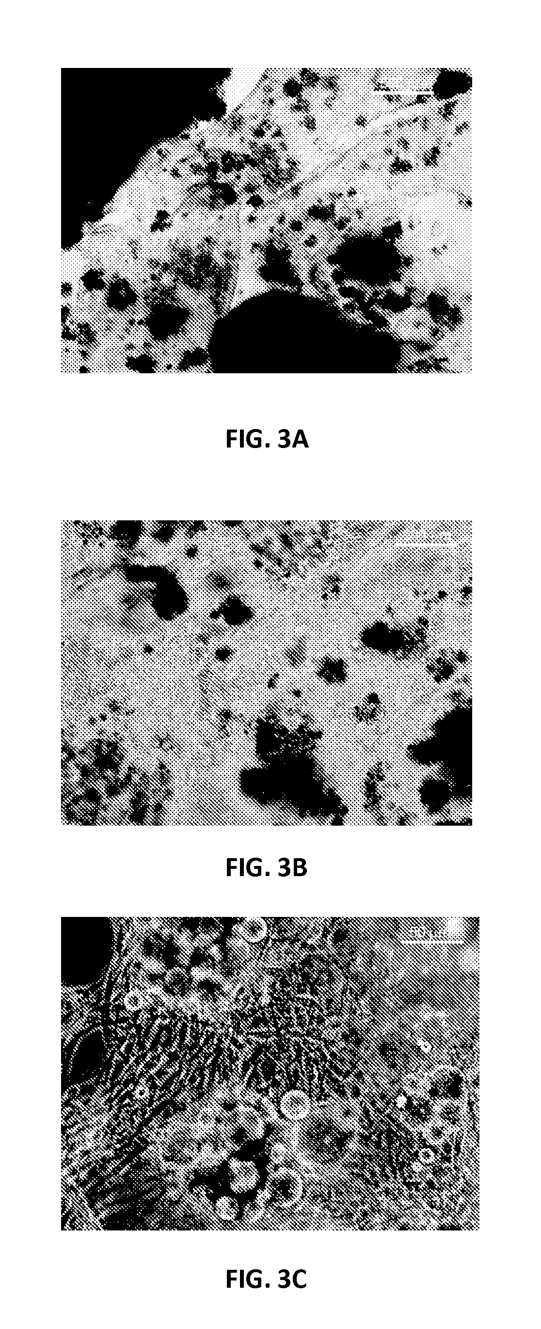
[0130]Mouse neo-natal fibroblasts (CRL-2097) were grown in DMEM / F12 medium supplemented with 10% fetal bovine serum (FBS) in an incubator at 37° C. and 5% CO2. Cells were first allowed to proliferate in the culture flasks. The cells were suspended in growth medium solution containing magnetic beads coated with collagen and then the mixture was transferred into a 10 mm diameter glass tube which had been coated with poly9-ethylene glycol) (PEG) to prevent non-specific adhesion of proteins and fibroblasts. A magnet was placed around the tube to create a ring of magnetic beads. Therefore if cells were attached to the cells they would form a ring as well after producing extra cellular matrix. The tubes with the magnet were kept in the 37° C. incubator and the medium was changed every 2 days. After 1 week the medium was replaced with DMEM / F12 with 10% FBS containing 4 ng / ml fibroblast growth factor (FGF), and the medium was replaced e...
example 3
Growing Smooth Muscle Cells with Magnetic Beads in a Glass Tube
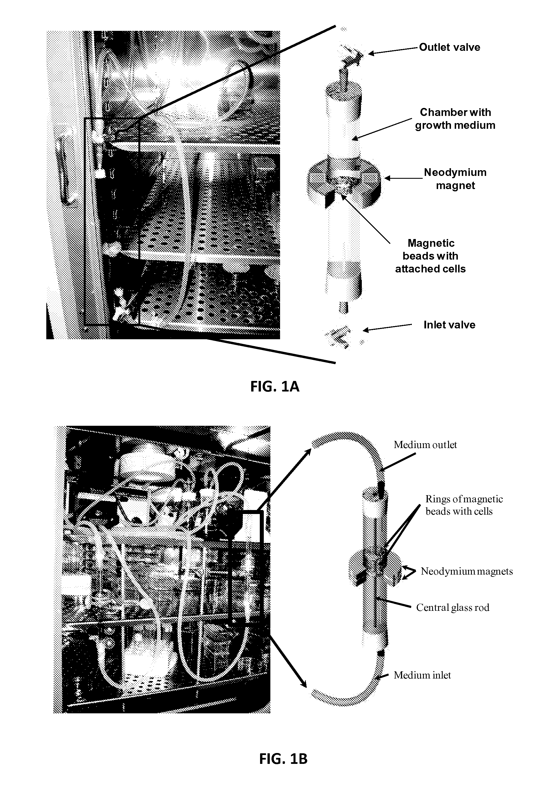
[0135]Rat aortic smooth muscle cells (RASMC) were grown in DMEM supplement with 10% FBS and 1% penicillin / streptomycin in 37° C. and 5% CO2 incubator. Agarose magnetic beads were activated and coated with collagen as mentioned above in the magnetic bead section. The collagen coated magnetic beads were finally rinsed with PBS and resuspended in the 0.5 ml of cell culture medium, containing 1 ml of DMEM+10% FBS+1% pen / strep. The 25 mm diameter×20 mm neodymium magnet was secured around the top edge of the 5 ml borosilicate glass tube. The bottom end of the PEG coated bioreactor (glass tubing of 5 mm diameter×100 mm) was sealed with rubber septum while leaving the top open, and it was inserted into the test tube to position the magnet around the bioreactor. About 1 ml of culture medium was added into the bioreactor, and then, the magnetic beads solution was slowly pipette into the bioreactor from the top so that the beads ar...
PUM
 Abstract
Description
Claims
Application Information