Renilla/gaussia transfected cells as a light source for in-situ photodynamic therapy of cancer

a cancer and in-situ technology, applied in the field of deep tissue tumor imaging and photodynamic therapy, can solve the problems of affecting the immune response of the subject, affecting the treatment effect, and affecting the survival rate of the subject, so as to stimulate the subject's own immune response

Inactive Publication Date: 2013-09-19
KANSAS STATE UNIV RES FOUND
View PDF1 Cites 7 Cited by
  • Summary
  • Abstract
  • Description
  • Claims
  • Application Information

AI Technical Summary

Benefits of technology

[0015]In one or more embodiments a further method for photodynamic therapy of cancerous tissue is provided. The method comprises (a) administering to a subject a therapeutically effective amount of tumor-trophic cells comprising a nucleic acid encoding for a luminescent protein; (b) administering a photosensitizing agent to the subject, wherein the photosensitizing agent is administered separately from the cells; (c) optionally administering a iron chelator to the subject; (d) administering a luminogenic substrate corresponding to the luminescent protein to the subject; and (e) repeating steps (a)-(d). Advantageously, the substrate reacts with the luminescent protein to produce light in situ (intracellularly), and the light activates the photosensitizing agent, which results in the damage and destruction of the cancerous tissue according to the various mechanisms for cell death described herein. By repeating the process more and more of the tissue is destroyed until the subject is preferably cancer-free. Repetition also helps to further stimulate the subject's own immune response.

Problems solved by technology

This energy transfer converts triplet oxygen to singlet oxygen, an extremely reactive species that is destructive to cells.
Despite these advantages, PDT's uses are severely limited due to the low penetration depth of light into tissue, with many light wavelengths only being able to penetrate ⅓-inch of skin or less.
Thus, PDT is generally limited to surface cancers, such as skin melanomas.
However, the light intensities required for the simultaneous absorption of two or several photons are very hard to achieve when treating tumors within the human / mammalian body, because the two- and three-photon absorption cross-sections are too low.
However, this results in a treatment protocol that is much more invasive than traditional PDT.
However, such methods also have drawbacks.
In particular, the deeper the penetration of the light, the less focused the light becomes and the more nonspecific the treatment becomes as an increasing amount of surrounding non-target tissue also gets irradiated and damaged during the process.
Thus, despite advances, PDT is still considered a treatment option primarily suitable for surface tissues and localized cancers, and not for use with deep-tissue tumors or cancers that have metastasized.

Method used


Image

  • Renilla/gaussia transfected cells as a light source for in-situ photodynamic therapy of cancer
  • Renilla/gaussia transfected cells as a light source for in-situ photodynamic therapy of cancer
  • Renilla/gaussia transfected cells as a light source for in-situ photodynamic therapy of cancer

Examples


example 1

Initial Investigations

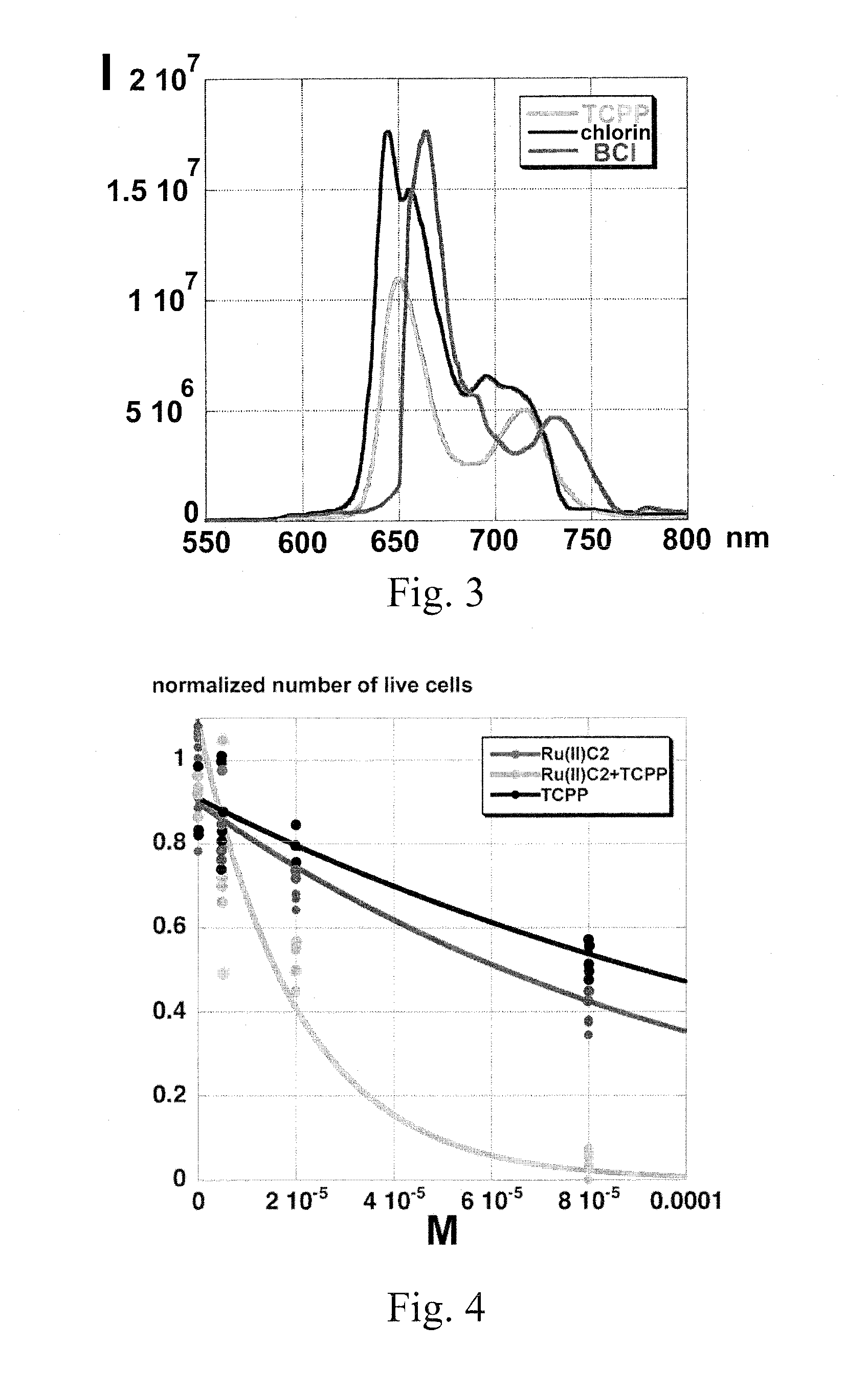
[0065]Using conventional transfection techniques according to the manufacturer's instructions, we have transfected C17.2. neural stem cells with a mammalian expression plasmid containing RLuc8 (Renilla luciferase pcDNA-RLuc8 plasmid, obtained from Dr. Loening; Stanford University, Stanford, Calif. 94305). The RLuc8 variant is approximately 4× brighter than native RLuc and about 200× more stable than RLuc in murine serum at 37° C. We hypothesized that the emitted light can be absorbed by RuC2 and then (partially) transferred to TCPP, TCPC, and TCPBC, or directly absorbed by the porphyrin, chlorin, or bactcriochlorin.

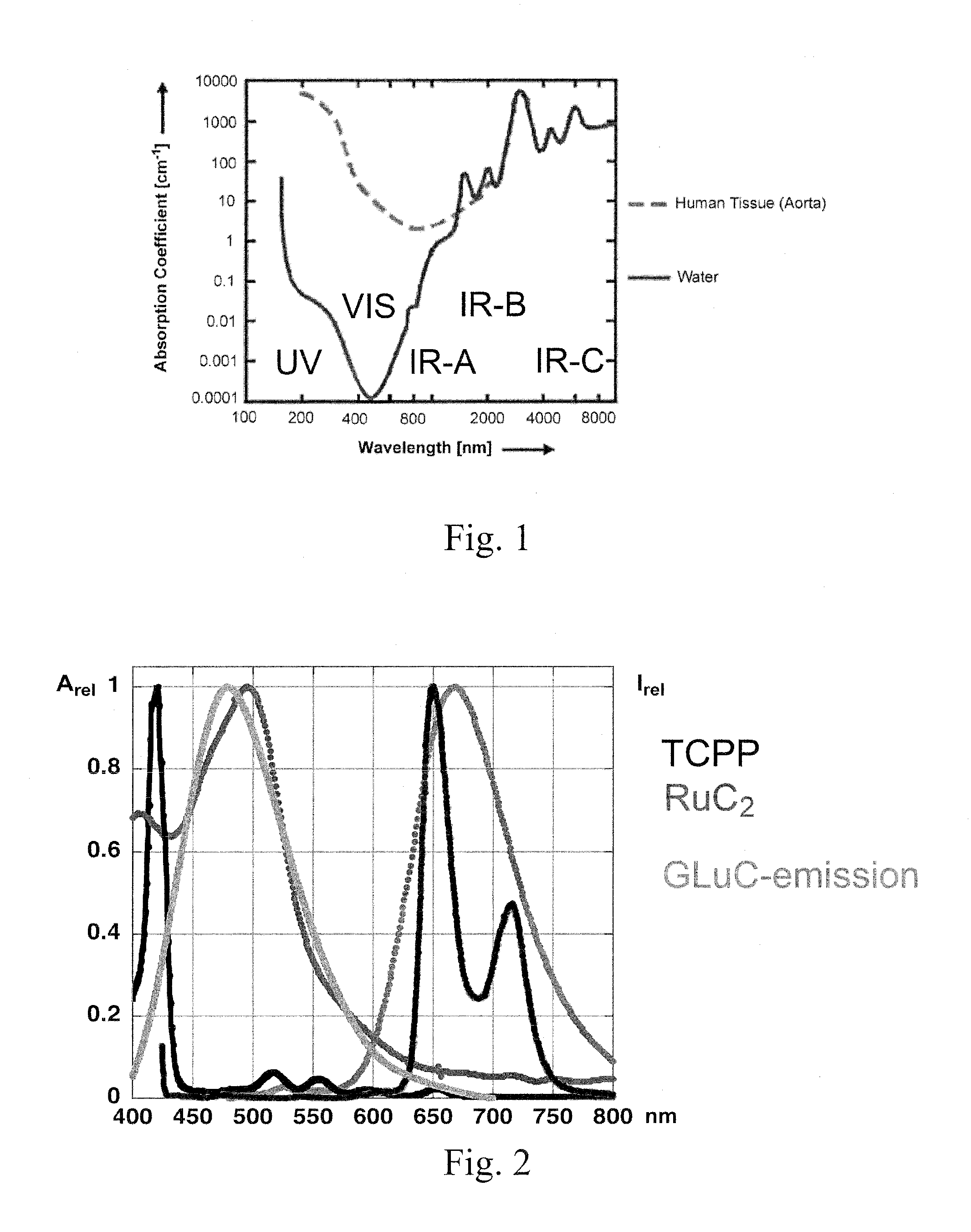
[0066]As shown in FIG. 2, the emission spectrum of Renilla luciferase and the 3MLCT-absorption band of RuC2 match perfectly. All measurements were taken at pH=6.8 in phosphate buffer using 4.0 mL quartz cuvettes (Helma) using a spectrofluorometer (Fluoromax 2) with dual monochromators and a diode array UV / Vis absorption spectrometer (HP 8453).

[0067]I...

example 2

Feasibility Study Using LED's

[0068]LED-irradiation experiments employing RL5-B12120 Superbright LED's, (Qmax=470 nm, light output 0.0114±0.0008 W) and a light-attenuation system consisting of a series of parallel optical filters was performed. After trypsinization, B16F10 mouse melanoma cells were counted using a hemacytometer and the trypan blue exclusion method, and plated in tissue culture plates (TPP brand, Midwest Scientific). Four hours post-plating, RuC2, TCPP, or a 1:1 mixture of RuC2 and TCPP was added to the plates, with the 1:1 mixture containing each agent at half their respective concentrations to permit comparisons at the same molar concentrations of PDT-agents. The cells were then stored in the dark for 24 hours. Next, the cells were irradiated with high or low intensity of LED light for 45 min. Twenty-four hours post-radiation, MTT cell proliferation reagent (thiazolyl blue) was added to the cells, which were then incubated four hours. Next, SDS-solubilization buffer...

example 3

In Vitro Study Using the Blue Luminescence from Gaussia Luciferase

[0071]A co-culture system was used to study the photodynamic effect of Gaussia luciferase-expressing neural stem cells (NSCs) or rat umbilical cord matrix stem cells (rUCMSCs) on B16F10 melanoma model cells. In previous work, stem cells have been used as stealth vehicles in a more classical cell-based gene therapy approach using, cytokine-transfected hUCMSCs, which significantly attenuated experimental lung metastasis of breast cancer cells without evidence of any damage in other normal tissues. As shown in FIG. 6a, dye-loaded IFN-expressing hUCMS cells were detected in small metastatic breast tumor tissue (see arrows), but not in the surrounding normal lung tissue. The image was recorded using a Nikon Diaphot inverted microscope on a TMD airtable equipped with Hoffman illumination, epifluorescence, and Leica and Eppendorf microinjection system. Similarly, previous work has established that stem cells preferentially a...


PUM

PropertyMeasurementUnit
light penetration depthaaaaaaaaaa

Abstract

A method for photodynamic therapy treatment of cancerous cells and tissue is provided. The method comprises administering tumor-trophic cells expressing a luminescent protein to a subject. A photosensitizing agent is then separately administered to the subject, followed by an optional iron chelator. On the day of treatment, a luminogenic substrate corresponding to the luminescent protein is administered to the subject. The substrate reacts with the luminescent protein in the vicinity of the cancerous tissue to produce light which activates the photosensitizing agent resulting in the selective destruction of the cancerous tissue.

Description

CROSS-REFERENCE TO RELATED APPLICATIONS[0001]The present application claims the priority benefit of U.S. Provisional Patent Application Ser. No. 61 / 384,957, filed Sep. 21, 2010, entitled Renilla / Gaussia Transfected Cells as Light Source for In-Situ Photodynamic Therapy of Cancer, incorporated by reference in its entirety herein.FEDERALLY SPONSORED RESEARCH OR DEVELOPMENT[0002]This invention was made with government support under Grant No. 933701, awarded by the National Science Foundation. The United States government has certain rights in the invention.SEQUENCE LISTING[0003]The following application contains a sequence listing in computer readable format (CRF), submitted as a text file in ASCII format entitled “SequenceListing,” created on Sep. 13, 2011, as 7 KB. The content of the CRF is hereby incorporated by reference.BACKGROUND OF THE INVENTION[0004]1. Field of the Invention[0005]The present invention relates to improved methods of photodynamic therapy (PDT) and imaging for dee...

Claims


Application Information

Patent Timeline
Patent Type & Authority Applications(United States)
IPC IPC(8): A61K41/00
CPCC08B37/0015C08L5/16A61K41/0057A61K45/06A61K41/0061B82Y5/00A61K48/005A61N5/062A61N2005/0653A61N2005/0656A61K47/48969A61K47/6951
Inventor BOSSMANN, STEFAN H.TROYER, DERYL L.BASEL, MATTHEW T.SHRESTHA, TEJ B.WANG, HONGWANG
Owner KANSAS STATE UNIV RES FOUND
Why Eureka
Try Eureka