Matrix scaffold with antimicrobial activity

Active Publication Date: 2013-12-26
WORCESTER POLYTECHNIC INSTITUTE
View PDF0 Cites 14 Cited by
  • Summary
  • Abstract
  • Description
  • Claims
  • Application Information

AI Technical Summary

Benefits of technology

[0019]In certain embodiments of the invention, a bioengineered protein construct includes an antimicrobial domain from human cathelicidin, a natural antimicrobial peptide produced by keratinocytes, fused with one of two collagen-binding domains, one derived from fibronectin, the other derived from collagenase. The construct was des

Problems solved by technology

These full-thickness wounds lead to impaired tissue regeneration and loss of barrier function.
Current scaffolds, however, are plagued by in

Method used


Image

  • Matrix scaffold with antimicrobial activity
  • Matrix scaffold with antimicrobial activity
  • Matrix scaffold with antimicrobial activity

Examples


example 1

Decellularization

A. Decellularization of Foreskins

[0050]Decellularization protocols were tested on foreskin samples as model demial tissue (foreskins collected from UMass Memorial University Hospital with IRB approval). Samples were prepared by scraping off fatty tissue and cutting the dermis to the size of 0.5 cm×0.25 cm.

[0051]1) Freeze-Thaw: Four samples were decellularized according to the protocol reported by Ngangan and McDevitt, 2009 (Biomaterials, 30(6):1143-49). The samples were immersed in liquid nitrogen for 2 min and then rotated in PBS at room temperature (RT) for 5 min. This cycle was repeated 3 times. Two of these samples were treated with DNase (1 mg / mL,

[0052]10 mM MgCl2) for 15 min following all freeze-thaw (FT) cycles. One was fixed immediately after treatment, and one was fixed 8 hours after treatment.

[0053]2) Sodium Dodecyl Sulfate: Six samples were decellularized using sodium dodecyl sulfate (SDS) according to the protocol by Elder et al., 2009 (Biomaterials, 30(...

example 2

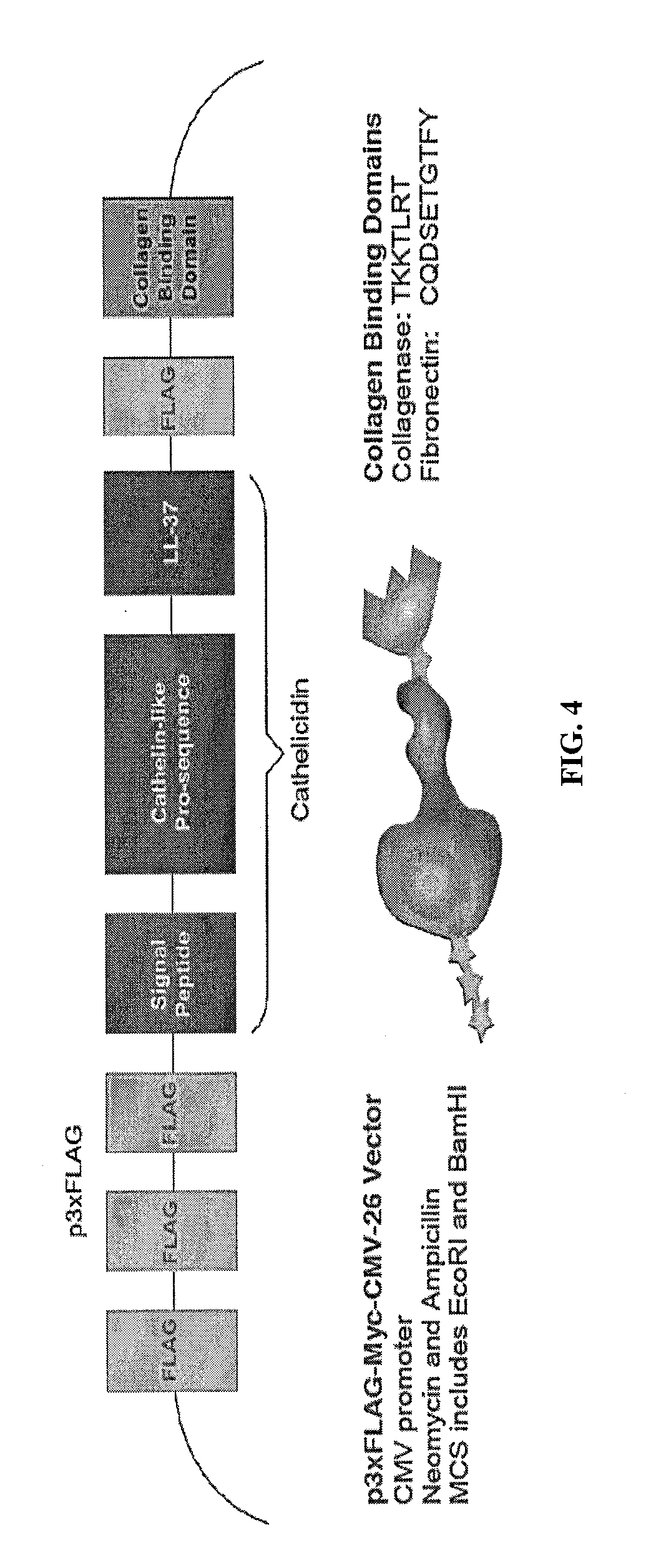
Chimeric Peptide Production

[0064]The 510 bp cathelicidin insert was isolated and amplified from pANT7 cGST (DNASU Plasmid Repository, Clone ID HsCD00357894, GenBank Acc. No. 570277) using PCR. Primers were designed to include and incorporate the recombinant features onto the cathelicidin insert. One forward and two reverse primers (corresponding to each CBD) were ordered from Integrated DNA Technologies (IDT). The sequences of the three primers are shown in FIG. 6.

[0065]For PCR, the samples were prepared by combining 10 μL of Promega's GoTac Green Mastermix, 1 μL of the template (pANT7-cGST), 1 μl forward primer, 1 μL reverse primer, and ddH2O to 20 μL. PCR controls included a sample with no primers, and two with primers and no template (the two corresponding to each of the reverse primers). The PCR thermocycler (Bio-Rad, MyCycler) was set to 4 min at 90° C. (30 s at 95° C., 30 s at 55° C., 1 min at 72° C)×30, 4 min at 72° C., and ∞ at 10° C. Agarose gel electrophoresis, pre-stained...

example 3

Localization of Chimeric Peptide

[0089]Transformation of E. coli and Verification Cathelicidin Construct. The pGEM vector with the recombinant cathelicidin DNA insert was amplified by E. coli transformation. Transformations were carried out with 50 μL of chemically competent E. coli and 2 μL of the pGEM plasmid with DNA insert in a cold Eppendorf tube. This mixture was incubated on ice for 20 minutes, heat shocked at 42° C. in a water bath for 45 s, before it was returned to ice for another 2 minutes. The E. coli received 450 μL of prewarmed LB media and was incubated at 37° C. under shaking at 100 rpm for one hour. 50 μL of the transformed E. coli were plated on to Pre-warmed LB agar Ampicillin plates. These plates were incubated overnight at 37° C.

[0090]The following day individual colonies were selected and inoculated in 3 mL of LB with 100 μg / mL of Ampicillin media and incubated overnight at 37° C. with shaking at 190 rpm. Macherey and Nagel's Nucleospin plasmid purification prot...


PUM

PropertyMeasurementUnit
Antimicrobial propertiesaaaaaaaaaa

Abstract

The invention provides a scaffold of extracellular matrix polymers with recombinant chimeric peptides tethered thereto. The invention also provides recombinant chimeric peptides of antimicrobial peptides and extracellular matrix binding domains. The invention also provides methods for treating chronic wounds using the scaffold and/or recombinant chimeric peptides.

Description

CROSS-REFERENCE TO RELATED APPLICATIONS[0001]This application claims priority to U.S. Provisional Application No. 61 / 664,386 filed Jun. 26, 2012 and entitled CELL-DERIVED MATRIX SCAFFOLD, the entire contents of which are incorporated herein by reference for all purposes.BACKGROUND OF THE INVENTION[0002]Skin is the body's main protective barrier against environmental insults. Significant wounds, caused by bums, physical trauma, surgery, and underlying pathologies, affect more than 35 million people annually in the United States. These full-thickness wounds lead to impaired tissue regeneration and loss of barrier function.[0003]Scaffolds have been explored as a therapeutic method for regenerating skin at the wound site. Scaffolds provide a three-dimensional support through which cells can migrate, adhere, and regenerate a functional new tissue. Current scaffolds, however, are plagued by inadequate biocompatibility and biodegradability, mechanical and structural mismatch with the nativ...

Claims


Application Information

Patent Timeline
IPC IPC(8): A61L27/54
CPCA61L27/54C07K14/78C07K14/4723C07K2319/00C12N15/62A61L2300/404
Inventor ROLLE, MARSHAPRIFTI, FIOLEDAMALCUIT, CHRISCAMESANO, TERRI ANNEDOMINKO, TANJA
Owner WORCESTER POLYTECHNIC INSTITUTE
Why Eureka
Try Eureka