Method for culturing pluripotency-maintained singly dispersed cells by means of laminar flow

Inactive Publication Date: 2014-09-04
ARKRAY INC
View PDF4 Cites 7 Cited by
  • Summary
  • Abstract
  • Description
  • Claims
  • Application Information

AI Technical Summary

Benefits of technology

[0013]In the culture method of one aspect of the present invention, pluripotent cells are cultured in a state where the cells are dispersed as single cells, to enable production of their cloned cells. Therefore, one

Problems solved by technology

In cases where these cells are dispersed into single cells by destruction of cell-cell adhesion under normal maintenance culture conditions, myosin hyperactivation occurs, and this le

Method used


Image

  • Method for culturing pluripotency-maintained singly dispersed cells by means of laminar flow
  • Method for culturing pluripotency-maintained singly dispersed cells by means of laminar flow
  • Method for culturing pluripotency-maintained singly dispersed cells by means of laminar flow

Examples


Example

Example 1

Preparation of Microfluidic Device

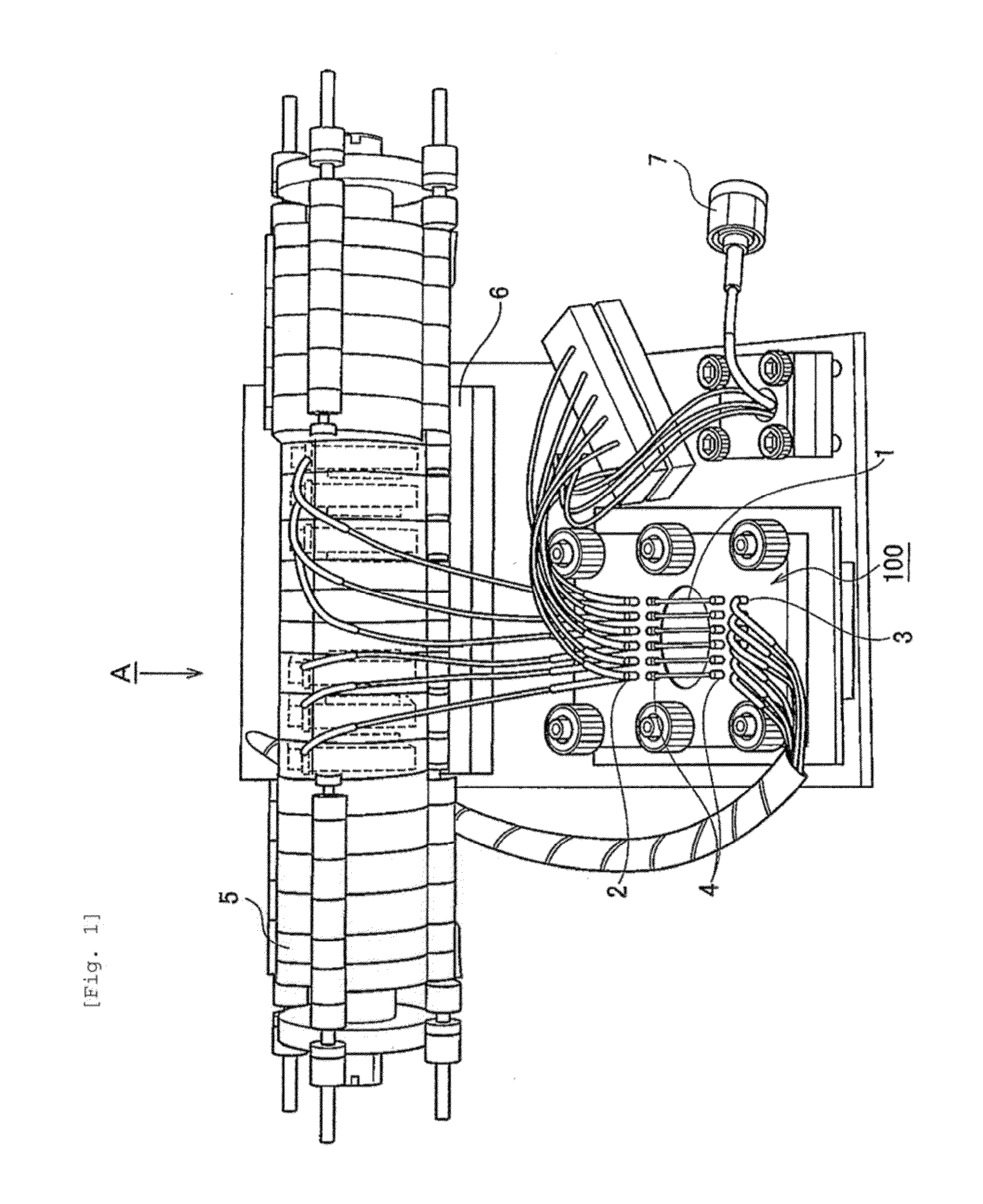
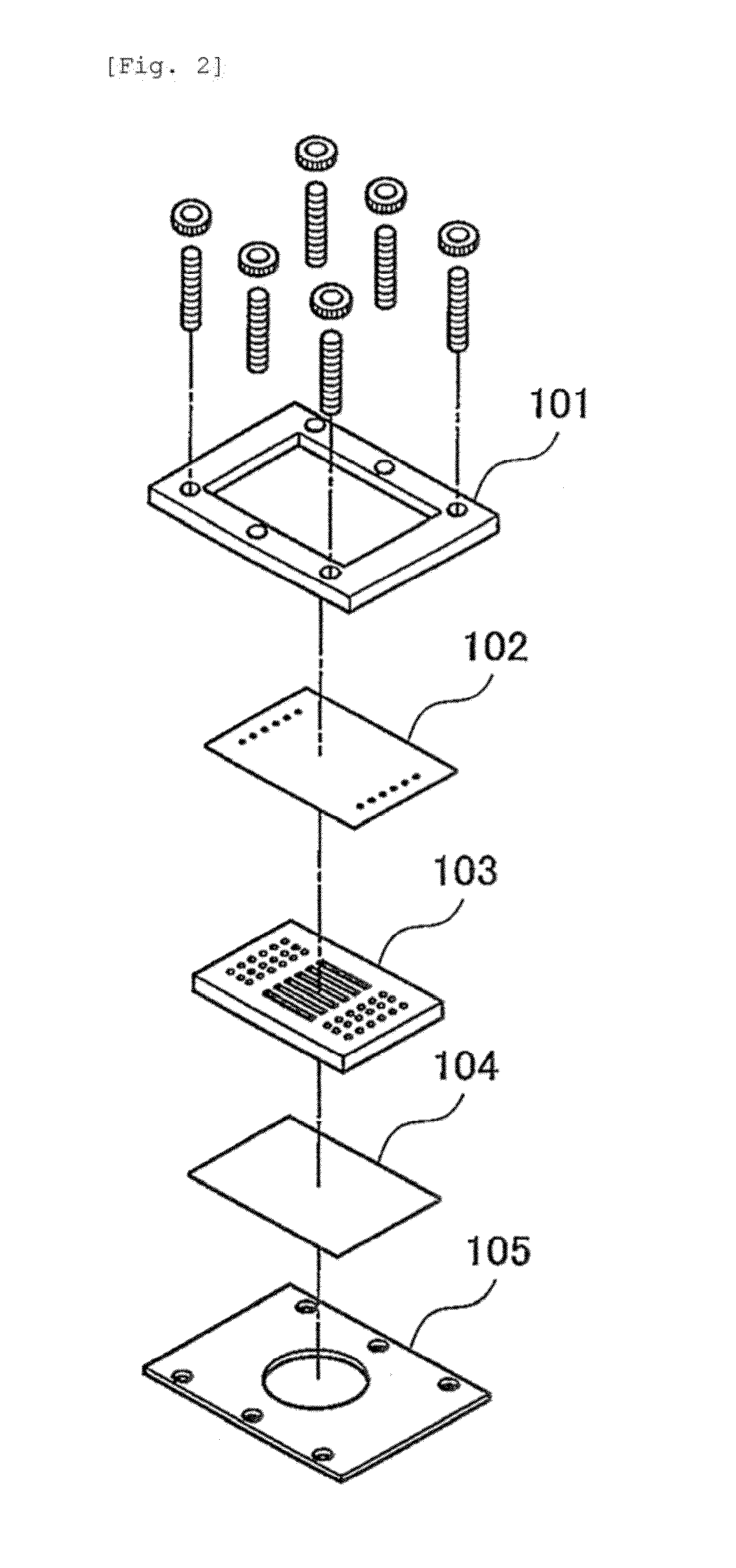
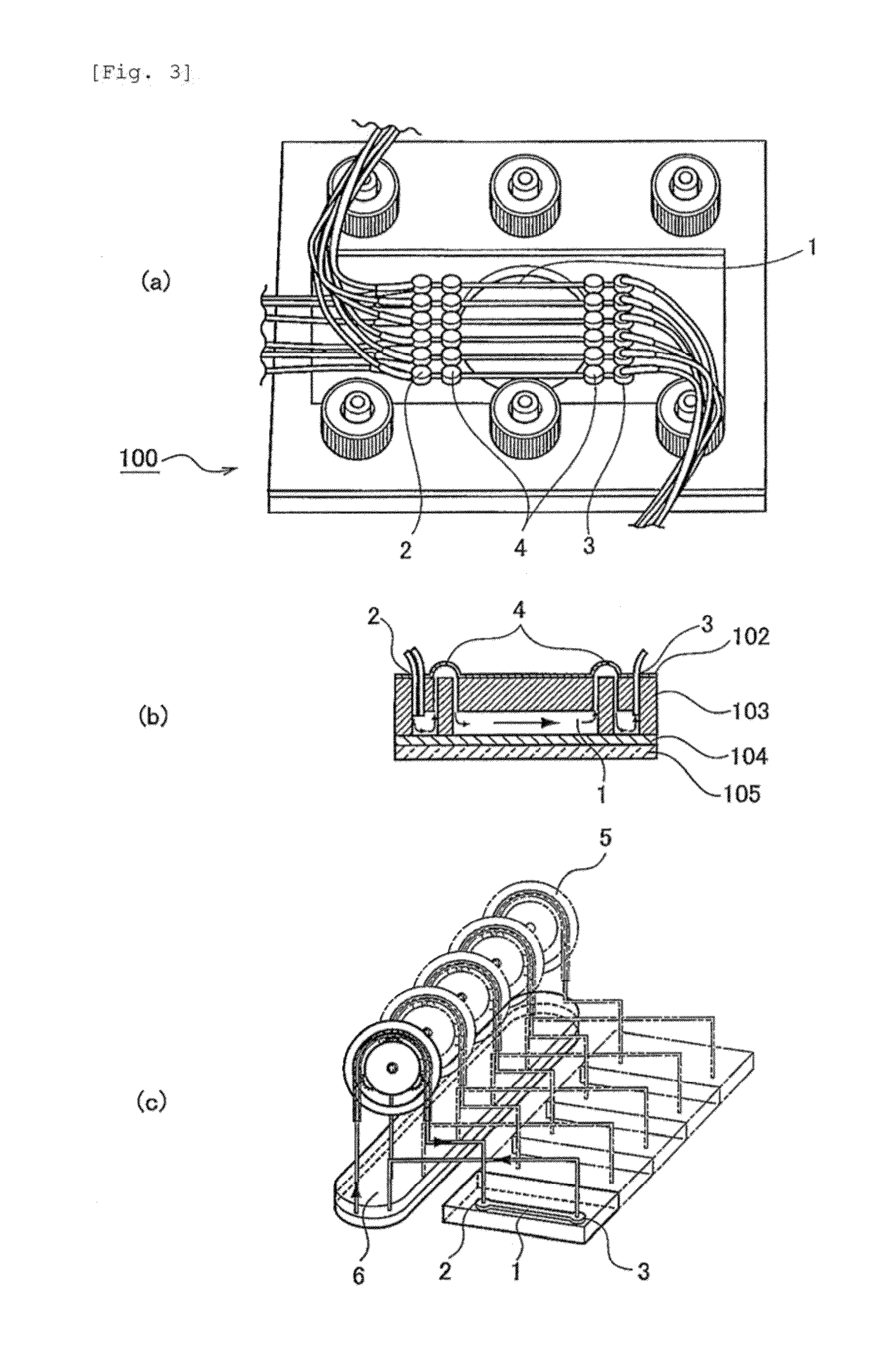
[0056]A polyethylene (PE) membrane 102, a polymethylsiloxane (PDMS) layer 103 and an SU8 layer 104, which is a glass plate to which SUS was applied, were sandwiched, between clamps 101 and 105, to construct a microfluidic chip 100 (FIG. 2). More specifically, the PDMS layer was prepared by pouring, to a thickness of 3 mm, a thermosetting PDMS (Slipot 184, Toray-Daw Corning, Japan) into a mold prepared by SU8 lithography for formation of 6 grooves (width, 0.5 mm; length, 20 mm; height, 0.5 mm), followed by curing at 80° C. for 4 hours in a drier. By removing the cured PDMS from the mold, a PDMS layer with 6 channels formed thereon was obtained. At both ends of the channels in the PDMS layer, holes were made as inlets and outlets using a stainless steel biopsy needle having a diameter of 1 mm. The SU8 layer was prepared by applying SU8 (SU-8 2010, Microchem) to a glass plate and curing the SU8 by UV irradiation. A cross mark was given by SU8 ...

Example

Example 2

Culture of iPS Cells Using Microfluidic Device, and Evaluation Thereof

[0058]OCT3 / 4, SOX2, KLF4 and c-MYC were introduced to human embryonic lung fibroblasts (TIG3) provided by the JCRB Cell Bank using a retrovirus, to induce an hiPSCs line. On a Matrigel-coated dish, TIG3-iPSC was cultured in an MEF (mouse embryonic fibroblast)-conditioned hES medium (DMEM / F12 supplemented with 20% knockout serum replacement (Invitrogen, Carlsbad, Calif., USA), L-glutamine, non-essential amino acids, 2-mercaptoethanol and 10 ng / ml bFGF (Peprotech, Rocky Hill, N.J., USA)).

[0059]The recovered cells were fixed using 4% PFA (paraformaldehyde) / PBS (phosphate buffered, saline) at room temperature for 10 minutes, and washed in PBST (PBS supplemented with 0.1% Triton X-100), followed by performing pretreatment at 4° C. overnight in a blocking solution (PBST supplemented with 3% BSA and 2% skim milk (DIFCO, USA)). Subsequently, immunoreaction was performed with primary antibody: anti-OCT4 (1:50, San...

Example

Example 3

Evaluation of Flow Field Based on Simulation and Hydromechanics

[0063]By confocal microscope analysis, hiPSCs on the channel were confirmed to have a volume of 550 μm3, diameter of 24.5 μm and height of 2.2 μm (FIG. 5a). The confocal microscope used had the following constitution: microscope, IX71 (Olympus); confocal scan unit, CSU-X1 (Yokogawa Electric Corporation); and objective lens, 100 UPlanSAPO NA1.4 (Olympus).

[0064]Using numeric codes obtained by the finite volume method (FVM) and the immersed boundary method, simulation based on numerical fluid dynamics was carried out. In terms of FVM, the continuity equation and Navier-Stokes equation were used after discretization. These were solved for the time-dependent, incompressible and three-dimensional conditions as described in Equations (1) and (2).

[Formula1]∂Ui∂xi=0(i=1,2,3)(1)ρfDUiDt=-∂P∂xi+∂∂xj(μ∂Ui∂xj)(2)

[0065]As the fluid and fin, properties of water and stainless steel were used, respectively. For discretization of ...


PUM


Abstract

The one aspect of the present invention aims to provide a novel cloning method for human embryonic stem cells (hESCs) and human induced pluripotent stem cells (hiPSCs), which method is based on culture of dispersed single cells without addition of an apoptotic agent. More specifically, the one aspect of the present invention aims to provide a cloning method for hESCs and hiPSCs based on culture of dispersed single cells utilizing shear stress. The above problem is solved by providing a method for culturing dispersed single pluripotent cells, wherein the dispersed single cells are cultured under laminar flow conditions.

Description

TECHNICAL FIELD[0001]The one aspect of the present invention relates to a method for culturing dispersed single pluripotent cells. More specifically, the one aspect of the present invention relates to a method for culturing dispersed single human embryonic stem cells (hESCs) or human induced pluripotent stem cells (hiPSCs).BACKGROUND ART[0002]hESCs (Non-patent Document 1) and hiPSCs (Non-patent Document 2) are cells whose use in regenerative medicine in the near future is expected. In cases where these cells are dispersed into single cells by destruction of cell-cell adhesion under normal maintenance culture conditions, myosin hyperactivation occurs, and this leads to formation of blebs on the cells, followed by occurrence of cell death through apoptosis. This caspase-dependent cell death is suppressed by suppression of Rho and the activity of Rac by signaling from E-cadherin-mediated cell-cell adhesion (Non-patent Document 3). Such extracellular signaling via cell-cell interaction ...

Claims


Application Information

Patent Timeline
IPC IPC(8): C12N5/0735C12N5/074
CPCC12N5/0606C12N2521/00C12N5/0696C12M23/16C12N2501/115C12N2501/602C12N2501/603C12N2501/604C12N2501/606C12N2502/13C12N2533/54
Inventor KOTERA, HIDETOSHITATSUMI, KAZUYAYOKOKAWA, RYUJITADA, TAKASHINAGATA, SHOGOOKITSU, TERUOKONOGI, ATSUHITONODA, YUICHIRONAKANISHI, NAOYUKIMATSUMURA, TAKUOSUMI, TAKASHI
Owner ARKRAY INC
Why Eureka
Try Eureka